



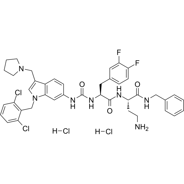
RWJ-56110 dihydrochloride

Based on 1 Customer Validation
RWJ-56110 dihydrochloride is a potent, selective, peptide-mimetic inhibitor of PAR-1 activation and internalization (binding IC50=0.44 uM) and shows no effect on PAR-2, PAR-3, or PAR-4. RWJ-56110 dihydrochloride inhibits the aggregation of human platelets induced by both SFLLRN-NH2 (IC50=0.16 μM) and thrombin (IC50=0.34 μM), quite selective relative to U46619 (HY-108566). RWJ-56110 dihydrochloride blocks angiogenesis and blocks the formation of new vessels in vivo. RWJ-56110 dihydrochloride induces cell apoptosis.
For research use only. We do not sell to patients.
- Purity : 99.87%
- CAS No.: 2387505-58-8
- Formula: C41H45Cl4F2N7O3
- Molecular Weight:863.65
-
Storage:
4°C, sealed storage, away from moisture and light
* In solvent : -80°C, 6 months; -20°C, 1 month (sealed storage, away from moisture and light)

To place orders, for customer services and technical support, please contact: MedChemExpress USA
Tel: 609-228-6898 E-mail: [email protected] [email protected]


-
 Biological Activity
-
Chemical Information
-
Solvent & Solubility
- Protocols
- Purity & Documentation
- References
-
 Help & FAQs


-
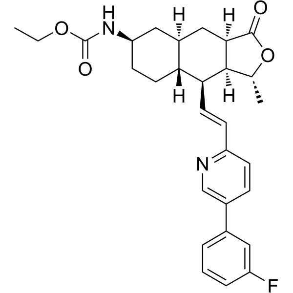
HY-10119
VorapaxarPAR-1 Antagonist -
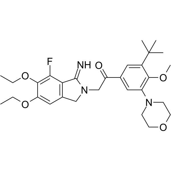
HY-18200
AtopaxarPAR-1 Antagonist -
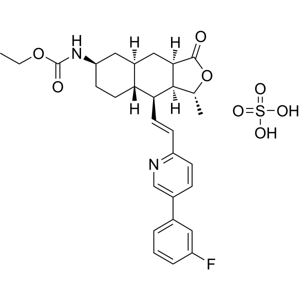
HY-10119A
Vorapaxar sulfatePAR-1 Antagonist -
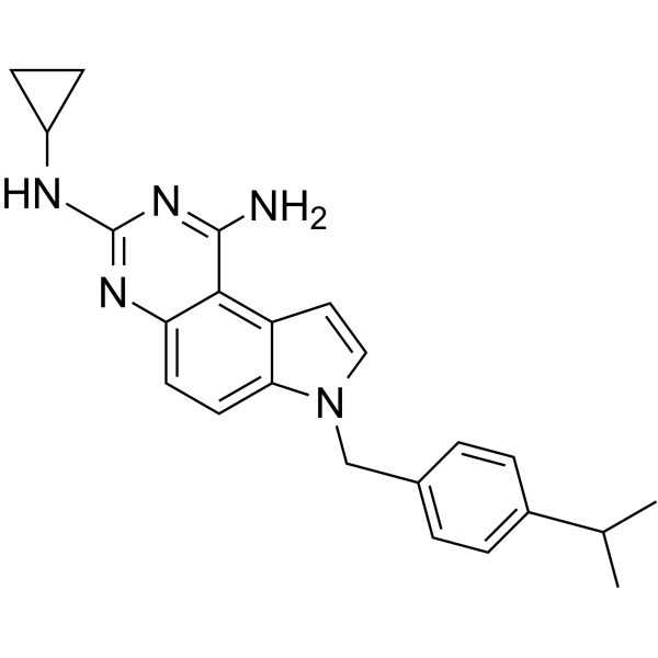
HY-14993
SCH79797PAR1 Antagonist -
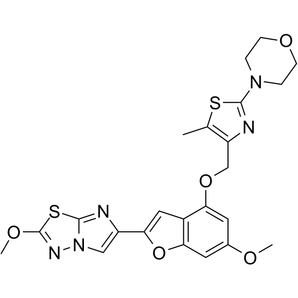
HY-19837
BMS-986120PAR4 Antagonist -

HY-10119R
Vorapaxar (Standard)PAR-1 Antagonist -
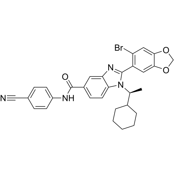
HY-112558
AZ3451PAR2 Antagonist -

HY-10119AR
Vorapaxar sulfate (Standard)PAR-1 Antagonist -

HY-108556AR
RWJ-56110 dihydrochloride (Standard)Reference Standard -
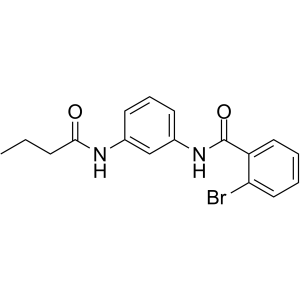
HY-13965
Parmodulin 2PAR1 Inhibitor -
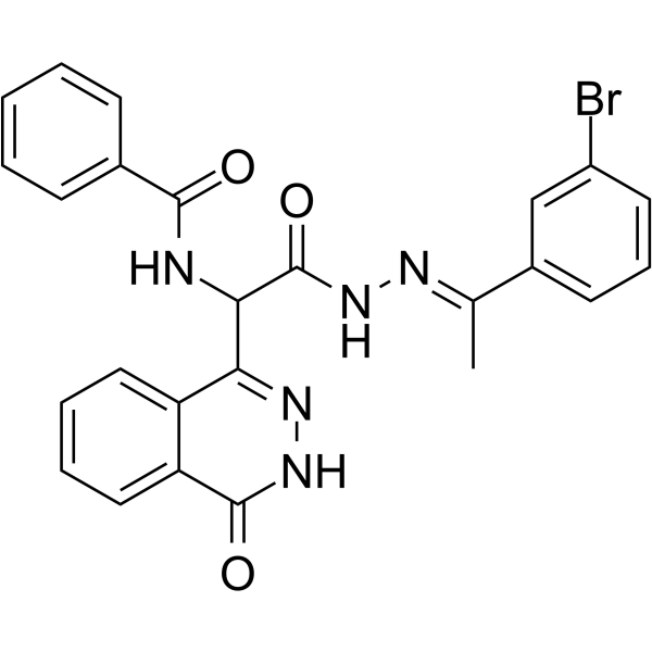
HY-14350
AC-55541PAR Agonist -
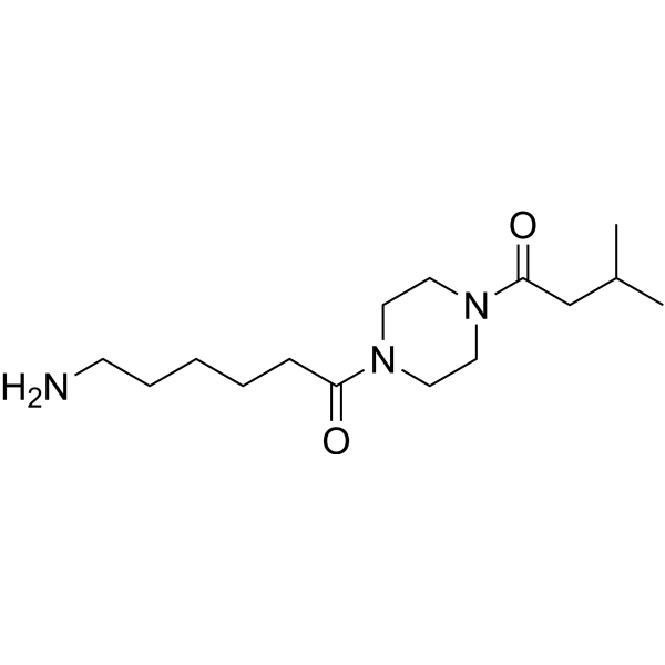
HY-124748
ENMD-1068PAR2 Antagonist


Biological Activity
Description
IC50 & Target
|
PAR1 .4 μM (IC50) |
In Vitro
Proteinase-activated receptors (PARs) are a family of G protein-coupled receptors activated by the proteolytic cleavage of their N-terminal extracellular domain, exposing a new amino terminal sequence that functions as a tethered ligand to activate the receptors. RWJ56110 inhibits the aggregation of human platelets induced by both SFLLRN-NH2 (IC50=0.16 μM) and thrombin (IC50=0.34 μM) while being quite selective relative to collagen and the thromboxane mimetic U46619 (HY-108566)[1].RWJ-56110 dihydrochloride is fully inhibits thrombin-induced RASMC proliferation with an IC50 value of 3.5 μM. RWJ-56110 dihydrochloride shows blockade of thrombin’s action with RASMC calcium mobilization (IC50=0.12 μM), as well as with HMVEC (IC50=0.13 μM) and HASMC calcium mobilization (IC50=0.17 μM)[1].RWJ56110 (0.1-10 μM; 24-96 hours) inhibits endothelial cell growth dose-dependently, with half-maximal inhibitory concentration of RWJ56110 is approximately 10 μM[2].RWJ56110 (0.1-10 μM; 6 hours) inhibits DNA synthesis of endothelial cells in a thymidine incorporation assays. Endothelial cells are in fast-growing state (50-60% confluence), RWJ56110 inhibits cell DNA synthesis in a dose-dependent manner, but when cells that are in the quiescent state (100% confluent), the inhibitory effect of PAR-1 antagonists is much less pronounced[2].RWJ56110 (0.1-10 μM; pretreatment for 15 min) inhibits thrombin-induced Erk1/2 activation in a concentration-dependent manner. However, when endothelial cells are stimulated by FBS (final concentration 4%), it reduces partially the activated levels of Erk1/2[2].RWJ56110 (30 μM; 24 hours) has an inhibitory effect on endothelial cell cycle progression. It reduces the percentage of cells in the S phase, while alterations in the percentages of G1 and G2/M cells are less pronounced[2].
MedChemExpress (MCE) has not independently confirmed the accuracy of these methods. They are for reference only. Further protocols information, click here.
-
Cell Line:Endothelial cells
-
Concentration:0 μM; 3 μM; 1 μM; 3 μM; 10 μM
-
Incubation Time:Pretreatment for 15 min
-
Result:Resulted in MAPK activation in Endothelial cells.
-
Cell Line:Endothelial cells
-
Concentration:0 μM; 3 μM; 1 μM; 3 μM; 10 μM
-
Incubation Time:Pretreatment for 15 min
-
Result:Reduced cell number in S phase.


Chemical Information
-
CAS No. 2387505-58-8
-
Appearance Solid
-
Molecular Weight 863.65
-
Formula C41H45Cl4F2N7O3
-
Color White to light yellow
-
SMILES
O=C(N[C@H](C(NCC1=CC=CC=C1)=O)CCN)[C@@H](NC(NC2=CC3=C(C=C2)C(CN4CCCC4)=CN3CC5=C(Cl)C=CC=C5Cl)=O)CC6=CC=C(F)C(F)=C6.[H]Cl.[H]Cl
-
Shipping
Room temperature in continental US; may vary elsewhere.
-
Storage
4°C, sealed storage, away from moisture and light
* In solvent : -80°C, 6 months; -20°C, 1 month (sealed storage, away from moisture and light)


Solvent & Solubility
In Vitro:
DMSO : 200 mg/mL (231.58 mM; Need ultrasonic; Hygroscopic DMSO has a significant impact on the solubility of product, please use newly opened DMSO)
Please refer to the solubility information to select the appropriate solvent. Once prepared, please aliquot and store the solution to prevent product inactivation from repeated freeze-thaw cycles.
Storage method and period of stock solution: -80°C, 6 months; -20°C, 1 month (sealed storage, away from moisture and light). When stored at -80°C, please use it within 6 months. When stored at -20°C, please use it within 1 month.
Please refer to the solubility information to select the appropriate solvent. Once prepared, please aliquot and store the solution to prevent product inactivation from repeated freeze-thaw cycles.
Storage method and period of stock solution: -80°C, 6 months; -20°C, 1 month (sealed storage, away from moisture and light). When stored at -80°C, please use it within 6 months. When stored at -20°C, please use it within 1 month.
Concentration (start) × Volume (start) = Concentration (final) × Volume (final)
In Vivo:
Select the appropriate dissolution method based on your experimental animal and administration route.
- For the following dissolution methods, please ensure to first prepare a clear stock solution using an In Vitro approach and then sequentially add co-solvents:
- To ensure reliable experimental results, the clarified stock solution can be appropriately stored based on storage conditions. As for the working solution for In Vivo experiments, it is recommended to prepare freshly and use it on the same day.
- The percentages shown for the solvents indicate their volumetric ratio in the final prepared solution. If precipitation or phase separation occurs during preparation, heat and/or sonication can be used to aid dissolution.
Add each solvent one by one: 10% DMSO 40% PEG300 5% Tween-80 45% Saline
Solubility: ≥ 5 mg/mL (5.79 mM); Clear solution
This protocol yields a clear solution of ≥ 5 mg/mL (saturation unknown).
Taking 1 mL working solution as an example, add 100 μL DMSO stock solution (50.0 mg/mL) to 400 μL PEG300, and mix evenly; then add 50 μL Tween-80 and mix evenly; then add 450 μL Saline to adjust the volume to 1 mL.
Preparation of Saline: Dissolve 0.9 g sodium chloride in ddH₂O and dilute to 100 mL to obtain a clear Saline solution.
Add each solvent one by one: 10% DMSO 90% (20% SBE-β-CD in Saline)
Solubility: ≥ 5 mg/mL (5.79 mM); Clear solution
This protocol yields a clear solution of ≥ 5 mg/mL (saturation unknown).
Taking 1 mL working solution as an example, add 100 μL DMSO stock solution (50.0 mg/mL) to 900 μL 20% SBE-β-CD in Saline, and mix evenly.
Preparation of 20% SBE-β-CD in Saline (4°C, storage for one week): 2 g SBE-β-CD powder is dissolved in 10 mL Saline, completely dissolve until clear.
In Vivo Dissolution Calculator
Please enter the basic information of animal experiments:
-
-
-
-
Recommended: Prepare an additional quantity of animals to account for potential losses during experiments.
Please enter your animal formula composition:
-
%DMSO +
Recommended: Keep the proportion of DMSO in working solution below 2% if your animal is weak.
-
%+
-
+%Tween-80 + +
-
%Saline +
The co-solvents required include: DMSO, . All of co-solvents are available by MedChemExpress (MCE). , Tween 80. All of co-solvents are available by MedChemExpress (MCE).
Working solution concentration: 0.22 mg/mL
Method for preparing stock solution: mg drug dissolved in μL DMSO. Stock solution concentration: mg/mL. * In solvent : -80°C, 6 months; -20°C, 1 month (sealed storage, away from moisture and light)
1. Take μL DMSO stock solution;
2. Add μL .
μL , mix evenly;
3. Then add μL Tween 80, mix evenly;
4. Then add μL
Please ensure that the stock solution in the first step is dissolved to a clear state, and add co-solvents in sequence. You can use ultrasonic heating (ultrasonic cleaner, recommended frequency 20-40 kHz), vortexing, etc. to assist dissolution.

Protocols
-
Human pluripotent stem cell endothelial-cell differentiation
Human pluripotent stem cell endothelial differentiation is based on stepwise developmental patterning: early activation of WNT/GSK3β inhibition promotes mesodermal or vascular progenitor entry, followed by endothelial specification using VEGF-related signaling, BMP4, FGF2, Notch modulation, or cAMP depending on the published protocol. Endothelial differentiation is read out by acquisition of CD31, CD34, VE-cadherin/CD144, KDR/VEGFR2, vWF, Tie2, NOS3, acetylated LDL uptake, tube/network formation, barrier function, and in vivo vessel-forming capacity where tested.
-
Apoptosis
Apoptosis, also called programmed cell death, is generally characterized by distinct morphological characteristics.
-
TUNEL staining for apoptotic DNA fragmentation
TUNEL staining detects DNA strand breaks by using terminal deoxynucleotidyl transferase to add labeled nucleotides to exposed 3′-OH DNA termini, generating either microscopic staining in fixed cells or tissue sections, or fluorescence/cytometric signal in cell suspensions. TUNEL positivity reflects DNA fragmentation but should not be interpreted alone as definitive apoptosis, because TUNEL can also label necrotic, autolytic, mechanically damaged, or DNA-repair-associated DNA breaks.
-
Cell Cytotoxicity Assay
Cytotoxicity assays are usually based on the assessment of cell membrane damage, which can also be indirectly detected by measuring cell viability. Detection methods include MTT assay, CKK-8 assay, LDH assay and ATP assay, etc.
-
Vascular/Branching Fractal Analysis
Vascular/branching fractal analysis quantifies the geometric complexity of vessel trees or vascular networks from segmented 2D images, commonly by converting vessels into binary and/or skeletonized maps and estimating fractal dimension using box-counting or related approaches. Fractal dimension is interpreted as an image-derived readout of vascular branching complexity, space filling, or density, and has been applied to retinal photographs, fluorescein angiography, OCT angiography, capillary perfusion maps, and in vitro Matrigel angiogenesis networks. The assay readout is generated from vessel-positive pixels after image preprocessing, vessel segmentation, binarization, and optional skeletonization; reported outputs include fractal dimension, vessel density, branchpoint density, endpoint density, vessel length density, tortuosity, and generation-based branching metrics when VESGEN-style analysis is used. The biological interpretation is limited to quantitative vascular patterning and s
-
Annexin V plus membrane-impermeant dye apoptosis staining
Annexin V-based apoptosis assays rely on the detection of phosphatidylserine (PS) externalization from the inner leaflet of the plasma membrane to the outer leaflet, an early biochemical hallmark of apoptosis. Fluorescently labeled Annexin V binds PS in a calcium-dependent manner, enabling identification of early apoptotic cells by flow cytometry or fluorescence microscopy. When combined with a membrane-impermeant DNA-binding dye (e. g. , propidium iodide), this approach allows discrimination between viable (Annexin V−/dye−), early apoptotic (Annexin V+/dye−), and late apoptotic or necrotic (Annexin V+/dye+) cell populations by assessing membrane integrity and PS exposure.
-
Endothelial Tube Formation Assay
Endothelial tube formation assay evaluates the ability of endothelial cells to attach, migrate, align, and organize into capillary-like networks when cultured on gelled basement membrane extract or Matrigel; the readout is the morphology and quantity of tube-like networks, which reflects an in vitro endothelial morphogenesis step related to angiogenesis. Basement membrane extract/Matrigel provides laminin-rich extracellular matrix cues that support endothelial differentiation into capillary-like structures, but it can contain biologically active growth factors, so growth-factor-reduced matrix is preferred when testing defined angiogenic stimulators or inhibitors.
-
Apoptosis Solutions
Apoptosis is a regulated, generally non-lytic cell-death pathway that removes unwanted, damaged, infected, or abnormal cells through coordinated morphological changes, caspase activation, DNA fragmentation, and membrane remodeling. The intrinsic apoptosis pathway is controlled mainly by mitochondrial outer membrane permeabilization, BCL-2 family proteins, cytochrome c release, apoptosome formation, caspase-9 activation, and downstream executioner caspase-3/7 activation. The extrinsic apoptosis pathway is initiated by death receptors such as Fas, TNFR, and TRAIL receptors, which recruit adaptor proteins and activate caspase-8 before engaging executioner caspases or mitochondrial amplification through BID cleavage. Apoptosis is linked to many phenotypes, including cancer cell killing, tissue homeostasis, immune regulation, neurodegeneration, infection response, and treatment-induced cytotoxicity; unresolved questions include how apoptosis interacts with necroptosis, pyroptosis, ferroptos
-
Endothelial Cell Migration/Angiogenic Sprouting Assay
Endothelial cell migration and angiogenic sprouting assays are in vitro (and partially ex vivo-adapted) functional models that quantify the ability of endothelial cells to undergo coordinated migration, extracellular matrix invasion, and multicellular organization into capillary-like sprouts in response to pro-angiogenic stimuli such as VEGF, bFGF, or conditioned microenvironments. These assays are used to model early angiogenic events including tip-cell formation, directional migration, and lumen-like sprout extension, which collectively reflect angiogenic activation and vascular morphogenesis processes observed in vivo.


Purity & Documentation
-
Data Sheet (278 KB)
-
SDS (252 KB)
- English - EN (252 KB)
- Français - FR (252 KB)
- Deutsch - DE (252 KB)
- Norwegian - NO (252 KB)
- Español - ES (252 KB)
- Swedish - SV (252 KB)
- Italian - IT (252 KB)
- Korean - KR (252 KB)
- Portuguese - PT (252 KB)
-
Handling Instructions (2659 KB)
References
[1]. Andrade-Gordon, et al.Design, synthesis, and biological characterization of a peptide-mimetic antagonist for a tethered-ligand receptor. oc Natl Acad Sci U S A. 1999 Oct 26;96(22):12257-62. [Content Brief]
[2]. Panagiota Zania, et al. Blockade of angiogenesis by small molecule antagonists to protease-activated receptor-1: association with endothelial cell growth suppression and induction of apoptosis. J Pharmacol Exp Ther. 2006 Jul;318(1):246-54. [Content Brief]


Complete Stock Solution Preparation Table
Please refer to the solubility information to select the appropriate solvent. Once prepared, please aliquot and store the solution to prevent product inactivation from repeated freeze-thaw cycles.
Storage method and period of stock solution: -80°C, 6 months; -20°C, 1 month (sealed storage, away from moisture and light). When stored at -80°C, please use it within 6 months. When stored at -20°C, please use it within 1 month.
| Optional Solvent | Concentration Solvent Mass | 1 mg | 5 mg | 10 mg | 25 mg |
|---|---|---|---|---|---|
| DMSO | 1 mM | 1.1579 mL | 5.7894 mL | 11.5788 mL | 28.9469 mL |
| 5 mM | 0.2316 mL | 1.1579 mL | 2.3158 mL | 5.7894 mL | |
| 10 mM | 0.1158 mL | 0.5789 mL | 1.1579 mL | 2.8947 mL | |
| 15 mM | 0.0772 mL | 0.3860 mL | 0.7719 mL | 1.9298 mL | |
| 20 mM | 0.0579 mL | 0.2895 mL | 0.5789 mL | 1.4473 mL | |
| 25 mM | 0.0463 mL | 0.2316 mL | 0.4632 mL | 1.1579 mL | |
| 30 mM | 0.0386 mL | 0.1930 mL | 0.3860 mL | 0.9649 mL | |
| 40 mM | 0.0289 mL | 0.1447 mL | 0.2895 mL | 0.7237 mL | |
| 50 mM | 0.0232 mL | 0.1158 mL | 0.2316 mL | 0.5789 mL | |
| 60 mM | 0.0193 mL | 0.0965 mL | 0.1930 mL | 0.4824 mL | |
| 80 mM | 0.0145 mL | 0.0724 mL | 0.1447 mL | 0.3618 mL | |
| 100 mM | 0.0116 mL | 0.0579 mL | 0.1158 mL | 0.2895 mL |


RWJ-56110 dihydrochloride Related Classifications
HY-108556A Related Classifications
 Powered by Bioz