



SB-267268

Based on 1 Customer Validation
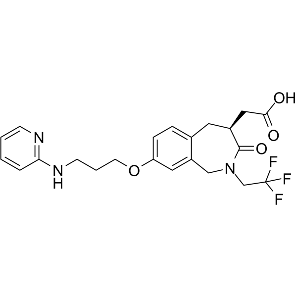
SB-267268 is a selective and nonpeptidic alpha(v)beta3 (αvβ3) and alpha(v)beta5 (αvβ5) integrins antagonist, with Kis of 0.9, 0.5 and 0.7 nM for human αvβ3, monkey αvβ3 and human αvβ5, respectively. SB-267268 inhibits human and mouse αvβ3 with IC50s of 0.68 and 0.29 nM, respectively. SB-267268 reduces angiogenesis and VEGF expression.
For research use only. We do not sell to patients.
- Purity : 99.39%
- CAS No.: 205678-26-8
- Formula: C22H24F3N3O4
- Molecular Weight:451.44
-
Storage:Powder -20°C, 3 years , 4°C, 2 years ; In solvent -80°C, 6 months , -20°C, 1 month

To place orders, for customer services and technical support, please contact: MedChemExpress USA
Tel: 609-228-6898 E-mail: [email protected] [email protected]


-
 Biological Activity
-
Chemical Information
-
Solvent & Solubility
- Protocols
- Purity & Documentation
- References
-
 Help & FAQs




Biological Activity
Description
Cellular Effect
|
Cell Line

|
Type | Value | Description | References |
|---|---|---|---|---|
| HEK293 | IC50 |
12 nM
Compound: 5
|
HEK cell adhesion mediated by alpha v beta 3 integrin
HEK cell adhesion mediated by alpha v beta 3 integrin
|
[PMID: 10633035] |
In Vitro
SB-267268 was much less potent for inhibition of human, mouse, and ratαvβ6 integrin. SB-267268 inhibits the attachment of bothαvβ3-transfected HEK293 cells to microtiter plate wells precoated with arginine-glycine-aspartic acid (RGD)-containing matrix proteins with IC50 values of 12 nM. SB-267268 also inhibits vitronectin-mediated human and rat aortic smooth-muscle-cell (SMC) migration with IC50 values of approximately 12.3 nM and 3.6 nM, respectively[1].
MedChemExpress (MCE) has not independently confirmed the accuracy of these methods. They are for reference only.
In Vivo
In ROP mice treated with SB-267268, VEGF and VEGFR-2 gene expression in the inner nuclear layer (INL) and the ganglion cell layer (GCL) is reduced[1].
MedChemExpress (MCE) has not independently confirmed the accuracy of these methods. They are for reference only.
-
Animal Model:Pregnant female C57BL/6 mice (ROP mice)[1]
-
Dosage:60 mg/kg
-
Administration:I.p.; bi-daily
-
Result:Reduced blood vessel profiles (BVPs) in the inner retina by 50%.


Chemical Information
-
CAS No. 205678-26-8
-
Appearance Solid
-
Molecular Weight 451.44
-
Formula C22H24F3N3O4
-
Color White to off-white
-
SMILES
O=C(O)C[C@H]1C(N(CC(F)(F)F)CC2=CC(OCCCNC3=NC=CC=C3)=CC=C2C1)=O
-
Shipping
Room temperature in continental US; may vary elsewhere.
-
Storage
Powder -20°C 3 years 4°C 2 years In solvent -80°C 6 months -20°C 1 month


Solvent & Solubility
In Vitro:
DMSO : 200 mg/mL (443.03 mM; Need ultrasonic; Hygroscopic DMSO has a significant impact on the solubility of product, please use newly opened DMSO)
Please refer to the solubility information to select the appropriate solvent. Once prepared, please aliquot and store the solution to prevent product inactivation from repeated freeze-thaw cycles.
Storage method and period of stock solution: -80°C, 6 months; -20°C, 1 month. When stored at -80°C, please use it within 6 months. When stored at -20°C, please use it within 1 month.
Please refer to the solubility information to select the appropriate solvent. Once prepared, please aliquot and store the solution to prevent product inactivation from repeated freeze-thaw cycles.
Storage method and period of stock solution: -80°C, 6 months; -20°C, 1 month. When stored at -80°C, please use it within 6 months. When stored at -20°C, please use it within 1 month.
Concentration (start) × Volume (start) = Concentration (final) × Volume (final)


Purity & Documentation
-
Data Sheet (273 KB)
-
SDS (251 KB)
- English - EN (251 KB)
- Français - FR (251 KB)
- Deutsch - DE (251 KB)
- Norwegian - NO (251 KB)
- Español - ES (251 KB)
- Swedish - SV (251 KB)
- Italian - IT (251 KB)
- Korean - KR (251 KB)
- Portuguese - PT (251 KB)
-
Handling Instructions (2659 KB)
References


Complete Stock Solution Preparation Table
Please refer to the solubility information to select the appropriate solvent. Once prepared, please aliquot and store the solution to prevent product inactivation from repeated freeze-thaw cycles.
Storage method and period of stock solution: -80°C, 6 months; -20°C, 1 month. When stored at -80°C, please use it within 6 months. When stored at -20°C, please use it within 1 month.
| Optional Solvent | Concentration Solvent Mass | 1 mg | 5 mg | 10 mg | 25 mg |
|---|---|---|---|---|---|
| DMSO | 1 mM | 2.2151 mL | 11.0757 mL | 22.1513 mL | 55.3783 mL |
| 5 mM | 0.4430 mL | 2.2151 mL | 4.4303 mL | 11.0757 mL | |
| 10 mM | 0.2215 mL | 1.1076 mL | 2.2151 mL | 5.5378 mL | |
| 15 mM | 0.1477 mL | 0.7384 mL | 1.4768 mL | 3.6919 mL | |
| 20 mM | 0.1108 mL | 0.5538 mL | 1.1076 mL | 2.7689 mL | |
| 25 mM | 0.0886 mL | 0.4430 mL | 0.8861 mL | 2.2151 mL | |
| 30 mM | 0.0738 mL | 0.3692 mL | 0.7384 mL | 1.8459 mL | |
| 40 mM | 0.0554 mL | 0.2769 mL | 0.5538 mL | 1.3845 mL | |
| 50 mM | 0.0443 mL | 0.2215 mL | 0.4430 mL | 1.1076 mL | |
| 60 mM | 0.0369 mL | 0.1846 mL | 0.3692 mL | 0.9230 mL | |
| 80 mM | 0.0277 mL | 0.1384 mL | 0.2769 mL | 0.6922 mL | |
| 100 mM | 0.0222 mL | 0.1108 mL | 0.2215 mL | 0.5538 mL |


SB-267268 Related Classifications
HY-19306 Related Classifications
 Powered by Bioz