



SB-590885 GMP is SB-590885 (HY-10966) produced by using GMP guidelines. GMP small molecules works appropriately as an auxiliary reagent for cell therapy manufacture. SB-590885 is a BRAF/c-Raf kinase inhibitor that selectively targets B-Raf, and it amplifies the ERK/MAPK signaling pathway in RAS-activated cells. SB-590885 effectively inhibits the malignant proliferation, transformation and tumorigenicity of oncogenic B-Raf cells; it also induces the proliferation of erythroid progenitor cells, delays their differentiation and promotes hemoglobin synthesis, thereby improving ineffective erythropoiesis and reducing apoptosis. SB-590885 exerts a synergistic effect with TGF-β inhibitors and glucocorticoids, significantly promoting the formation of erythroid colonies in cells from patients with Diamond-Blackfan anemia (DBA). SB-590885 is mainly used in relevant studies on DBA, cisplatin-induced myelosuppression-related anemia, and pan-cancers such as melanoma and colorectal cancer.
Nur für Forschungszwecke. Wir verkaufen nicht an Patienten.
- CAS. Nr.: 405554-55-4
- Formel: C27H27N5O2
- Molecular Weight:453.54
-
Speicherung:
Please store the product under the recommended conditions in the Certificate of Analysis.


To place orders, for customer services and technical support, please contact: MedChemExpress USA
Tel: 609-228-6898 E-mail: [email protected] [email protected]


-
 Biologische Aktivität
-
Chemical Information
- Protokoll
- Reinheit & Dokumentation
- Verweise
-
 Help & FAQs


-
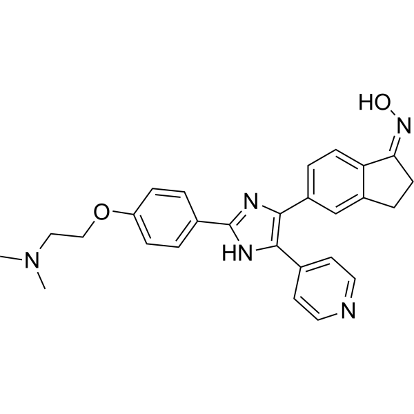
HY-10966
SB-590885BRAF/c-Raf Inhibitor -
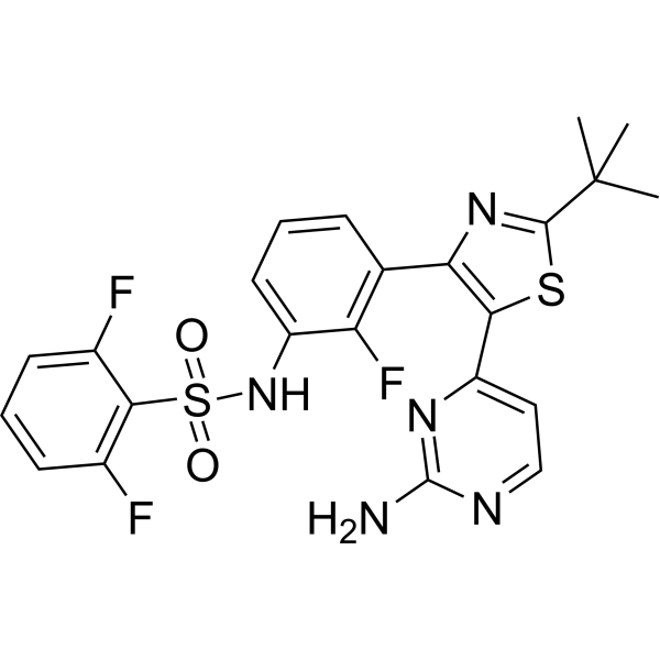
HY-14660
DabrafenibBRAF Inhibitor -

HY-10966R
SB-590885 (Standard)Reference Standard -
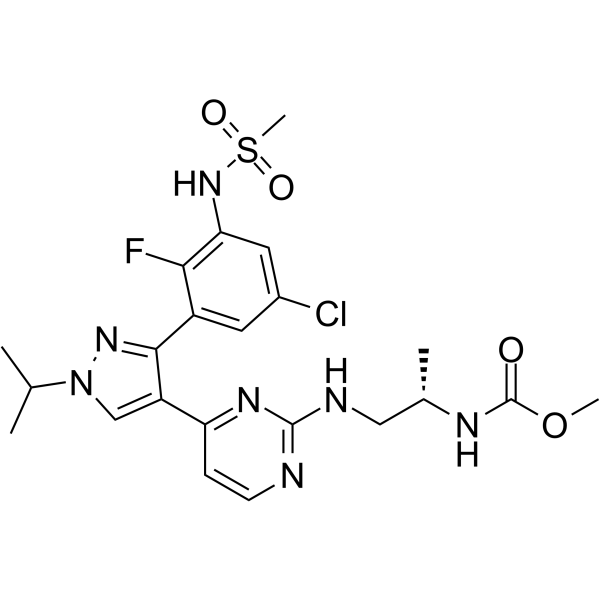
HY-15605
EncorafenibBRAF Inhibitor -
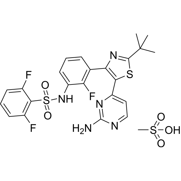
HY-14660A
Dabrafenib MesylateRaf Inhibitor -
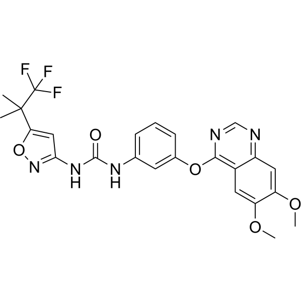
HY-15200
AgerafenibBRAF Inhibitor -
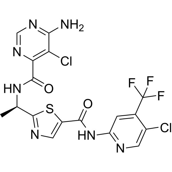
HY-15246
TovorafenibRaf Kinase Inhibitor -
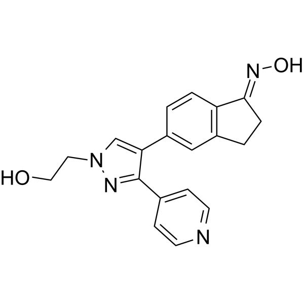
HY-50864
GDC-0879B-Raf Inhibitor -
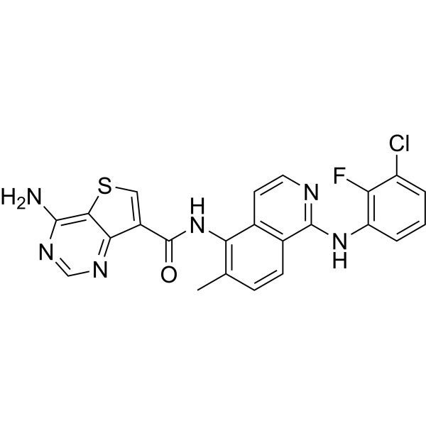
HY-109080
BelvarafenibRAF Inhibitor -
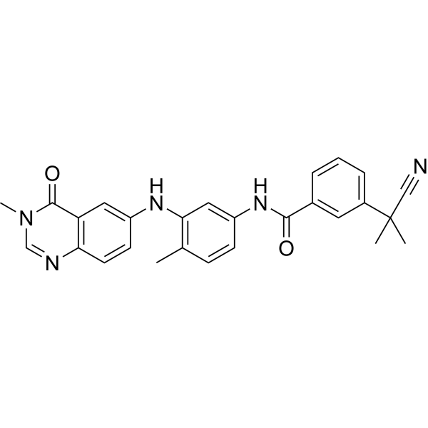
HY-11004
AZ 628Apoptosis Inducer -
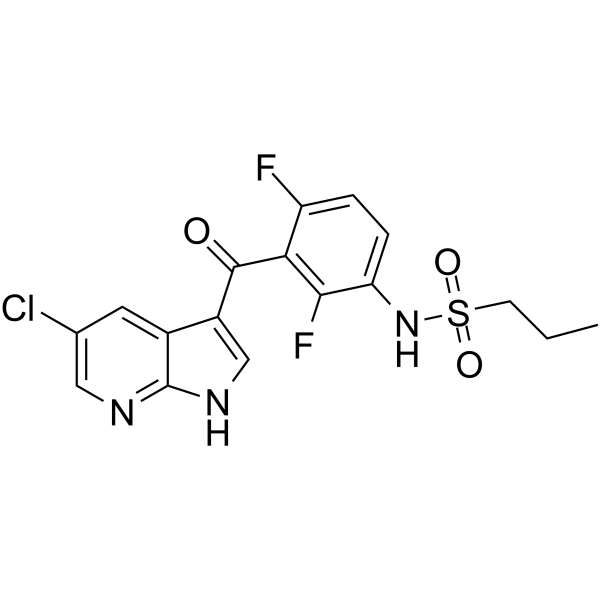
HY-51424
PLX-4720B-Raf Inhibitor -
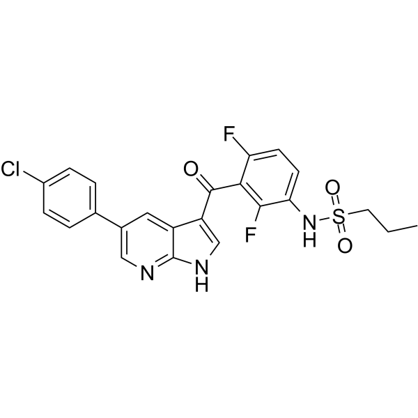
HY-12057
VemurafenibB-Raf Inhibitor


Biologische Aktivität
Beschreibung
IC50 & Target
|
Braf |
In Vitro
SB-590885 GMP (1 μM; 12 d) increases the cell number by more than 10-fold compared with the control group in erythroid differentiation culture of UCB-CD34+ hematopoietic stem/progenitor cells, while transiently delaying the erythroid differentiation process[1].
SB-590885 GMP (1 μM; 14 d) significantly increases the area of BFU-E erythroid colonies and the total number of erythroid cells, without affecting the proportion of colonies of other lineages. It also alleviates ineffective erythropoiesis caused by cytokine deprivation, reduces apoptosis, and promotes cell proliferation[1].
MedChemExpress (MCE) has not independently confirmed the accuracy of these methods. They are for reference only. Further protocols information, click here.
-
Cell Line:Primary human umbilical cord blood CD34+ hematopoietic stem and progenitor cells (UCB-CD34+ HSPCs); Primary human peripheral blood mononuclear cells (PBMCs) from healthy donors
-
Concentration:1 μM
-
Incubation Time:14 days
-
Result:After 7 days of treatment in UCB-CD34+ HSPCs erythroid differentiation culture, it significantly promoted the proliferation of erythroid progenitor cells and enhanced cell viability.
After 9 days of treatment in healthy donor PBMCs erythroid differentiation culture, it markedly promoted erythroid proliferation.
After 12 days of treatment in UCB-CD34+ HSPCs culture, it increased the cell number by more than 10-fold compared with the control group, and temporarily delayed erythroid differentiation.
After 14 days of treatment in UCB-CD34+ HSPCs culture, it achieved 281516-fold cell expansion, while the control group only achieved 16573-fold expansion; and also alleviated ineffective erythropoiesis and promoted cell proliferation under cytokine-restricted conditions.


Chemical Information
-
CAS. Nr. 405554-55-4
-
Molecular Weight 453.54
-
Formel C27H27N5O2
-
SMILES
O/N=C1CCC2=CC(C3=C(NC(C4=CC=C(C=C4)OCCN(C)C)=N3)C5=CC=NC=C5)=CC=C/12
-
Versand
Room temperature in continental US; may vary elsewhere.
-
Speicherung
Please store the product under the recommended conditions in the Certificate of Analysis.

Protokoll
-
Carcinogenicity Bioassay
A carcinogenicity bioassay detects whether long-term exposure to a test substance increases benign or malignant tumor incidence, changes tumor spectrum, or shortens tumor latency in experimental animals; the classical rodent design exposes rats and/or mice to multiple dose levels for most of their lifespan, followed by complete necropsy and histopathologic diagnosis of neoplastic and non-neoplastic lesions. The readout is tumor incidence by organ, sex, species, dose group, and survival status; interpretation requires concurrent controls, dose-response assessment, survival-adjusted tumor statistics, and pathology review because mortality, spontaneous tumor background, and body-weight effects can influence apparent tumor rates.
-
Detection of Apoptosis
Apoptosis, also called programmed cell death, is generally characterized by distinct morphological characteristics.
-
TUNEL staining for apoptotic DNA fragmentation
TUNEL staining detects DNA strand breaks by using terminal deoxynucleotidyl transferase to add labeled nucleotides to exposed 3′-OH DNA termini, generating either microscopic staining in fixed cells or tissue sections, or fluorescence/cytometric signal in cell suspensions. TUNEL positivity reflects DNA fragmentation but should not be interpreted alone as definitive apoptosis, because TUNEL can also label necrotic, autolytic, mechanically damaged, or DNA-repair-associated DNA breaks.
-
Annexin V plus membrane-impermeant dye apoptosis staining
Annexin V-based apoptosis assays rely on the detection of phosphatidylserine (PS) externalization from the inner leaflet of the plasma membrane to the outer leaflet, an early biochemical hallmark of apoptosis. Fluorescently labeled Annexin V binds PS in a calcium-dependent manner, enabling identification of early apoptotic cells by flow cytometry or fluorescence microscopy. When combined with a membrane-impermeant DNA-binding dye (e. g. , propidium iodide), this approach allows discrimination between viable (Annexin V−/dye−), early apoptotic (Annexin V+/dye−), and late apoptotic or necrotic (Annexin V+/dye+) cell populations by assessing membrane integrity and PS exposure.
-
Apoptosis Solutions
Apoptosis is a regulated, generally non-lytic cell-death pathway that removes unwanted, damaged, infected, or abnormal cells through coordinated morphological changes, caspase activation, DNA fragmentation, and membrane remodeling. The intrinsic apoptosis pathway is controlled mainly by mitochondrial outer membrane permeabilization, BCL-2 family proteins, cytochrome c release, apoptosome formation, caspase-9 activation, and downstream executioner caspase-3/7 activation. The extrinsic apoptosis pathway is initiated by death receptors such as Fas, TNFR, and TRAIL receptors, which recruit adaptor proteins and activate caspase-8 before engaging executioner caspases or mitochondrial amplification through BID cleavage. Apoptosis is linked to many phenotypes, including cancer cell killing, tissue homeostasis, immune regulation, neurodegeneration, infection response, and treatment-induced cytotoxicity; unresolved questions include how apoptosis interacts with necroptosis, pyroptosis, ferroptos
-
Soft Agar Colony Formation Assay
Soft agar colony formation assay measures anchorage-independent growth, in which transformed or tumorigenic cells proliferate as colonies in a semisolid agar matrix while many non-transformed adherent cells fail to proliferate without attachment; classic studies showed that growth in semisolid medium correlates with tumorigenicity in nude mice, and later protocol papers describe the method as a stringent in vitro assay for malignant transformation. The readout is the number, size, morphology, or signal intensity of colonies formed within agar after incubation; published formats include manual colony counting after staining, 96-well or 384-well quantitative formats, DNA-binding dye detection, MTT/tetrazolium-based detection, digital image analysis, and PCR-based marker detection from soft agar cultures.
-
Cell Viability Determination by MTT Colorimetric Assay
The following protocol uses the MTT colorimetric assay as a classic literature-established method for assessing cell viability/metabolic activity in cultured mammalian cells. MTT[3-(4,5-dimethylthiazol-2-yl)-2,5-diphenyltetrazolium bromide] is reduced by metabolically active cells to a colored formazan product; the amount of formazan is quantified spectrophotometrically and provides an indirect measure of metabolically active viable cells. Importantly, MTT reduction reflects cellular oxidoreductase/metabolic activity rather than an absolute direct count of living cells, so changes in cellular metabolism can alter the signal independently of cell number.


Reinheit & Dokumentation
Verweise
[1]. Wu S, et al. BRAF inhibitors enhance erythropoiesis and treat anemia through paradoxical activation of MAPK signaling. Signal Transduct Target Ther. 2024;9(1):338. Published 2024 Dec 2. [Content Brief]
[2]. King AJ, et al. Demonstration of a genetic therapeutic index for tumors expressing oncogenic BRAF by the kinase inhibitor SB-590885. Cancer Res. 2006;66(23):11100-11105. [Content Brief]
[3]. Chen Z, et al. Pan-Cancer Analysis of the TRP Family, Especially TRPV4 and TRPC4, and Its Expression Correlated with Prognosis, Tumor Microenvironment, and Treatment Sensitivity. Biomolecules. 2023;13(2):282. Published 2023 Feb 2. [Content Brief]
[4]. Ruan D, et al. Establishment of human expanded potential stem cell lines via preimplantation embryo cultivation and somatic cell reprogramming. Nat Protoc. 2025;20(10):2698-2734. [Content Brief]

Calculators
Konzentration (Stammlösung) × Volumen (Stammlösung) = Konzentration (Ziellösung) × Volumen (Ziellösung)


SB-590885 (GMP) Related Classifications
HY-10966G Related Classifications
 Powered by Bioz