



Scyllo-Inositol

Based on 1 Customer Validation
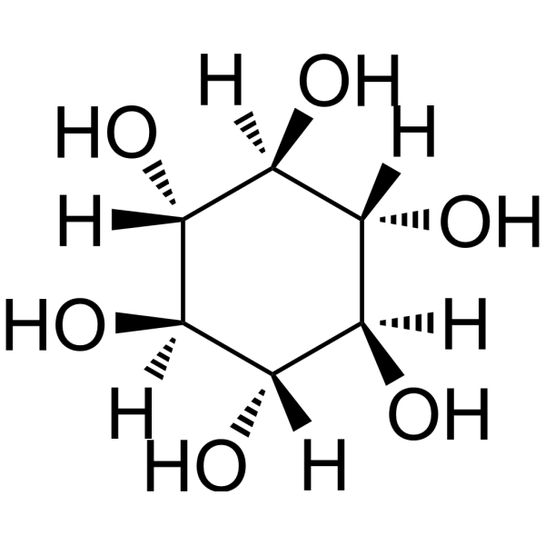
Scyllo-Inositol is an inhibitor that targets the aggregation of misfolded proteins (such as α-synuclein and Amyloid-β), is orally effective, and can cross the blood-brain barrier. Scyllo-Inositol can selectively bind to and stabilize non-toxic oligomers, preventing them from converting into toxic fibers, exerting protein homeostasis regulation and neuroprotective activity. Scyllo-Inositol binds to the hydrophobic region of pathogenic proteins, inhibits protein aggregation, and promotes lysosome- and proteasome-mediated degradation pathways, thereby reducing neurotoxicity. Scyllo-Inositol can be used in the study of neurodegenerative diseases such as Parkinson's disease, Alzheimer's disease, and Huntington's disease.
For research use only. We do not sell to patients.
- Purity : 99.99%
- CAS No.: 488-59-5
- Formula: C6H12O6
- Molecular Weight:180.16
-
Storage:
4°C, protect from light
* In solvent : -80°C, 6 months; -20°C, 1 month (protect from light)

To place orders, for customer services and technical support, please contact: MedChemExpress USA
Tel: 609-228-6898 E-mail: [email protected] [email protected]


-
 Biological Activity
-
Chemical Information
-
Solvent & Solubility
- Protocols
- Purity & Documentation
- References
-
 Help & FAQs


-
HY-113409
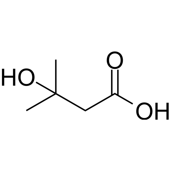
3-Hydroxyisovaleric acidEndogenous Metabolite -

HY-W010041R
Scyllo-Inositol (Standard)α-synuclein/Amyloid-β Aggregation Inhibitor -
HY-103240
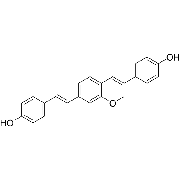
Methoxy-X04Fluorescent Dye -
HY-D0218
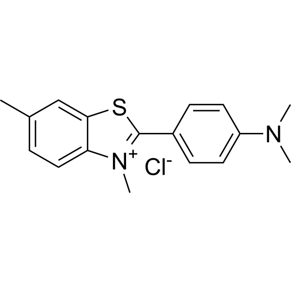
Thioflavin Tβ-amyloid Dye -
HY-19370
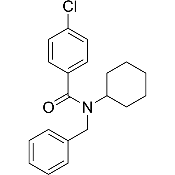
FPS-ZM1Amyloid-β Inhibitor -
HY-N0615
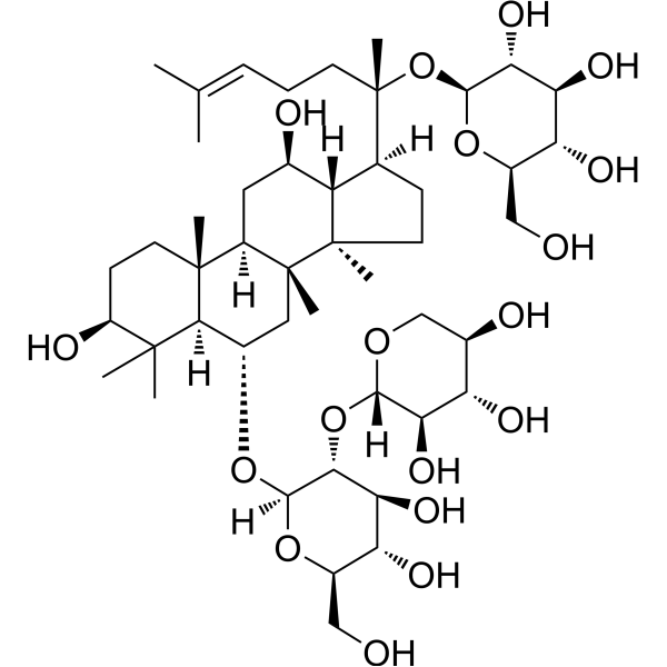
Notoginsenoside R1Apoptosis Inhibitor -
HY-50682
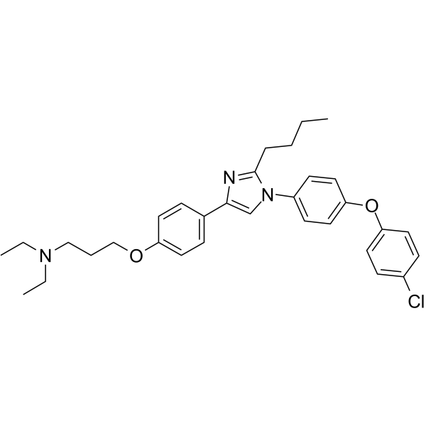
AzeliragonRAGE Inhibitor -
HY-N0373
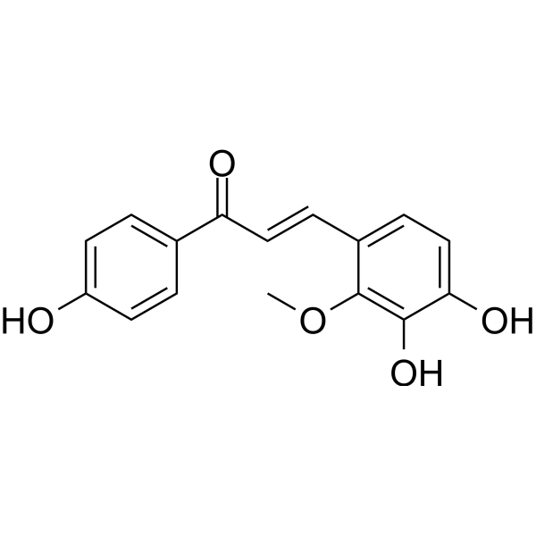
Licochalcone BApoptosis Inducer -
HY-14602
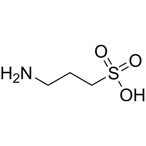
TramiprosateNeuroprotection Agent -
HY-N0249
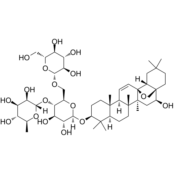
Saikosaponin CMMP-2 Inducer -
HY-103242
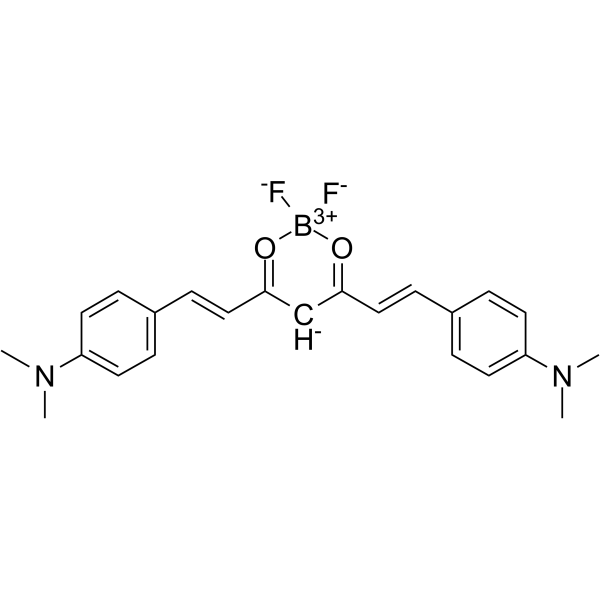
CRANAD-2Fluorescent Probe -
HY-N0602
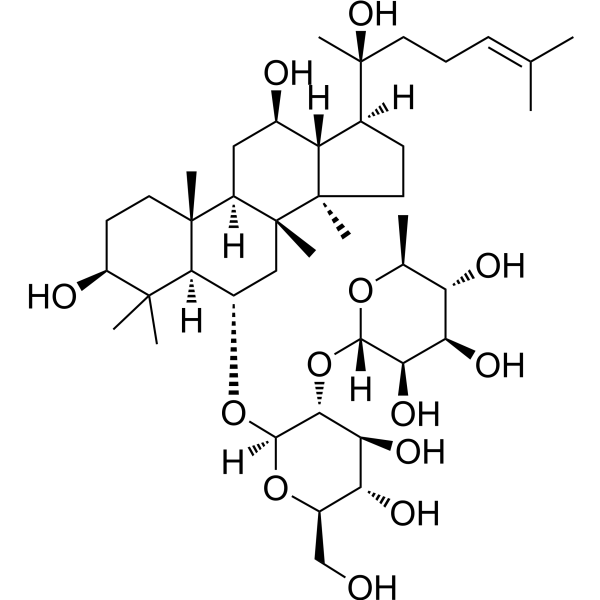
Ginsenoside Rg2NF-κB Inhibitor
All α-synuclein Isoforms
More

Biological Activity
Description
IC50 & Target
|
Human Endogenous Metabolite |
In Vitro
Scyllo-Inositol (1:1 (w/v%) mixture; 1-21 d) inhibits fibril formation of human and mouse α-synuclein and was more potent than chiro-inositol (HY-N3021)[1].
Scyllo-Inositol (1-100 μM; 24 h) dose-dependently reduces polyQ-Htt protein levels and misfolded protein aggregates in PC12 cells. Scyllo-Inositol also activates the proteasome and lysosomal degradation pathways without affecting the levels of autophagy markers (LC3, p62)[2].
MedChemExpress (MCE) has not independently confirmed the accuracy of these methods. They are for reference only. Further protocols information, click here.
-
Cell Line:PC12 cells overexpressing polyQ-Htt
-
Concentration:1, 10, 25, 50, 100 μM
-
Incubation Time:24 h
-
Result:Dose-dependently reduced polyQ-Htt protein levels and misfolded protein aggregates.
Activated proteasome and lysosomal degradation pathways, restored cathepsin B activity and reduced ubiquitinated protein accumulation.
Did not affect the levels of autophagy markers (LC3, p62).
In Vivo
MedChemExpress (MCE) has not independently confirmed the accuracy of these methods. They are for reference only.
Clinical Trial
| NCT Number | Sponsor | Condition | Start Date |
Phase

|
|---|---|---|---|---|
| NCT01329991 | Plexxikon| | 2011-05 | PHASE1 |


Chemical Information
-
CAS No. 488-59-5
-
Appearance Solid
-
Molecular Weight 180.16
-
Formula C6H12O6
-
Color White to off-white
-
SMILES
O[C@H]1[C@@H]([C@H]([C@@H]([C@H]([C@@H]1O)O)O)O)O
-
Structure Classification
-
Initial Source
-
Shipping
Room temperature in continental US; may vary elsewhere.
-
Storage
4°C, protect from light
* In solvent : -80°C, 6 months; -20°C, 1 month (protect from light)


Solvent & Solubility
In Vitro:
H2O : 10 mg/mL (55.51 mM; Need ultrasonic)
Please refer to the solubility information to select the appropriate solvent. Once prepared, please aliquot and store the solution to prevent product inactivation from repeated freeze-thaw cycles.
Storage method and period of stock solution: -80°C, 6 months; -20°C, 1 month (protect from light). When stored at -80°C, please use it within 6 months. When stored at -20°C, please use it within 1 month.
* Note: If you choose water as the stock solution, please dilute it to the working solution, then filter and sterilize it with a 0.22 μm filter before use.
Please refer to the solubility information to select the appropriate solvent. Once prepared, please aliquot and store the solution to prevent product inactivation from repeated freeze-thaw cycles.
Storage method and period of stock solution: -80°C, 6 months; -20°C, 1 month (protect from light). When stored at -80°C, please use it within 6 months. When stored at -20°C, please use it within 1 month.
* Note: If you choose water as the stock solution, please dilute it to the working solution, then filter and sterilize it with a 0.22 μm filter before use.
Concentration (start) × Volume (start) = Concentration (final) × Volume (final)

Protocols
-
Neurotoxicity Study
This protocol assesses in vitro neurotoxicity by combining neuronal viability, mitochondrial/metabolic activity, neurite outgrowth, and optional neuronal network function readouts. Calcein-AM or resazurin/PrestoBlue readouts estimate viable or metabolically active cells; βIII-tubulin immunofluorescence detects neuronal morphology and neurite networks; TMRE detects mitochondrial membrane potential; and MEA recordings detect functional changes in neuronal network activity.
-
Human pluripotent stem cell midbrain dopaminergic neuron differentiation
Human pluripotent stem cells are directed toward midbrain dopaminergic neurons by first inducing a neural floor-plate-like progenitor state, then patterning cells with ventralizing SHH signaling and midbrain/WNT-FGF cues, and finally maturing progenitors into neurons expressing dopaminergic markers such as TH, NURR1/NR4A2, PITX3, DAT/SLC6A3, VMAT2/SLC18A2, GIRK2/KCNJ6, FOXA2, LMX1A, and EN1. The main readouts are loss of pluripotency, acquisition of FOXA2+/LMX1A+ midbrain floor-plate progenitors, emergence of βIII-tubulin+/MAP2+ neurons, and production of TH+ dopaminergic neurons with molecular, dopamine-release, and electrophysiological features of midbrain dopaminergic identity.
-
Lysosome and acidic-vesicle live-cell staining
Lysosome and acidic-vesicle live-cell staining detects acidic intracellular compartments by using membrane-permeant acidotropic probes that accumulate in low-pH vesicles, including lysosomes, late endosomes, autolysosomes, and acidic phagosomes. LysoTracker staining is commonly used as an intensity-based readout of acidic lysosomal compartment abundance or enlargement, while acridine orange produces green fluorescence in less concentrated compartments and red fluorescence after concentration-dependent accumulation in acidic vesicular organelles. Loss or reduction of acridine-orange red signal can be used as a readout of lysosomal membrane permeabilization or reduced acidic-vesicle integrity. This protocol is designed for live cultured cells and can be adapted for fluorescence microscopy, high-content imaging, plate-reader readout, or flow cytometry when the selected literature supports the readout. Because these dyes report acidotropic accumulation rather than lysosome identity alone,
-
Amyloid: Congo Red Amyloid Staining
Congo red amyloid staining is a histochemical method used to detect extracellular amyloid deposits in tissue sections based on the affinity of Congo red dye for β-pleated sheet-rich protein aggregates. When bound to amyloid, Congo red produces characteristic apple-green birefringence under polarized light microscopy, which is widely regarded as a diagnostic feature of amyloid deposition in histopathology. The diagnostic principle relies on the combination of dye binding (congophilia) and optical anisotropy under polarized illumination, which distinguishes amyloid from most non-amyloid eosinophilic extracellular deposits in routine histological evaluation. Amyloid identification by Congo red staining remains a cornerstone in diagnostic pathology despite the availability of adjunct methods such as immunohistochemistry and mass spectrometry, particularly because of its ability to localize deposits directly within tissue architecture. The specificity of Congo red-positive deposits is incre
-
Alzheimer’s Disease Modeling
Alzheimer’s Disease (AD) is a neurodegenerative disorder characterized by a progressive decline in cognitive functions and loss of specific types of neurons and synapses. Alzheimer's symptoms can be simulated in mice by injecting drugs (such as Aβ) or genetically modified.


Purity & Documentation
-
Data Sheet (282 KB)
-
SDS (393 KB)
- English - EN (393 KB)
- Français - FR (393 KB)
- Deutsch - DE (393 KB)
- Norwegian - NO (393 KB)
- Español - ES (393 KB)
- Swedish - SV (393 KB)
- Italian - IT (393 KB)
- Korean - KR (393 KB)
- Portuguese - PT (393 KB)
-
Handling Instructions (2659 KB)
References
[1]. Ibrahim T, et al. α-Synuclein aggregation, seeding and inhibition by scyllo-inositol. Biochem Biophys Res Commun. 2016 Jan 15;469(3):529-34. [Content Brief]
[2]. Lai AY, et al. scyllo-Inositol promotes robust mutant Huntingtin protein degradation. J Biol Chem. 2014 Feb 7;289(6):3666-76. [Content Brief]
[3]. Choi JK, et al. Detection of increased scyllo-inositol in brain with magnetic resonance spectroscopy after dietary supplementation in Alzheimer's disease mouse models. Neuropharmacology. 2010 Sep-Oct;59(4-5):353-7. [Content Brief]


Complete Stock Solution Preparation Table
Please refer to the solubility information to select the appropriate solvent. Once prepared, please aliquot and store the solution to prevent product inactivation from repeated freeze-thaw cycles.
Storage method and period of stock solution: -80°C, 6 months; -20°C, 1 month (protect from light). When stored at -80°C, please use it within 6 months. When stored at -20°C, please use it within 1 month.
| Optional Solvent | Concentration Solvent Mass | 1 mg | 5 mg | 10 mg | 25 mg |
|---|---|---|---|---|---|
| H2O | 1 mM | 5.5506 mL | 27.7531 mL | 55.5062 mL | 138.7655 mL |
| 5 mM | 1.1101 mL | 5.5506 mL | 11.1012 mL | 27.7531 mL | |
| 10 mM | 0.5551 mL | 2.7753 mL | 5.5506 mL | 13.8766 mL | |
| 15 mM | 0.3700 mL | 1.8502 mL | 3.7004 mL | 9.2510 mL | |
| 20 mM | 0.2775 mL | 1.3877 mL | 2.7753 mL | 6.9383 mL | |
| 25 mM | 0.2220 mL | 1.1101 mL | 2.2202 mL | 5.5506 mL | |
| 30 mM | 0.1850 mL | 0.9251 mL | 1.8502 mL | 4.6255 mL | |
| 40 mM | 0.1388 mL | 0.6938 mL | 1.3877 mL | 3.4691 mL | |
| 50 mM | 0.1110 mL | 0.5551 mL | 1.1101 mL | 2.7753 mL |
* Note: If you choose water as the stock solution, please dilute it to the working solution, then filter and sterilize it with a 0.22 μm filter before use.


Scyllo-Inositol Related Classifications
HY-W010041 Related Classifications
 Powered by Bioz