897 Results for "

cell-membrane

" in MedChemExpress (MCE) Product Catalog:
Products (897)

897 Results for "cell-membrane" in MCE Product Catalog:

Cat. No.: HY-D3127
CAS No.: 455251-97-5
Target:  

Fluorescent Dye

Research Areas:  

Others

DCA-Mln is a Fluorescent probe for ferric iron (Fe³⁺) detection. The probe works via an internal charge transfer (ICT) process from its diethylamino group to its dicyano group, which produces bright pink fluorescence in its unbound state; when Fe³⁺ is present, two molecules of DCA-Mln chelate with one Fe³⁺ ion, with one cyano group of each DCA-Mln participating in the complexation, and this binding triggers a photoinduced electron transfer (PET) process that provides a nonradiative deactivation pathway, resulting in fluorescence quenching; additionally, the probe exhibits a visible color change from purple to peach pink upon Fe³⁺ binding, enabling dual-channel detection. The probe has an excitation wavelength of 570 nm and an emission wavelength of 670 nm, and it shows a rapid response, with fluorescence stabilizing within 15 seconds of Fe³⁺ addition. DCA-Mln is cell-membrane-permeable, nontoxic at the imaging concentration of 10 μM, and can be used for Fe³⁺ detection in actual water samples, Fe³⁺ imaging in living cells, and as an anti-counterfeiting ink[1].
loading...
    loading...
Cat. No.: HY-P5107
CAS No.: 121052-30-0
Synonyms: LMWP; TDSP5
Target:  

VEGFR

Research Areas:  

Cancer

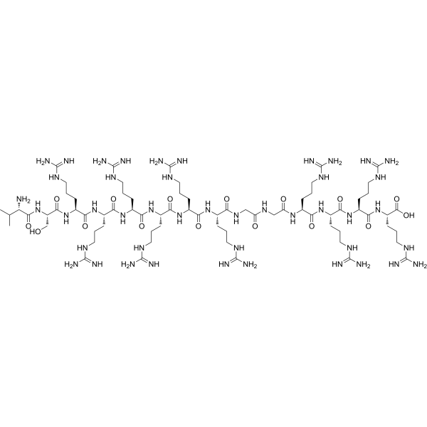
Low molecular weight protamine (LMWP;TDSP5) is a truncated arginine-rich protamine peptide, as well as a heparin/low-molecular-weight heparin antidote and a cell-penetrating delivery carrier. Low molecular weight protamine neutralizes heparin-induced anticoagulant activities, including aPTT, anti-Xa and anti-IIa activities, and also neutralizes anti-Xa activity of commercially available low-molecular-weight heparin preparations. Low molecular weight protamine translocates across mammalian cell membranes, delivers conjugated impermeable molecules across tumor tissues, enhances skin permeability of conjugated epidermal growth factor, and accelerates wound healing when conjugated with epidermal growth factor. Low molecular weight protamine retains the in vitro cell proliferation activity of conjugated EGF, and also enables site-specific conjugation with peptides or proteins via genetic recombination. Low molecular weight protamine can be used in studies related to colon adenocarcinoma, skin wounds and diabetic skin wounds .
loading...
    loading...
Cat. No.: HY-W783351
CAS No.: 1416808-87-1
Synonyms: Coppersensor 790 acetoxymethyl ester
Target:  

Fluorescent Dye

Research Areas:  

Metabolic Disease

CS790AM (Coppersensor 790 acetoxymethyl ester) is a cell-permeable, Cu +-targeted near-infrared fluorescent probe (λabs=760 nm, λem=790 nm) applicable to live cells. CS790AM can cross lipophilic cell membranes, and is converted into negatively charged CS790 under the action of intracellular esterases to be retained, thus enabling highly sensitive, reversible "turn-on" detection of labile Cu + pools in live cells and mice. CS790AM possesses excellent biocompatibility and selectivity, avoids interference from other metal ions, shows no obvious toxicity, and can be rapidly cleared. CS790AM allows long-term longitudinal monitoring of individual mice, visualizes copper levels in internal organs and isolated livers, and effectively evaluates abnormal copper accumulation in Wilson's disease models (Atp7b -/-) as well as dynamic changes after chelator treatment. CS790AM can be used for research on Wilson's disease and related copper metabolic disorders .
loading...
    loading...
Cat. No.: HY-P70482
Purity:  ≥ 95%, as determined by reducing SDS-PAGE.
Synonyms: HAVCR2; FLJ14428; Hepatitis A Virus cellular Receptor 2; HAVcr-2; TIMD3; TIMD-3; TIM3; T-cell Immunoglobulin And Mucin Domain 3; Tim-3; T cell Immunoglobulin Mucin 3; CD366; Kidney Injury Molecule-3; T-cell Immunoglobulin And Mucin Domain-Containing Protein 3; CD366 Antigen; T-cell Immunoglobulin Mucin Family Member 3; KIM-3; T-cell Immunoglobulin Mucin Receptor 3; SPTCL; T-cell membrane Protein 3; TIM-3
Species:  
Human
Source:  
HEK293
loading...
    loading...
Cat. No.: HY-15136R
CAS No.: 193275-84-2
Synonyms: Sch66336 (Standard)
Lonafarnib (Standard) (Sch66336 (Standard)) is the analytical standard of Lonafarnib (HY-15136). This product is intended for research and analytical applications. Lonafarnib (Sch66336) is an orally active, blood-brain barrier penetrant farnesyltransferase (FPTase) inhibitor. Lonafarnib increases the phosphorylation levels of Akt, CaMKII and CREB, upregulates BDNF expression in the hippocampus, elevates the content of α7nAChR on cell membranes, and blocks the isoprenylation of H-Ras. Lonafarnib repairs synapses and reverses spatial memory deficits in Aβ1-42 model mice. Lonafarnib inhibits RSV fusion and replication, and alleviates virus-induced lung injury. Lonafarnib activates the lysosomal and autophagic pathways, and reduces the phosphorylation and aggregation of tau. Lonafarnib acts synergistically with Sorafenib (HY-10201) to promote apoptosis, and inhibits the proliferation and invasion of melanoma cells. Lonafarnib inhibits the farnesylation of progerin, repairs nuclear membrane defects, and activates the cGAS-STING-STAT1 signaling pathway. Lonafarnib can be used in research related to diseases including Alzheimer's disease, viral infections, melanoma, and progeria .
loading...
    loading...
Cat. No.: HY-79602S
CAS No.: 1219795-34-2
Synonyms: p-Tosylamide-d4
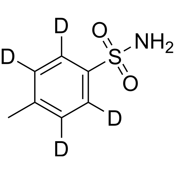
4-Tolyl-d4-sulfonamide (p-Tosylamide-d4) is the d4-labeled p-Toluenesulfonamide (HY-79602). p-Toluenesulfonamide is a small-molecule anticancer agent and plasticizer. p-Toluenesulfonamide exerts antitumor activity by inducing lysosomal membrane permeabilization, cathepsin B release and lysosome-mediated cell death. p-Toluenesulfonamide modulates cholesterol distribution in lipid rafts of tumor cell membranes and the Akt/mTOR/p70S6K pathway. p-Toluenesulfonamide shows activity against various cancers including hepatocellular carcinoma, non-small cell lung cancer and tongue squamous cell carcinoma; intrapleural injection effectively reduces malignant pleural effusion without causing pleural adhesion. p-Toluenesulfonamide is also the main degradation product of the disinfectant Chloramine-T (HY-B0959) in water. p-Toluenesulfonamide facilitates the localization of fluorescent probes to the endoplasmic reticulum. p-Toluenesulfonamide can be used in cancer-related research .
loading...
    loading...
Cat. No.: HY-D3438
Target:  

Fluorescent Dye

Research Areas:  

Others

Photoswitchable PM8 is a fluorescent probe used for super-resolution imaging of the plasma membrane. Photoswitchable PM8 functions through a light-induced Z/E isomerization mechanism: in the hydrophobic cell membrane environment, the Z isomer forms a planar intramolecular charge transfer state stabilized by an intramolecular hydrogen bond, which restricts conformational freedom, reduces nonradiative decay, and produces bright fluorescence; in contrast, the E isomer lacks this hydrogen bond, has increased conformational freedom, and exhibits a fluorescence-quenched state. The fluorescence of Photoswitchable PM8 can be switched "on/off" by visible light: activation with 405 nm light forms the fluorescent Z isomer, and switching with 561 nm light converts it to the nonfluorescent E isomer. In the membrane environment, Photoswitchable PM8 enables single-molecule localization for super-resolution imaging. Due to solvent-induced quenching and aggregation, Photoswitchable PM8 has negligible fluorescence in aqueous solution, but upon binding to the hydrophobic membrane environment it produces bright fluorescence, enabling high signal-to-noise ratio imaging without washing .
loading...
    loading...
Cat. No.: HY-134096
CAS No.: 78323-98-5
Synonyms: DNS-M
Target:  

Fluorescent Dye

Research Areas:  

Others

Dansyl-morpholine (DNS-M) is a Fluorescent probe for lipid droplet imaging, cancer cell discrimination, and real-time tracking of lipid droplet dynamics. As a solvatochromic probe with a donor-π-acceptor structure, it relies on hydrophobic interaction for its mechanism of action: its good lipophilicity, confirmed by an oil-water partition coefficient LogP = 2.35, allows it to rapidly penetrate cell membranes, and it specifically localizes to the hydrophobic core of lipid droplets; its fluorescence is strongly enhanced in the nonpolar environment of lipid droplets, while it emits very weak fluorescence in polar environments like PBS buffer, and it exhibits a bathochromic shift in emission wavelength with increasing solvent polarity. It has negligible cytotoxicity, with cell viability remaining over 95% after 24-hour incubation with 100 μM of the probe, and it possesses excellent photostability, retaining over 97% of initial fluorescence intensity after 60 continuous laser scans. For cell imaging applications, its excitation/emission wavelengths for lipid droplet labeling are Ex/Em = 405/480−540 nm, and in a simulative lipid environment O/W emulsion, it has an excitation wavelength of ~346 nm and emission wavelength of ~500 nm, giving a Stokes shift of 154 nm[1].
loading...
    loading...
Cat. No.: HY-D3128
CAS No.: 2348351-60-8
Target:  

Fluorescent Dye

Research Areas:  

Others

Mito-RhFe is a Fluorescent probe for mitochondrial labile Fe³⁺ monitoring via imaging and flow cytometry. This probe is a rhodamine-based construct with a spirolactam fluorescence signaling group and an N2-hydroxyethyldiethylenetriamine chelator; its delocalized positive charge enables mitochondria-targeting ability in live cells, and it exhibits fine cell membrane permeability. In its native state, it exists in the non-fluorescent spirolactam form, but upon binding to Fe³⁺, it undergoes a ring-opening conversion to the fluorescent rhodamine form, triggering a turn-on fluorescent response; this process is reversible, as the addition of the metal chelator TPEN removes Fe³⁺ and converts the probe back to its non-fluorescent spirolactam form, and re-addition of Fe³⁺ restores fluorescence. The probe shows high selectivity for Fe³⁺ over most other metal cations present in living systems, with a ~90-fold fluorescence enhancement upon binding to 20 equiv of Fe³⁺. Mito-RhFe has excitation/emission wavelengths of Ex/Em = 540/578 nm, with an ~8 nm bathochromic shift in emission upon Fe³⁺ binding, and it can also be excited at 543 nm for confocal imaging with emission detected at 570-620 nm[1].
loading...
    loading...
Cat. No.: HY-P11242
Cm-CATH2 is an antimicrobial peptide discovered from Chelonia mydas. Cm-CATH2 has a potent, broad-spectrum and rapid bactericidal ability by rapidly destroying the integrity of bacterial cell membranes. It shows strong activity against Gram-positive bacteria (such as VREF, Staphylococcus aureus), Gram-negative bacteria (such as Escherichia coli, Klebsiella pneumoniae), and fungi (such as Candida albicans) with MICs ranges from 1.17 to 18.75 μg/mL. Cm-CATH2 is also effective against various aquatic pathogenic bacteria. Cm-CATH2 not only inhibits biofilm formation but can also remove the formed biofilms. Cm-CATH2 has immunomodulatory functions and chemotactic effects on immune cells, and can inhibit the production of pro-inflammatory cytokines by macrophages stimulated by LPS (HY-D1056). Cm-CATH2 prevents the activation of NF-κB by inhibiting the degradation of IκBα, and also inhibits the phosphorylation of MAPK signaling pathways (p38, JNK, ERK). Cm-CATH2 demonstrates strong anti-infective ability in mouse peritonitis models and pneumonia models .
loading...
    loading...
Cat. No.: HY-L001P
32,925 compounds

Bioactive compounds are a general term for a class of substances that can cause certain biological effects in the body, which are the main source of small molecule drugs. These compounds generally penetrate cell membranes, act on specific target proteins in cells, regulate intracellular signaling pathways, and cause some changes in cell phenotype.

MCE owns a unique collection of 32,925 compounds with confirmed biological activities and clear targets. These compounds include natural products, innovative compounds, approved compounds, and clinical compounds. This library is a useful tool for signal pathway research, drug discovery and drug repurposing, etc.

Bioactive Compound Library Plus, with more powerful screening capability, further complements Bioactive Compound Library (HY-L001) by adding some compounds with low solubility or solution stability (Part B) and some novel, rare or exclusive compounds (Part C) to this library. Overall, bioactive compound library plus (HY-L001P) includes tree parts: Part A, Part B and Part C. Compounds in Part A are equal to the products in HY-L001, which can be supplied in solution or solid form. Compounds in Part B and C are only supplied in solid form.

Cat. No.: HY-P73609
Purity:  ≥ 90%, as determined by reducing SDS-PAGE.
Synonyms: HAVCR1; T-cell membrane Protein 1; Hepatitis A Virus cellular Receptor 1; TIMD-1; TIM-1; KIM-1; TIMD1; TIM; TIM1; T-cell Immunoglobulin And Mucin Domain-Containing Protein 1; KIM1; T cell Immunoglobin Domain And Mucin Domain Protein 1; Kidney Injury Molecule 1; Hepatitis A Virus cellular Receptor 1a; HAVCR-1; Hepatitis A Virus cellular Receptor 1b; HAVCR; Hepatitis A Virus cellular Receptor 1c; CD365; CD365 Antigen; T-cell Immunoglobulin Mucin Family Member 1; HAVcr-1; T-cell Immunoglobulin Mucin Receptor 1
Species:  
Canine
Source:  
HEK293
loading...
    loading...
Cat. No.: HY-L938
8350 compounds

Currently,the incidence and mortality rates of clinical fungal infections remain high. Existing antifungal drugs are limited in variety and associated with numerous adverse effects, creating an urgent demand for the development of novel antifungal agents. Antifungal compound libraries can support the screening and development of new antifungal drugs.

The mechanisms of action of antifungal drugs cover key processes such as fungal cell membrane synthesis, cell wall synthesis, and cell division. They exert fungicidal or fungistatic effects by specifically targeting different molecular pathways. This library includes a variety of core analogs of antifungal drugs, making it adaptable to antifungal research in diverse scenarios. It can be used for the high-throughput screening of novel antifungal drug candidates, enabling the rapid identification of compounds with potential antifungal activity and facilitating the elucidation of drug-target interactions and resistance mechanisms. Additionally, it supports the screening of compounds and combinations that reverse drug resistance, thereby uncovering the novel antifungal potential of existing compounds.

The library comprises 8350 compounds with a well-defined screening strategy. The core sources of the compounds include analogs of known antifungal active moleculeswith a similarity score of ≥ 0.6 MCE has collected more than 500 antifungal molecules.All screened compounds conform to lead-like physicochemical properties, exhibiting both structural diversity and drug-like characteristics, and providing valuable support for the research and development of novel antifungal drugs.

Cat. No.: HY-112624K
CAS No.: 9004-54-0
Synonyms: Dextran 5; Dextran D5; Dextran T5(MW 4500-5500)
Dextran T5 (MW 5,000) is a sulfated polysaccharide anti-apoptotic and autophagic agent. Dextran T5 (MW 5,000) has sulfated groups and interacts with cell membranes by mimicking endogenous glycosaminoglycans, inhibiting the mitochondrial apoptotic pathway and delaying DNA fragmentation to exert anti-apoptotic activity. Dextran T5 (MW 5,000) also promotes the conversion of LC3-I to LC3-II and the formation of autophagosomes to activate the autophagic pathway. Dextran T5 (MW 5,000) can prolong the survival cycle of CHO cells and increase the production of recombinant erythropoietin (EPO). The Dextran series of compounds are also natural polysaccharide drug carriers that can be connected to drugs through covalent bonding methods such as ester bonds, amide bonds or click chemistry, or self-assembled to form carriers such as nanoparticles and hydrogels. Dextran is biodegradable and biocompatible, and can achieve targeted delivery and controlled release of drugs. Dextran derivatives can prolong drug half-life, increase local concentration and reduce immune clearance activity. The Dextran series of compounds are also natural polysaccharide drug carriers that can be connected to drugs through covalent bonding methods such as ester bonds, amide bonds or click chemistry, or self-assembled to form carriers such as nanoparticles and hydrogels. Dextran is biodegradable and biocompatible, and can achieve targeted delivery and controlled release of drugs. Dextran derivatives can prolong the half-life of drugs, increase local concentrations, and reduce the activity of immune clearance .
loading...
    loading...
Cat. No.: HY-126359
CAS No.: 27098-24-4
Purity:  ≥98.0%
Synonyms: SLPC; 18:0-18:2 PC
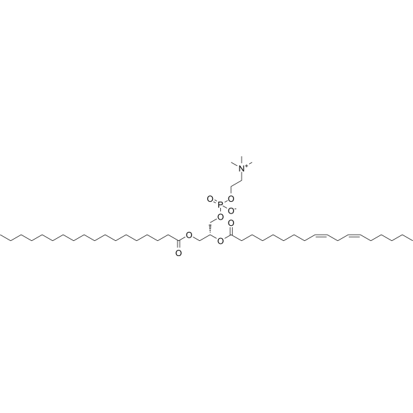
1-Stearoyl-2-linoleoyl-sn-glycero-3-phosphocholine (SLPC; 18:0-18:2 PC) is an endogenous phospholipid marker molecule in the glycerophospholipid metabolic pathway. 1-Stearoyl-2-linoleoyl-sn-glycero-3-phosphocholine is a core component of the phospholipid bilayer of biological membranes and a key responsive lipid for radiation injury and cardiometabolic diseases. 1-Stearoyl-2-linoleoyl-sn-glycero-3-phosphocholine constitutes the phospholipid bilayers of cell membranes and high-density lipoprotein (HDL), and regulates the core activity of lipoprotein functional homeostasis. The content of 1-Stearoyl-2-linoleoyl-sn-glycero-3-phosphocholine in mouse serum shows a significant dose-dependent decrease with increasing ionizing radiation dose, and its level in human HDL also decreases significantly in metabolic syndrome. 1-Stearoyl-2-linoleoyl-sn-glycero-3-phosphocholine can serve as a biological dosimeter marker for ionizing radiation injury, and is used for rapid and accurate assessment of radiation absorbed dose in exposed individuals. 1-Stearoyl-2-linoleoyl-sn-glycero-3-phosphocholine can also act as a lipidomics research target for cardiometabolic diseases such as lipid metabolic syndrome and early-onset coronary heart disease .
loading...
    loading...
Cat. No.: HY-L923
9000 compounds

Ion channels are key proteins on the cell membrane that regulate the flow of ions across membranes. They participate in nearly all physiological processes, including nerve conduction, muscle contraction, heart rhythm, and pain perception. Abnormalities in their function can lead to various serious diseases such as arrhythmia, epilepsy, hypertension, neuropathic pain, and cancer. Therefore, ion channels are highly valuable drug targets—over 15% of approved drugs target ion channels currently, demonstrating their irreplaceable therapeutic value in cardiovascular, neurological, and analgesic fields.

MCE has collected a library of over 5,000 reported ion channel-related bioactive compounds targeting major sites such as Na+ channels, K+ channels, Ca2+ channels, GABA receptors, iGluRs, and others. Using AI models, these compounds are characterized through both 2D representations (molecular fingerprints, pharmacophores) and 3D representations (3D conformation) to screen for a collection of lead-like compounds highly similar to known active molecules. Additionally, an hERG channel prediction algorithm integrating XGB and ISE mapping strategy is employed to assess and exclude potential cardiotoxicity in the library.. This step significantly reduces safety risks in subsequent screenings, particularly for ion channel drug development related to cardiovascular systems (e.g., Nav1.5, Cav1.2), effectively minimizing failures due to hERG inhibition and serving as a valuable tool for ion channel drug screening.

Cat. No.: HY-W127487
CAS No.: 479050-96-9
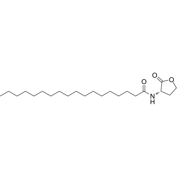
Quorum sensing is a regulatory system used by bacteria to control gene expression in response to increased cell density. This regulatory process manifests itself in a variety of phenotypes, including biofilm formation and virulence factor production. Coordinated gene expression is achieved through the production, release and detection of small diffusible signaling molecules called autoinducers. N-acylated homoserine lactones (AHLs) comprise a class of such autoinducers, each of which generally consists of a fatty acid coupled to a homoserine lactone (HSL). Modulation of bacterial quorum-sensing signaling systems to suppress pathogenesis represents a new approach to antimicrobial research for infectious diseases. AHLs differ in acyl length (C4-C18), C3 substitution (hydrogen, hydroxyl, or oxo group), and the presence or absence of one or more carbon-carbon double bonds in the fatty acid chain. These differences confer signaling specificity through the affinity of the LuxR family of transcriptional regulators. C18-HSL, one of four lipophilic long acyl side chain AHLs produced by the LuxI AHL synthase homolog SinI, is involved in quorum-sensing signaling in strains of Rhizobium meliloti (a nitrogen-fixing bacterial symbiont of the legume M. sativa) . C18-HSL and other hydrophobic AHLs tend to localize in the relatively lipophilic environment of bacterial cells and cannot diffuse freely across the cell membrane. Long-chain N-acyl homoserine lactones can be exported from cells by efflux pumps, or can be transported between communicating cells by extracellular outer membrane vesicles.
loading...
    loading...

