6 Results for "

fixed-cell imaging

" in MedChemExpress (MCE) Product Catalog:
Products (6)

6 Results for "fixed-cell imaging" in MCE Product Catalog:

2
2 Cited Publications
Cat. No.: HY-156404
CAS No.: 1449483-78-6
Purity:  99.04%
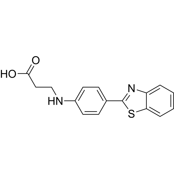
PM-1, a derivative of Thioflavin-T (ThT; HY-D0218), is a small but highly specific plasma membrane (PM) fluorescent dye for specific and long-time membrane imaging of living and fixed cells. PM-1 is embedded directly into the cell membrane and exhibits a very long retention time on the plasma membrane with a half-life of approximately 15 h. PM-1 can be used in combination with protein labeling probes to study ectodomain shedding and endocytosis processes of cell surface proteins .
loading...
    loading...
Cat. No.: HY-D1991
CAS No.: 906664-68-4
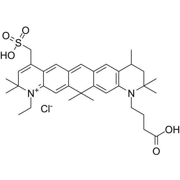
ATTO 647 is a carborhodamine fluorophore and imaging tracer with photostable properties. ATTO 647 serves as a fluorescent probe to investigate cell membrane structure and diffusion characteristics. When conjugated with wheat germ agglutinin, ATTO 647 specifically binds to N-acetyl-β-D-glucosamine and sialic acid residues on membrane glycoproteins, enabling single-molecule tracing of glycoprotein diffusion. ATTO 647 exhibits highly stable fluorescence properties with significantly reduced blinking in mounting media such as ROXS (AA/MV) and ROXS (TX/TQ), whereas its brightness properties vary in Ibidi-MM and Vectashield. ATTO 647 can also be used to label histone H2B-GFP in fixed cells for confocal microscopy photobleaching experiments .
loading...
    loading...
Cat. No.: HY-D3446
CAS No.: 2358677-26-4
PA-SiR-Halo is a photoactivatable fluorophore targeting HaloTag. When bound to HaloTag, PA-SiR-Halo stabilizes its fluorescent form under physiological pH. After conjugation and photoactivation, it exhibits enhanced resistance to nucleophiles, and enables live-cell confocal imaging, fixed-cell single-molecule localization microscopy imaging, live-cell single-particle tracking, as well as live-cell single-molecule localization microscopy imaging of dynamic mitochondrial structures without the need for washing .
loading...
    loading...
Cat. No.: HY-D3362
Research Areas:  

Others

RL420 is an RNA-selective fluorescent covalent probe that binds covalently to the 2′-hydroxyl group of RNA via an acylimidazole-mediated reaction. RL420 has excitation/emission wavelengths of 420/452 nm, exhibits a 150-fold selectivity for RNA over DNA, and shows high labeling efficiency. RL420 can be used in studies related to in vitro RNA labeling, gel imaging, and blue fluorescent imaging of RNA in fixed cells (e.g., nucleoli) .
loading...
    loading...
Cat. No.: HY-D3363
Target:  

Fluorescent Dye mRNA

Research Areas:  

Others

RL450 is an RNA-selective fluorescent covalent probe that binds covalently to the 2′-hydroxyl group of RNA via an acylimidazole-mediated reaction. RL450 has excitation/emission wavelengths of 450/497 nm, exhibits a 970-fold selectivity for RNA over DNA, and features high labeling efficiency. RL450 can be used in studies related to in vitro RNA labeling, gel imaging, and cyan fluorescent imaging of RNA in fixed cells (e.g., nucleoli) .
loading...
    loading...
Cat. No.: HY-D1737
RADA is a fluorescent D-amino acid (FDAA) with high photostability and thermostability, which emits yellow-to-orange fluorescence. RADA shows low outer membrane permeability in wild-type Gram-negative Escherichia coli, but it targets penicillin-binding proteins and L,D-transpeptidases, mimics the interaction between acyl acceptors and enzyme intermediates, and integrates into peptidoglycan during biosynthesis. As a peptidoglycan labeling reagent, RADA metabolically integrates into the nascent peptidoglycan of live bacterial cells, labels the peptidoglycan at the poles and lateral walls of mycobacteria, and enables visualization of peptidoglycan synthesis and remodeling processes. RADA serves as a non-specific stain for fixed cells, is non-toxic to bacterial cells, and its red-shifted excitation/emission spectra reduce phototoxicity. RADA also supports virtual pulse-chase labeling experiments and stochastic optical reconstruction microscopy for sub-diffraction-limited imaging of bacterial cell walls .
loading...
    loading...

