



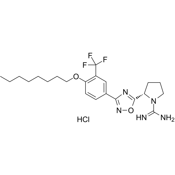
SLM6031434 hydrochloride

Based on 1 Customer Validation
SLM6031434 hydrochloride is the hydrochloride salt form of SLM6031434 (HY-120268). SLM6031434 is a highly selective sphingosine kinase 2 (SphK2) inhibitor with an IC50 value of 0.4 μM for SphK2. SLM6031434 exerts anti-fibrotic effects by increasing sphingosine accumulation and Smad7 expression. SLM6031434 demonstrates effective anti-fibrotic efficacy in a unilateral ureteral obstruction (UUO)-induced tubulointerstitial fibrosis mouse model. SLM6031434 can be used for the study of proteinuric kidney diseases or chronic kidney disease (CKD).
For research use only. We do not sell to patients.
- Purity : 99.1%
- CAS No.: 1897379-34-8
- Formula: C22H31ClF3N5O2
- Molecular Weight:489.96
-
Storage:
4°C, sealed storage, away from moisture
* In solvent : -80°C, 6 months; -20°C, 1 month (sealed storage, away from moisture)

To place orders, for customer services and technical support, please contact: MedChemExpress USA
Tel: 609-228-6898 E-mail: [email protected] [email protected]


-
 Biological Activity
-
Chemical Information
-
Solvent & Solubility
- Protocols
- Purity & Documentation
- References
-
 Help & FAQs


-
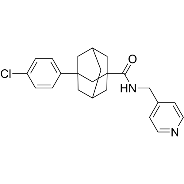
HY-16015
Opaganib -
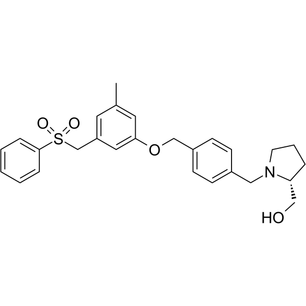
HY-15425
PF-543SPHK1 Inhibitor -
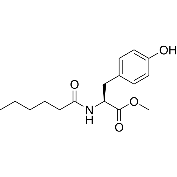
HY-101572
MHPSPHK1 Activator -
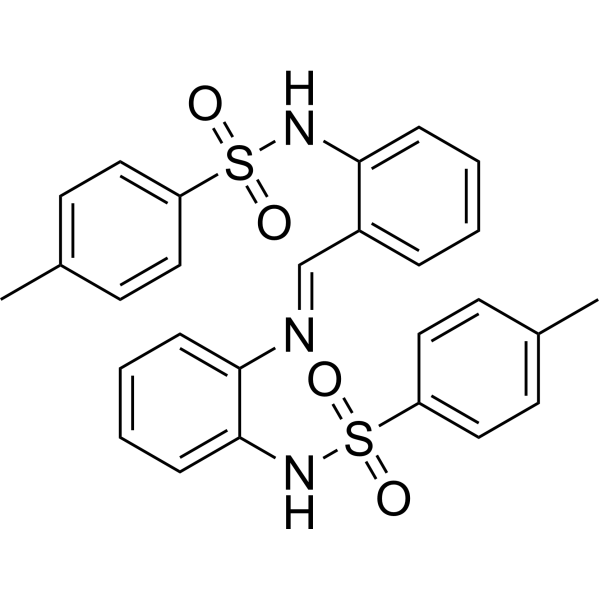
HY-19794
MP-A08SphK Inhibitor -
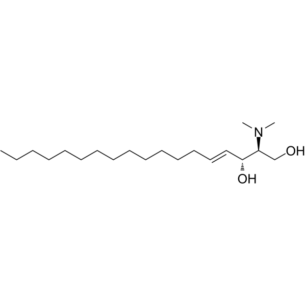
HY-108491
N,N-DimethylsphingosineSphingosine Kinase Inhibitor -
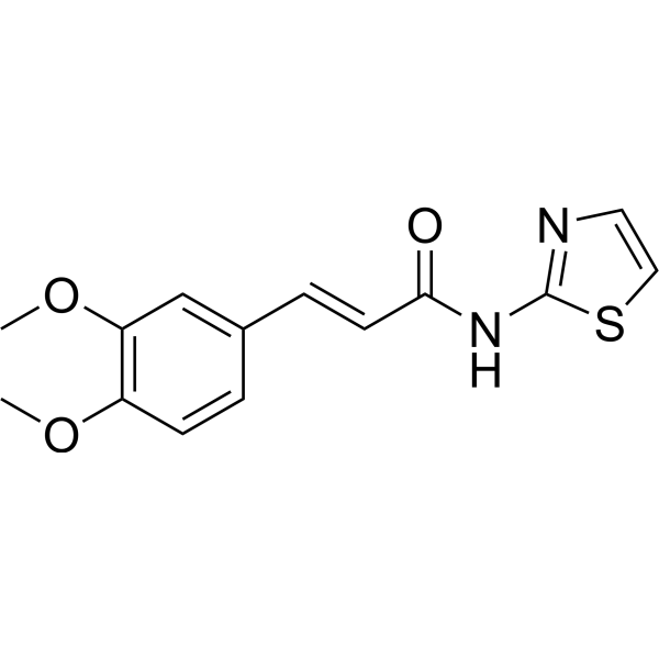
HY-148707
SphK1&2-IN-1SphK Inhibitor -
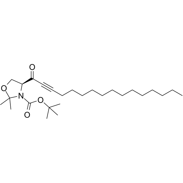
HY-117563
CAY10621SPHK1 Inhibitor -
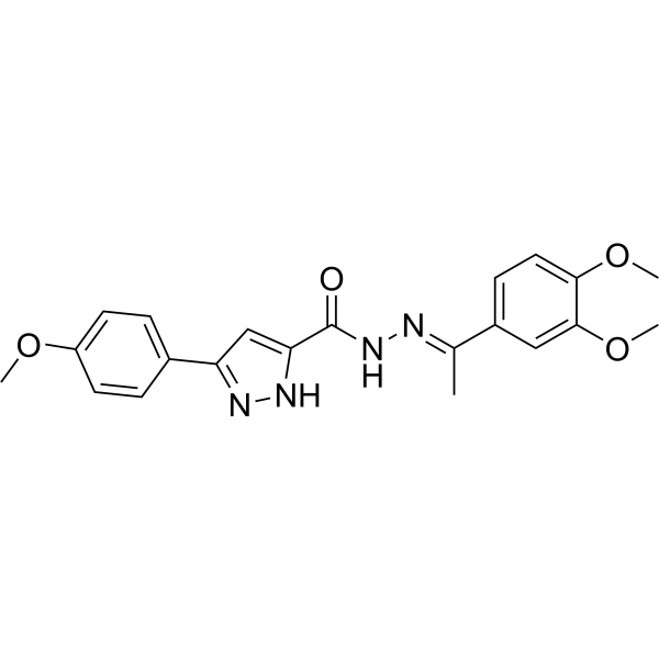
HY-12892
SKI-178SphK1/SphK2 Inhibitor -

HY-124042
K6PC-5SPHK1 Activator -
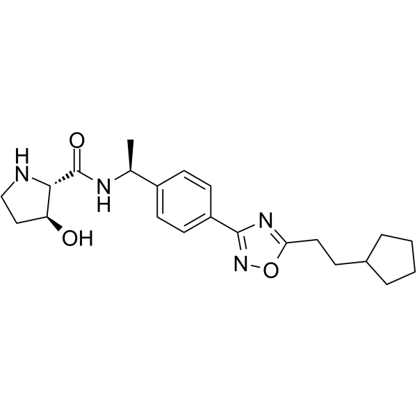
HY-101805
SK1-IN-1SPHK1 Inhibitor -

HY-101572R
MHP (Standard)SPHK1 Activator -
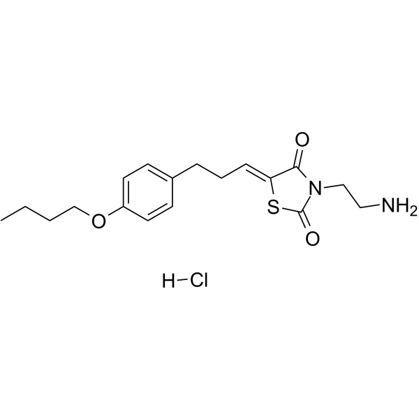
HY-15779A
K145 hydrochlorideSphK2 Inhibitor


Biological Activity
Description
IC50 & Target
[1]|
SphK2 0.4 μM (IC50) |
SphK1 16 μM (IC50) |
In Vitro
SLM6031434 (3 μM, 16 h) reduces TGFβ-induced expression of profibrotic markers (Col1, FN-1, CTGF) in primary mouse renal fibroblasts[1].
SLM6031434 (0.3-10 μM, 16 h) dose-dependently increases Smad7 protein expression in primary mouse renal fibroblasts[1].
SLM6031434 (1 μM, 20 h) significantly increases cellular sphingosine levels in human podocytes[2].
SLM6031434 (1 μM, 24 h) upregulates nephrin and Wilm’s tumor suppressor gene 1 (WT1) protein expressions and mRNA expressions in wildtype human podocytes[2].
MedChemExpress (MCE) has not independently confirmed the accuracy of these methods. They are for reference only. Further protocols information, click here.
In Vivo
MedChemExpress (MCE) has not independently confirmed the accuracy of these methods. They are for reference only.
-
Animal Model:UUO-induced renal interstitial fibrosis mouse models were established by performing unilateral ureteral ligation surgery on 10-12 week-old adult male mice[1]
-
Dosage:5 mg/kg
-
Administration:i.p., daily, 9 days
-
Result:Reduced collagen accumulation and ECM deposition.
Decreased α-SMA expression to reduce myofibroblast activation.
Downregulated mRNA and protein levels of Col1, FN-1, CTGF.
Increased sphingosine accumulation and Smad7 expression, while reduced Smad2 phosphorylation.
Reduced F4/80-positive macrophage infiltration and downregulated iNOS, Mcr1 mRNA.


Chemical Information
-
CAS No. 1897379-34-8
-
Appearance Solid
-
Molecular Weight 489.96
-
Formula C22H31ClF3N5O2
-
Color White to off-white
-
SMILES
N=C(N1[C@H](C2=NC(C(C=C3)=CC(C(F)(F)F)=C3OCCCCCCCC)=NO2)CCC1)N.Cl
-
Shipping
Room temperature in continental US; may vary elsewhere.
-
Storage
4°C, sealed storage, away from moisture
* In solvent : -80°C, 6 months; -20°C, 1 month (sealed storage, away from moisture)


Solvent & Solubility
In Vitro:
DMSO : 5 mg/mL (10.20 mM; Need ultrasonic; Hygroscopic DMSO has a significant impact on the solubility of product, please use newly opened DMSO)
Please refer to the solubility information to select the appropriate solvent. Once prepared, please aliquot and store the solution to prevent product inactivation from repeated freeze-thaw cycles.
Storage method and period of stock solution: -80°C, 6 months; -20°C, 1 month (sealed storage, away from moisture). When stored at -80°C, please use it within 6 months. When stored at -20°C, please use it within 1 month.
Please refer to the solubility information to select the appropriate solvent. Once prepared, please aliquot and store the solution to prevent product inactivation from repeated freeze-thaw cycles.
Storage method and period of stock solution: -80°C, 6 months; -20°C, 1 month (sealed storage, away from moisture). When stored at -80°C, please use it within 6 months. When stored at -20°C, please use it within 1 month.
Concentration (start) × Volume (start) = Concentration (final) × Volume (final)

Protocols
-
Fibrosis/Collagen Morphometry
Fibrosis and collagen morphometry is based on the quantitative visualization of fibrillar collagen deposition in tissue sections using histochemical stains such as Sirius Red (Picrosirius Red) or Masson's trichrome, followed by image-based or polarization-enhanced analysis to estimate collagen proportional area as a surrogate of extracellular matrix accumulation during fibrotic remodeling. Sirius Red combined with polarized light microscopy enhances detection of collagen fibers due to birefringence properties, enabling more specific visualization of collagen type I and III fibrils compared to conventional bright-field histology, while whole-section or region-restricted digital morphometry reduces field-selection bias in fibrosis assessment. Alternative quantitative approaches include second harmonic generation (SHG) and two-photon excited fluorescence microscopy, which enable label-free detection of fibrillar collagen and have been validated against histological staining and biochemica
-
RNA extraction experimental
By lysing cells, releasing RNA, and removing impurities such as proteins and DNA, high-purity RNA products are finally obtained. The commonly used traditional method is the guanidine isothiocyanate/phenol/chloroform method (Trizol), which is suitable for a variety of animal materials including animal tissues, microorganisms, cultured cells, etc., and most plant materials.
-
Collagen: Sirius Red Staining
Sirius Red or picrosirius red staining is a histochemical method for visualizing collagen-rich extracellular matrix in tissue sections, and collagen fibers are detected as red-stained structures under bright-field microscopy with enhanced birefringence under polarized light. Picrosirius red is useful for assessing total collagen organization, distribution, and fibrosis burden, but polarized color should not be interpreted as a definitive collagen type I versus type III readout because color is affected by fiber orientation, thickness, and packing.
-
Cell Cytotoxicity Assay
Cytotoxicity assays are usually based on the assessment of cell membrane damage, which can also be indirectly detected by measuring cell viability. Detection methods include MTT assay, CKK-8 assay, LDH assay and ATP assay, etc.
-
Connective Tissue: Masson's Trichrome/Collagen Trichrome Staining
Masson’s Trichrome (collagen/trichrome staining) is a histological technique that differentially stains tissue compartments using sequential acidic dyes to distinguish collagen from muscle and cytoplasmic components based on dye affinity and tissue permeability differences, enabling visualization of fibrosis and connective tissue architecture in histological sections. The classical formulation typically uses Weigert's iron hematoxylin for nuclear staining, Biebrich scarlet-acid fuchsin for cytoplasm and muscle, and aniline blue (or light green variants) for collagen, producing a characteristic blue/green collagen signal contrasted against red cytoplasm and dark nuclei. The staining principle relies on selective displacement of smaller dye molecules by larger anionic dyes in collagen-rich regions under controlled acidified conditions, which enhances collagen-specific dye retention. This property makes the method widely used for fibrosis assessment in organs such as heart, liver, lung, a
-
How to Choose the Right Model Animal
Choosing the right model animal is a validity-driven decision in which the species, strain, sex, age, genetic background, disease-induction method, outcome measures, and welfare burden must match the scientific question rather than laboratory tradition or convenience. A model should be selected by judging face validity, construct validity, and predictive validity: whether it resembles the human phenotype, whether it reproduces relevant mechanisms, and whether results are likely to predict human biology or treatment response. Animal studies often fail to translate because of species differences, weak disease resemblance, poor experimental design, inadequate reporting, publication bias, and underuse of randomization, blinding, and sample-size justification. Unresolved questions include how to rank competing models objectively, how much human-disease complexity must be reproduced for a given objective, and when non-animal systems such as organoids, ex vivo tissue, or computational models


Purity & Documentation
-
Data Sheet (272 KB)
-
SDS (394 KB)
- English - EN (394 KB)
- Français - FR (394 KB)
- Deutsch - DE (394 KB)
- Norwegian - NO (394 KB)
- Español - ES (394 KB)
- Swedish - SV (394 KB)
- Italian - IT (394 KB)
- Korean - KR (394 KB)
- Portuguese - PT (394 KB)
-
Handling Instructions (2659 KB)
References
[1]. Schwalm S, et al. Validation of highly selective sphingosine kinase 2 inhibitors SLM6031434 and HWG-35D as effective anti-fibrotic treatment options in a mouse model of tubulointerstitial fibrosis. Cell Signal. 2021 Mar;79:109881. [Content Brief]
[2]. Imeri F, et al. Loss of sphingosine kinase 2 enhances Wilm's tumor suppressor gene 1 and nephrin expression in podocytes and protects from streptozotocin-induced podocytopathy and albuminuria in mice. Matrix Biol. 2021 Apr;98:32-48. [Content Brief]


Complete Stock Solution Preparation Table
Please refer to the solubility information to select the appropriate solvent. Once prepared, please aliquot and store the solution to prevent product inactivation from repeated freeze-thaw cycles.
Storage method and period of stock solution: -80°C, 6 months; -20°C, 1 month (sealed storage, away from moisture). When stored at -80°C, please use it within 6 months. When stored at -20°C, please use it within 1 month.
| Optional Solvent | Concentration Solvent Mass | 1 mg | 5 mg | 10 mg | 25 mg |
|---|---|---|---|---|---|
| DMSO | 1 mM | 2.0410 mL | 10.2049 mL | 20.4098 mL | 51.0246 mL |
| 5 mM | 0.4082 mL | 2.0410 mL | 4.0820 mL | 10.2049 mL | |
| 10 mM | 0.2041 mL | 1.0205 mL | 2.0410 mL | 5.1025 mL |


SLM6031434 hydrochloride Related Classifications
HY-120268A Related Classifications
 Powered by Bioz