



Spironolactone
Based on 7 publication(s) in Google Scholar
Spironolactone is an aldosterone antagonist that acts on the aldosterone mineralocorticoid receptor (IC50=24 nM) and androgen receptor (IC50=77 nM), promotes podocyte autophagy and regulates pain. Spironolactone improves hypertension-related vascular hypertrophy and remodeling by reducing angiotensin II (Ang II)-induced inflammation, reduces aldosterone-induced vascular and soft tissue calcification through PIT1-dependent signaling, and alleviates vascular dysfunction in type II diabetic mice by reducing oxidative stress and restoring NO/GC signaling; at low concentrations, it and its metabolites can interfere with aldosterone biosynthesis in the adrenal cortex and inhibit voltage-dependent Ca2+ channels to exert antihypertensive effects.
For research use only. We do not sell to patients.
- Purity : 99.81%
- CAS No.: 52-01-7
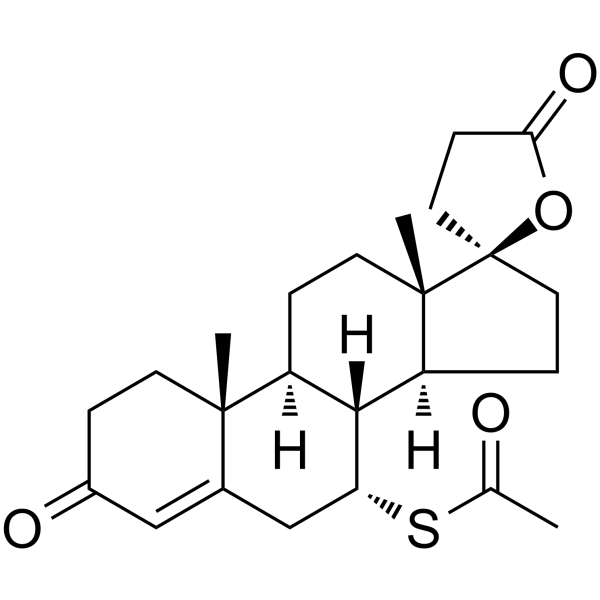
- Formula: C24H32O4S
- Molecular Weight:416.57
-
Storage:Powder -20°C, 3 years , 4°C, 2 years ; In solvent -80°C, 1 year , -20°C, 6 months

To place orders, for customer services and technical support, please contact: MedChemExpress USA
Tel: 609-228-6898 E-mail: [email protected] [email protected]


-
 Biological Activity
-
Chemical Information
-
Solvent & Solubility
- Protocols
- Purity & Documentation
- References
-
 Help & FAQs


-
HY-B0251
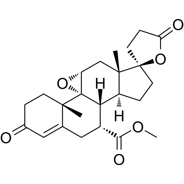
EplerenoneAldosterone Blocker -
HY-13683
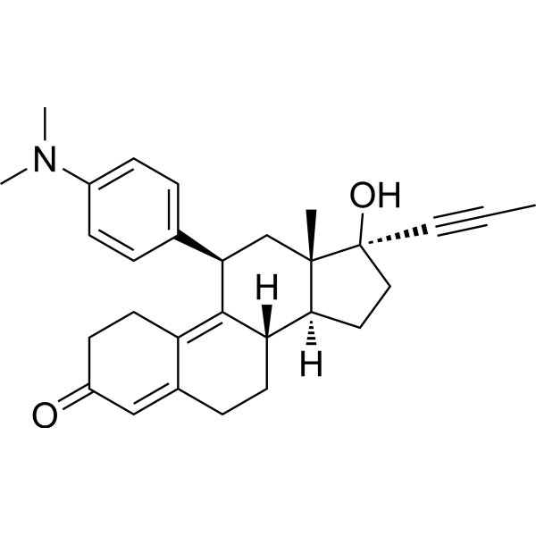
MifepristoneProgesterone Receptor Antagonist -
HY-14249
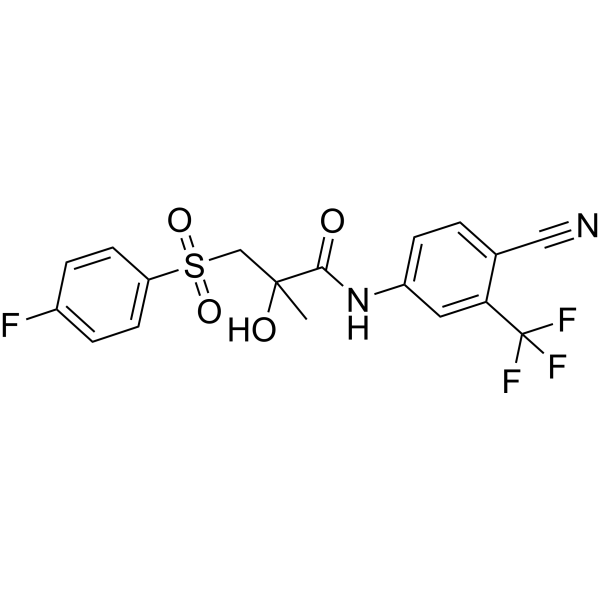
BicalutamideAndrogen Receptor Inhibitor -
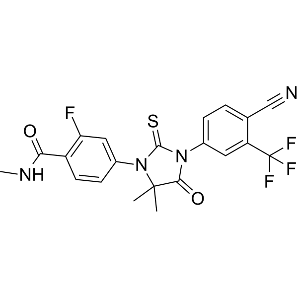
HY-70002
EnzalutamideAndrogen Receptor Antagonist -
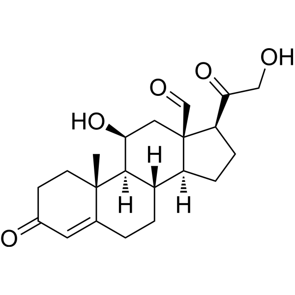
HY-113313
AldosteroneMineralocorticoid Steroid -

HY-14648
DexamethasoneGR Agonist -

HY-13259
MG-132Proteasome Inhibitor -
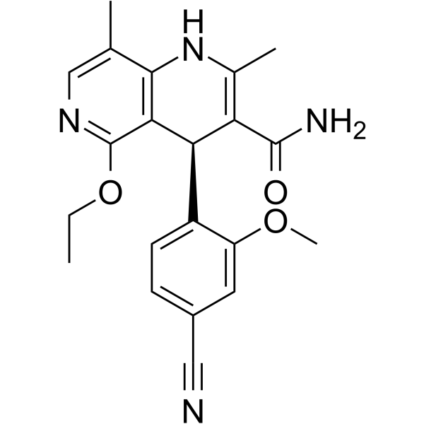
HY-111372
FinerenoneMineralocorticoid Receptor Antagonist -
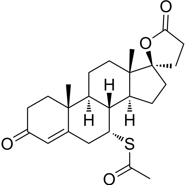
HY-B0561R
Spironolactone (Standard) -
HY-B1438
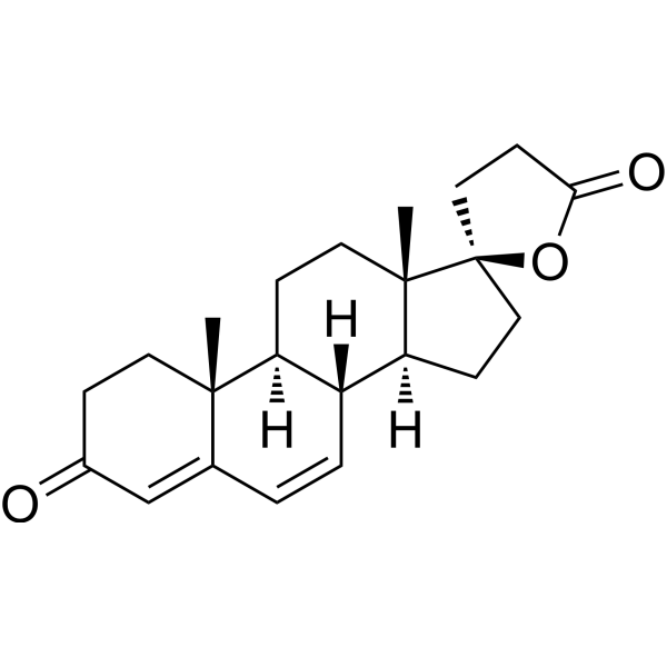
CanrenoneEndogenous Metabolite -
HY-13604
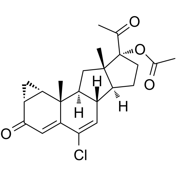
Cyproterone acetateAndrogen Receptor Antagonist -
HY-B0154
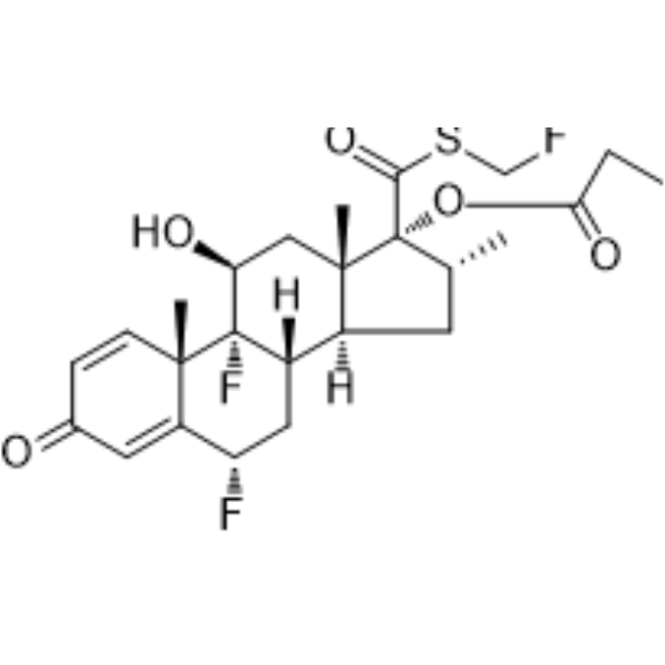
Fluticasone propionateGlucocorticoid Receptor Agonist
Publications Citing Use of MedChemExpress (MCE) Spironolactone
More- Circulation. 2022 Oct 4;146(14):1082-1095. [Abstract]
- Adv Sci (Weinh). 2025 Aug 23:e03409. [Abstract]
- J Orthop Surg Res. 2024 Feb 19;19(1):147. [Abstract]
- Theriogenology. 2026 Apr 15:255:117836. [Abstract]
- Gene Rep. 2025 Oct 4.
- Hong Kong Polytechnic University. 2025.
- J Healthc Eng. 2021 Dec 16:2021:3843830. [Abstract]
 Customer Validation & Images

-
In Vivo Efficacy Study
-
Histological Imaging/Staining
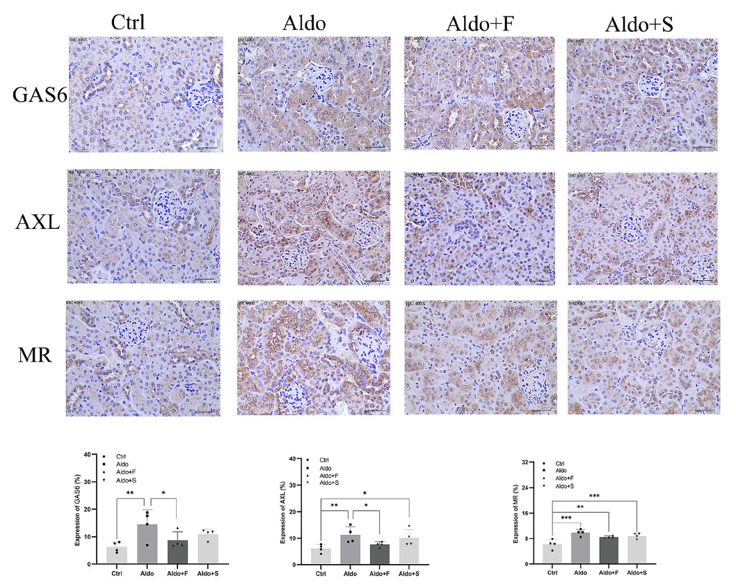
-
In Vivo Efficacy Study
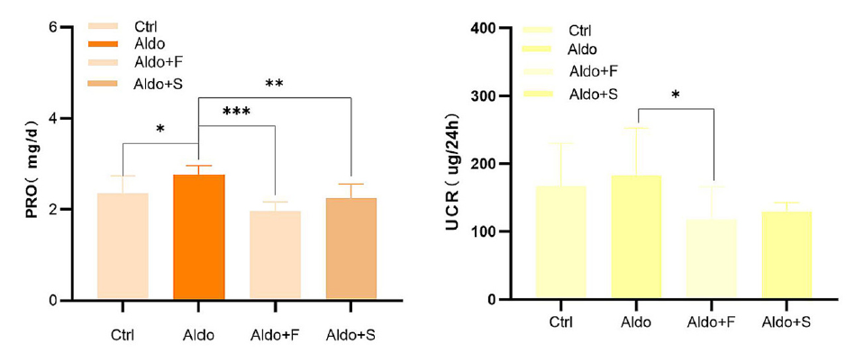
-
Cell Proliferation/Viability Assay
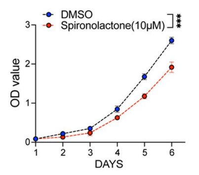
-
ELISA
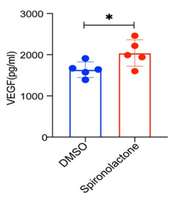
All Calcium Channel Isoforms
More

Biological Activity
Description
Cellular Effect
|
Cell Line

|
Type | Value | Description | References |
|---|---|---|---|---|
| CHO-K1 | IC50 |
0.67 μM
Compound: B, SPL
|
Antagonist activity at human androgen receptor expressed in CHOK1 cells assessed as inhibition of D(-)-norgestrel-induced protein interaction with steroid receptor co-activator peptide after 24 hrs by beta-galactosidase reporter gene assay
Antagonist activity at human androgen receptor expressed in CHOK1 cells assessed as inhibition of D(-)-norgestrel-induced protein interaction with steroid receptor co-activator peptide after 24 hrs by beta-galactosidase reporter gene assay
|
[PMID: 25941555] |
| COS-1 | IC50 |
60 nM
Compound: Spironolactone
|
Antagonist activity at human MR transfected in human COS1 cells after 1 day by luciferase reporter gene assay
Antagonist activity at human MR transfected in human COS1 cells after 1 day by luciferase reporter gene assay
|
[PMID: 22074142] |
| COS-1 | IC50 |
60 nM
Compound: Spironolactone
|
Antagonist activity at human mineralocorticoid receptor expressed in COS1 cells after 1 day by luciferase reporter gene assay
Antagonist activity at human mineralocorticoid receptor expressed in COS1 cells after 1 day by luciferase reporter gene assay
|
[PMID: 25187277] |
| COS-7 | IC50 |
1.6 nM
Compound: Spironolactone
|
Antagonist activity at mineralocorticoid receptor ligand binding domain expressed in african green monkey COS7 cells co-transfected with Gal4-LBD by luciferase reporter gene assay
Antagonist activity at mineralocorticoid receptor ligand binding domain expressed in african green monkey COS7 cells co-transfected with Gal4-LBD by luciferase reporter gene assay
|
[PMID: 19863083] |
| HEK293 | IC50 |
18.6 μM
Compound: spironolactone
|
Inhibition of human MATE1-mediated ASP+ uptake expressed in HEK293 cells after 1.5 mins by fluorescence assay
Inhibition of human MATE1-mediated ASP+ uptake expressed in HEK293 cells after 1.5 mins by fluorescence assay
|
[PMID: 23241029] |
| HEK293 | IC50 |
49 nM
Compound: Spironolactone
|
Displacement of [3H]aldosterone from cytosolic human MR expressed in HEK293 cells after 16 hrs by scintillation counting
Displacement of [3H]aldosterone from cytosolic human MR expressed in HEK293 cells after 16 hrs by scintillation counting
|
[PMID: 22074142] |
| Huh-7 | IC50 |
13 nM
Compound: spironolactone
|
Antagonist activity at Gal4-tagged mineralocorticoid receptor expressed in human Huh7 cells by luciferase reporter gene assay
Antagonist activity at Gal4-tagged mineralocorticoid receptor expressed in human Huh7 cells by luciferase reporter gene assay
|
[PMID: 20672822] |
| NIH-3T3-G185 | IC50 |
23.6 μM
Compound: Spironolactone
|
TP_TRANSPORTER: inhibition of Daunorubicin efflux in NIH-3T3-G185 cells
TP_TRANSPORTER: inhibition of Daunorubicin efflux in NIH-3T3-G185 cells
|
[PMID: 11743742] |
In Vitro
Spironolactone significantly inhibits Ang II-mediated ERK and AKT phosphorylation in A7r5 cells without affecting EGFR phosphorylation[1].
Spironolactone increases PIT1 expression in human aortic smooth muscle cells in a dose-dependent manner and reverses the effects of aldosterone[2].
Spironolactone (0, 100 nM, 1 μM, 10 μM, 30 min) inhibits Ang II-induced inflammation[5].
Spironolactone increases the expression of podocyte-specific markers WT1 and NPHS2 and autophagy markers Beclin1 and LC3B, promoting cell autophagy[10].
MedChemExpress (MCE) has not independently confirmed the accuracy of these methods. They are for reference only. Further protocols information, click here.
-
Cell Line:HAoSMCs (induced by 100 nM aldosterone)
-
Concentration:10 μM
-
Incubation Time:24 h
-
Result:Inhibited the mRNA expression of PITI, TNFA, MSX2, CBFA1 and ALPL.
-
Cell Line:Angiotensin II-induced hu-PBMC
-
Concentration:0, 100 nM, 1 µM, 10 µM
-
Incubation Time:30 min
-
Result:Inhibited the expression of monocyte chemoattractant protein-1 (MCP-1) and tumor necrosis factor-α (TNF-α).
In Vivo
Spironolactone (80 mg/L, p.o.) extends the lifespan of mesothelin mutant mice and reduces aldosterone-induced vascular and soft tissue calcification[2].
Spironolactone (10-80 mg/mL, subcutaneous injection) increases pain behavior in a dose-dependent manner in Male Swiss albino mice thermal and electrical pain models, but reduces inflammatory visceral pain caused by intraperitoneal acetic acid and chemical pain caused by plantar capsaicin[3].
Spironolactone (50 mg/kg, once a day for 6 weeks, oral administration) can reduce diabetes-related vascular oxidative stress and prevent vascular dysfunction by increasing the expression of antioxidant enzymes and soluble guanidyl cyclase (sGC)[4].
Spironolactone (40 mg/kg, daily, 8 weeks, i.g.) reduces urinary albumin excretion, blood lipids and fasting blood glucose levels, alleviates renal damage, promotes autophagy and reduces podocyte loss[10].
MedChemExpress (MCE) has not independently confirmed the accuracy of these methods. They are for reference only.
-
Animal Model:Hypertensive transgenic mice that overproduce angiotensin II (AngII)[1]
-
Dosage:20 mg/kg; Once day; 4 weeks
-
Administration:i.h.
-
Result:Eliminated intimal hyperplasia and medial hypertrophy, inhibited the expression of osteopontin (OPN), and had an inhibitory effect on AngII.
-
Animal Model:Klotho-hypomorphic mice (kl/kl mice)[2]
-
Dosage:80 mg/L
-
Administration:Oral gavage (p.o.)
-
Result:Increased mouse lifespan, did not significantly improve hypercalcemia, hyperphosphatemia or excess 1,25(OH)2D3 and FGF23 plasma concentrations, reduced tissue calcification in kl/kl mice, reduced PIT1, osteoblast differentiation Promoter for expression of Tnfa, Alpl, Msx2, Cbfa1, Osx.
-
Animal Model:Male Swiss albino mice[3]
-
Dosage:10, 20, 40 or 80 mg/kg
-
Administration:Subcutaneous injection (s.c.) for Hot-plate assay, Tail electric stimulation test, Capsaicin-induced hind paw licking; Oral gavage (p.o.) for Acetic acid-induced writhing; i.p. for Rotarod testing
-
Result:Reduced response latency in the hot plate test. Lowering the nociceptive threshold of electrically induced pain in mice. Caused significant anti-pain effects in acetic acid-induced writhing experiments in mice and reduced capsaicin-induced chemical pain at doses of 20-160 mg/kg. Reduced locomotor activity in mice and produced significant impairment in the rotation test at doses of 40 or 80 mg/kg.
-
Animal Model:Leptin receptor knockout (db/db) mice, a model of DM2[4]
-
Dosage:50 mg/kg , daily, 6 weeks
-
Administration:Oral gavage (p.o.)
-
Result:Eliminated endothelial dysfunction in the arteries of db/db mice, increased endothelial nitric oxide synthase (eNOS) phosphorylation (Ser1177), increased the expression of superoxide dismutase-1 and catalase in the arteries of db/db mice, improved relaxation induced by sodium nitroprusside and BAY 41-2277, and increased the expression of soluble corona yl cyclase (sGC) β subunit.
-
Animal Model:SD rat diabetic nephropathy (DN) model[10]
-
Dosage:40mg/kg, daily, 8weeks
-
Administration:i.g.
-
Result:Decreased blood lipid and blood glucose levels, improved liver and kidney function, reduced urinary albumin excretion, restored podocyte morphology to relative normality, and autophagic vacuoles could be seen in podocytes.


Chemical Information
-
CAS No. 52-01-7
-
Appearance Solid
-
Molecular Weight 416.57
-
Formula C24H32O4S
-
Color White to off-white
-
SMILES
C[C@@]12[C@](OC3=O)(CC3)CC[C@@]1([H])[C@@]([C@@H](CC4=CC5=O)SC(C)=O)([H])[C@]([C@]4(CC5)C)([H])CC2
-
Synonyms
SC9420
-
Shipping
Room temperature in continental US; may vary elsewhere.
-
Storage
Powder -20°C 3 years 4°C 2 years In solvent -80°C 1 year -20°C 6 months


Publications (7)
-
Journal Impact Factor
-
Most Recent
-
Circulation
Prenatal Lipopolysaccharides Exposure Induces Transgenerational Inheritance of Hypertension. [Abstract]2022 Oct 4;146(14):1082-1095. PMID: 36004643 -
Adv Sci (Weinh)
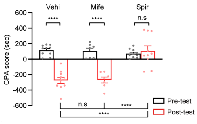
Corticosterone Contributes to Context-Triggered Retrieval of Morphine Withdrawal Memories by Acting on Basolateral Amygdala Neurons Projecting to Nucleus Accumbens Core. [Abstract]2025 Aug 23:e03409. PMID: 40847673
Spironolactone purchased from MedChemExpress. Usage Cited in: Adv Sci (Weinh). 2025 Aug 23:e03409. [Abstract]
The MR inhibitor Spironolactone (Spir) (50 ng) significantly decreased the average CPA scores and inhibited CTR-MWM.
-
J Orthop Surg Res
Several first-line anti-hypertensives act on fibrosarcoma progression and PD1ab blockade therapy. [Abstract]2024 Feb 19;19(1):147. PMID: 38373964
Spironolactone purchased from MedChemExpress. Usage Cited in: J Orthop Surg Res. 2024 Feb 19;19(1):147. [Abstract]
In vitro, Spironolactone (5 mg/kg; i.p.; every other day) significantly inhibited the proliferation ability of MCA-205.
Spironolactone purchased from MedChemExpress. Usage Cited in: J Orthop Surg Res. 2024 Feb 19;19(1):147. [Abstract]
Spironolactone (5 mg/kg; i.p.; every other day) increased the amounts of VEGF of MCA-205 in the supernatant.
-
Theriogenology
Aldosterone regulates in vitro maturation of porcine oocytes derived from small follicles via Wnt/ β-catenin. [Abstract]2026 Apr 15:255:117836. PMID: 41564797 -
Spironolactone purchased from MedChemExpress. Usage Cited in: Gene Rep. 2025 Oct 4.
Spironolactone (20 mg/kg; i.g.; once daily for 4 weeks) significantly increased the expression levels of GAS6 AXL, and MR in mice with aldosterone-induced renal injury.
Spironolactone purchased from MedChemExpress. Usage Cited in: Gene Rep. 2025 Oct 4.
Treatment with MR antagonists Spironolactone (20 mg/kg; i.g.; once daily for 4 weeks) significantly reduced urinary protein (PRO) and urinary creatinine (UCR) levels.
-
-
J Healthc Eng
Spironolactone Inhibits Cardiomyocyte Hypertrophy by Regulating the Ca2+/Calcineurin/p-NFATc3 Pathway. [Abstract]2021 Dec 16:2021:3843830. PMID: 34956570


Solvent & Solubility
In Vitro:
DMSO : ≥ 50 mg/mL (120.03 mM; Hygroscopic DMSO has a significant impact on the solubility of product, please use newly opened DMSO)
H2O : < 0.1 mg/mL (insoluble)
* "≥" means soluble, but saturation unknown.
Please refer to the solubility information to select the appropriate solvent. Once prepared, please aliquot and store the solution to prevent product inactivation from repeated freeze-thaw cycles.
Storage method and period of stock solution: -80°C, 1 year; -20°C, 6 months. When stored at -80°C, please use it within 1 year. When stored at -20°C, please use it within 6 months.
Please refer to the solubility information to select the appropriate solvent. Once prepared, please aliquot and store the solution to prevent product inactivation from repeated freeze-thaw cycles.
Storage method and period of stock solution: -80°C, 1 year; -20°C, 6 months. When stored at -80°C, please use it within 1 year. When stored at -20°C, please use it within 6 months.
Concentration (start) × Volume (start) = Concentration (final) × Volume (final)
In Vivo:
Select the appropriate dissolution method based on your experimental animal and administration route.
- For the following dissolution methods, please ensure to first prepare a clear stock solution using an In Vitro approach and then sequentially add co-solvents:
- To ensure reliable experimental results, the clarified stock solution can be appropriately stored based on storage conditions. As for the working solution for In Vivo experiments, it is recommended to prepare freshly and use it on the same day.
- The percentages shown for the solvents indicate their volumetric ratio in the final prepared solution. If precipitation or phase separation occurs during preparation, heat and/or sonication can be used to aid dissolution.
Add each solvent one by one: 10% DMSO 40% PEG300 5% Tween-80 45% Saline
Solubility: ≥ 2.5 mg/mL (6.00 mM); Clear solution
This protocol yields a clear solution of ≥ 2.5 mg/mL (saturation unknown).
Taking 1 mL working solution as an example, add 100 μL DMSO stock solution (25.0 mg/mL) to 400 μL PEG300, and mix evenly; then add 50 μL Tween-80 and mix evenly; then add 450 μL Saline to adjust the volume to 1 mL.
Preparation of Saline: Dissolve 0.9 g sodium chloride in ddH₂O and dilute to 100 mL to obtain a clear Saline solution.
Add each solvent one by one: 10% DMSO 90% (20% SBE-β-CD in Saline)
Solubility: ≥ 2.5 mg/mL (6.00 mM); Clear solution
This protocol yields a clear solution of ≥ 2.5 mg/mL (saturation unknown).
Taking 1 mL working solution as an example, add 100 μL DMSO stock solution (25.0 mg/mL) to 900 μL 20% SBE-β-CD in Saline, and mix evenly.
Preparation of 20% SBE-β-CD in Saline (4°C, storage for one week): 2 g SBE-β-CD powder is dissolved in 10 mL Saline, completely dissolve until clear.
In Vivo Dissolution Calculator
Please enter the basic information of animal experiments:
-
-
-
-
Recommended: Prepare an additional quantity of animals to account for potential losses during experiments.
Please enter your animal formula composition:
-
%DMSO +
Recommended: Keep the proportion of DMSO in working solution below 2% if your animal is weak.
-
%+
-
+%Tween-80 + +
-
%Saline +
The co-solvents required include: DMSO, . All of co-solvents are available by MedChemExpress (MCE). , Tween 80. All of co-solvents are available by MedChemExpress (MCE).
Working solution concentration: 0.22 mg/mL
Method for preparing stock solution: mg drug dissolved in μL DMSO. Stock solution concentration: mg/mL.
1. Take μL DMSO stock solution;
2. Add μL .
μL , mix evenly;
3. Then add μL Tween 80, mix evenly;
4. Then add μL
Please ensure that the stock solution in the first step is dissolved to a clear state, and add co-solvents in sequence. You can use ultrasonic heating (ultrasonic cleaner, recommended frequency 20-40 kHz), vortexing, etc. to assist dissolution.

Protocols
-
Cell Cytotoxicity Assay
Cytotoxicity assays are usually based on the assessment of cell membrane damage, which can also be indirectly detected by measuring cell viability. Detection methods include MTT assay, CKK-8 assay, LDH assay and ATP assay, etc.
-
ROS/oxidative-stress fluorescent staining
ROS/oxidative-stress fluorescent staining uses cell-permeant fluorogenic probes that become fluorescent after oxidation inside cells or tissues; commonly used examples include DCFH-DA/DCFDA for broad cellular oxidant detection, DHE for superoxide-related signal detection, MitoSOX for mitochondrial superoxide-related signal detection, and CellROX probes for oxidative-stress-associated fluorescence readouts. The assay detects probe oxidation rather than a single ROS species unless the probe and analysis method have been chemically validated for that species. DCFH-DA enters cells, is deacetylated by intracellular esterases to DCFH, and produces fluorescent DCF after oxidation, so the readout is used as an operational measure of total cellular oxidative stress rather than a species-specific ROS measurement. DHE and MitoSOX can report superoxide-related oxidation, but red fluorescence alone can include non-specific ethidium-like oxidation products; HPLC or optimized spectral approaches are
-
Research Protocol for Endocrine Diseases
Endocrine diseases often arise from disrupted hormone production, hormone signaling, or target-tissue responsiveness; for diabetes-focused endocrine disease models, insulin signaling regulates glucose uptake, hepatic glucose output, lipid metabolism, and β-cell compensation. Type 2 diabetes develops through interacting defects in insulin resistance, β-cell dysfunction, adipose inflammation, hepatic glucose overproduction, altered incretin signaling, and ectopic lipid metabolism. A major unresolved question is whether endocrine dysfunction is driven primarily by target-tissue insulin resistance, intrinsic β-cell failure, immune/inflammatory stress, or combined multi-organ failure that differs by disease stage.
-
Autophagy
Autophagy is a process in which eukaryotic cells use lysosomes to degrade their own cytoplasmic proteins and damaged organelles under the regulation of autophagy related gene (Atg). Microtubule-associated proteins light chain 3 (LC3) is recognized as autophagy marker, which transfers from cytoplasmic LC3 (LC3-I) to membrane type (LC3-II). LC3-II/I ratio could be detected by Western Blot and fluorescence microscopy.
-
Research Protocol for Cardiovascular Diseases
Cardiovascular disease can be modeled as maladaptive cardiac remodeling, where ischemic injury or pressure overload activates inflammatory signaling, fibroblast activation, extracellular-matrix deposition, cardiomyocyte hypertrophy, vascular remodeling, and progressive ventricular dysfunction. The TGF-β/SMAD axis is a central profibrotic pathway after myocardial injury and pressure overload, while innate immune and cytokine pathways regulate leukocyte recruitment, scar formation, and adverse remodeling. Key unresolved questions include which inflammatory signals are reparative versus harmful, when fibrosis is protective versus maladaptive, and whether pathway inhibition improves function without weakening necessary infarct healing or compensatory remodeling.
-
Lysosome and acidic-vesicle live-cell staining
Lysosome and acidic-vesicle live-cell staining detects acidic intracellular compartments by using membrane-permeant acidotropic probes that accumulate in low-pH vesicles, including lysosomes, late endosomes, autolysosomes, and acidic phagosomes. LysoTracker staining is commonly used as an intensity-based readout of acidic lysosomal compartment abundance or enlargement, while acridine orange produces green fluorescence in less concentrated compartments and red fluorescence after concentration-dependent accumulation in acidic vesicular organelles. Loss or reduction of acridine-orange red signal can be used as a readout of lysosomal membrane permeabilization or reduced acidic-vesicle integrity. This protocol is designed for live cultured cells and can be adapted for fluorescence microscopy, high-content imaging, plate-reader readout, or flow cytometry when the selected literature supports the readout. Because these dyes report acidotropic accumulation rather than lysosome identity alone,
-
Research Protocol for Inflammation-related Diseases
The NLRP3 inflammasome is a cytosolic innate immune signaling platform that integrates priming signals and danger-signal activation to promote caspase-1 activation, maturation of IL-1β and IL-18, and gasdermin D-mediated pyroptotic cell death. The core experimental logic is to determine whether inflammatory disease phenotypes are driven by increased NLRP3 expression, ASC-containing inflammasome assembly, caspase-1 cleavage, GSDMD cleavage, and extracellular release of IL-1β/IL-18 rather than by nonspecific cell injury alone. The pathway is strongly linked to inflammation-related disease phenotypes because monosodium urate crystals activate NALP3/NLRP3 inflammasome signaling in gout-like crystal inflammation, cholesterol crystals activate NLRP3 inflammasomes in atherogenesis models, and DSS-induced intestinal inflammation has been reported to involve NLRP3 inflammasome activity. However, experimental colitis studies also show context-dependent protective effects of NLRP3 inflammasome co
-
Ca2+ Staining Technique
Ca2+ staining is an experimental technique that utilizes specific fluorescent probes (such as Fluo-4 AM, Fura-2, etc.) to qualitatively or quantitatively detect dynamic changes in intracellular Ca2+ concentrations; this is achieved by monitoring the changes in fluorescent signals generated when these probes bind to free intracellular calcium ions. The underlying principle relies primarily on the presence of chelating groups within the probe's molecular structure that possess high affinity for calcium ions.
-
Macroautophagy Solutions
Macroautophagy is a conserved lysosome-dependent degradation pathway in which cytoplasmic material is sequestered into double-membrane autophagosomes and delivered to lysosomes for degradation and recycling. The pathway supports cellular homeostasis during nutrient limitation, organelle stress, protein-aggregate accumulation, infection, differentiation, and tissue remodeling by coupling cargo sequestration, autophagosome maturation, lysosomal fusion, and degradation of cargo-derived macromolecules. The core molecular sequence includes initiation by nutrient- and stress-regulated autophagy machinery, autophagosome nucleation, LC3/ATG8-family conjugation to autophagosomal membranes, cargo selection through receptors such as SQSTM1/p62, autophagosome-lysosome fusion, and lysosomal degradation. LC3 was identified as a mammalian homolog of yeast Atg8 that localizes to autophagosomal membranes after processing, and p62/SQSTM1 was shown to connect ubiquitinated cargo with autophagic degradati


Purity & Documentation
-
Data Sheet (292 KB)
-
SDS (419 KB)
- English - EN (419 KB)
- Français - FR (419 KB)
- Deutsch - DE (419 KB)
- Norwegian - NO (419 KB)
- Español - ES (419 KB)
- Swedish - SV (419 KB)
- Italian - IT (419 KB)
- Korean - KR (419 KB)
- Portuguese - PT (419 KB)
-
Handling Instructions (2659 KB)
References
[1]. Sachiko Sakurabayashi-Kitade , et al. Aldosterone blockade by Spironolactone improves the hypertensive vascular hypertrophy and remodeling in angiotensin II overproducing transgenic mice. Atherosclerosis. 2009 Sep;206(1):54-60. [Content Brief]
[2]. Jakob Voelkl , et al. Spironolactone ameliorates PIT1-dependent vascular osteoinduction in klotho-hypomorphic mice. J Clin Invest. 2013 Feb;123(2):812-22. [Content Brief]
[3]. Omar M E Abdel-Salam, et al. Effect of spironolactone on pain responses in mice. EXCLI J. 2010 Feb 25:9:46-57. [Content Brief]
[4]. Marcondes A B Silva, et al. Spironolactone treatment attenuates vascular dysfunction in type 2 diabetic mice by decreasing oxidative stress and restoring NO/GC signaling. Front Physiol. 2015 Oct 5:6:269. [Content Brief]
[5]. Ryuzea Miura, et al. Anti-inflammatory effect of spironolactone on human peripheral blood mononuclear cells. J Pharmacol Sci. 2006 Jul;101(3):256-9. [Content Brief]
[6]. S C Cheng, et al. Effects of Spironolactone, Canrenone and Canrenoate-K on Cytochrome P450, and 11β- and 18-Hydroxylation in Bovine and Human Adrenal Cortical Mitochondria1. Endocrinology. 1976 Oct;99(4):1097-106. [Content Brief]
[7]. R Sorrentino. Effect of Spironolactone and Its Metabolites on Contractile Property of Isolated Rat Aorta Rings. J Cardiovasc Pharmacol. 2000 Aug;36(2):230-5. [Content Brief]
[8]. Kim GK, et al. Oral Spironolactone in Post-teenage Female Patients with Acne Vulgaris: Practical Considerations for the Clinician Based on Current Data and Clinical Experience. J Clin Aesthet Dermatol. 2012;5(3):37-50. [Content Brief]
[9]. Fagart J, et al. A new mode of mineralocorticoid receptor antagonism by a potent and selective nonsteroidal molecule. J Biol Chem. 2010;285(39):29932-29940. [Content Brief]
[10]. Dong D, et al. Spironolactone alleviates diabetic nephropathy through promoting autophagy in podocytes. Int Urol Nephrol. 2019;51(4):755-764. [Content Brief]


Complete Stock Solution Preparation Table
Please refer to the solubility information to select the appropriate solvent. Once prepared, please aliquot and store the solution to prevent product inactivation from repeated freeze-thaw cycles.
Storage method and period of stock solution: -80°C, 1 year; -20°C, 6 months. When stored at -80°C, please use it within 1 year. When stored at -20°C, please use it within 6 months.
| Optional Solvent | Concentration Solvent Mass | 1 mg | 5 mg | 10 mg | 25 mg |
|---|---|---|---|---|---|
| DMSO | 1 mM | 2.4006 mL | 12.0028 mL | 24.0056 mL | 60.0139 mL |
| 5 mM | 0.4801 mL | 2.4006 mL | 4.8011 mL | 12.0028 mL | |
| 10 mM | 0.2401 mL | 1.2003 mL | 2.4006 mL | 6.0014 mL | |
| 15 mM | 0.1600 mL | 0.8002 mL | 1.6004 mL | 4.0009 mL | |
| 20 mM | 0.1200 mL | 0.6001 mL | 1.2003 mL | 3.0007 mL | |
| 25 mM | 0.0960 mL | 0.4801 mL | 0.9602 mL | 2.4006 mL | |
| 30 mM | 0.0800 mL | 0.4001 mL | 0.8002 mL | 2.0005 mL | |
| 40 mM | 0.0600 mL | 0.3001 mL | 0.6001 mL | 1.5003 mL | |
| 50 mM | 0.0480 mL | 0.2401 mL | 0.4801 mL | 1.2003 mL | |
| 60 mM | 0.0400 mL | 0.2000 mL | 0.4001 mL | 1.0002 mL | |
| 80 mM | 0.0300 mL | 0.1500 mL | 0.3001 mL | 0.7502 mL | |
| 100 mM | 0.0240 mL | 0.1200 mL | 0.2401 mL | 0.6001 mL |


Spironolactone Related Classifications
HY-B0561 Related Classifications
 Powered by Bioz