



TAT-cyclo-CLLFVY TFA
Based on 2 publication(s) in Google Scholar
TAT-cyclo-CLLFVY TFA is a cyclic peptide inhibitor of HIF-1 heterodimerization that inhibits hypoxia signaling in cancer cells. TAT-cyclo-CLLFVY TFA disrupts HIF-1α/HIF-1β protein-protein interaction with an IC50 of 1.3 μM.
For research use only. We do not sell to patients.
- Formula: C111H188N42O24S2.xC2HF3O2
- Molecular Weight:2559.08 (free acid)
-
Storage:
Please store the product under the recommended conditions in the Certificate of Analysis.

To place orders, for customer services and technical support, please contact: MedChemExpress USA
Tel: 609-228-6898 E-mail: [email protected] [email protected]


-
 Biological Activity
-
Chemical Information
-
Solvent & Solubility
- Protocols
- Purity & Documentation
- References
-
 Help & FAQs


Publications Citing Use of MedChemExpress (MCE) TAT-cyclo-CLLFVY TFA
More

Biological Activity
Description
In Vitro
TAT-cyclo-CLLFVY inhibits HIF-1 activity in a mammalian cell luciferase reporter assay[1].
Hypoxia (1% O2) results in a ~12-fold increase in the luciferase signal, which is inhibited in a dose-dependent manner by TAT-cyclo-CLLFVY (IC50 of 19±2 μM)[1].
To assess the cell-specificity of TAT-cyclo-CLLFVY, the experiment is repeated in MCF-7 breast cancer cells with similar results (TAT-cyclo-CLLFVY IC50 of 16±1 μM)[1].
MedChemExpress (MCE) has not independently confirmed the accuracy of these methods. They are for reference only. Further protocols information, click here.


Chemical Information
-
Molecular Weight 2559.08 (free acid)
-
Formula C111H188N42O24S2.xC2HF3O2
-
Sequence Shortening
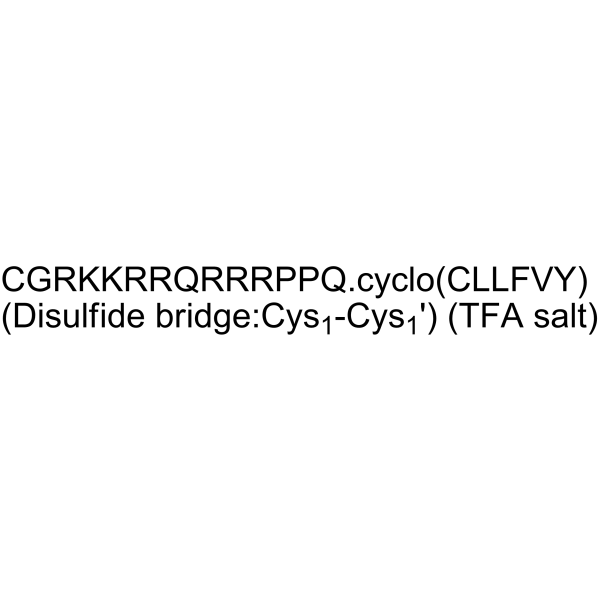
CGRKKRRQRRRPPQ.cyclo(CLLFVY) (Disulfide bridge:Cys1-Cys1')
-
Shipping
Room temperature in continental US; may vary elsewhere.
-
Storage
Please store the product under the recommended conditions in the Certificate of Analysis.


Publications (2)
-
Journal Impact Factor
-
Most Recent
-
Biochem Pharmacol
The 'ABC' of split-nanoluciferase HIF heterodimerization bioassays: Applications, Benefits & Considerations. [Abstract]2024 Nov:229:116478. PMID: 39128589 -


Solvent & Solubility
In Vitro
H2O
Peptide Solubility and Storage Guidelines:
1. Calculate the length of the peptide.
2. Calculate the overall charge of the entire peptide according to the following table:
| Contents | Assign value | |
|---|---|---|
| Acidic amino acid | Asp (D), Glu (E), and the C-terminal -COOH. | -1 |
| Basic amino acid | Arg (R), Lys (K), His (H), and the N-terminal -NH2 | +1 |
| Neutral amino acid | Gly (G), Ala (A), Leu (L), Ile (I), Val (V), Cys (C), Met (M), Thr (T), Ser (S), Phe (F), Tyr (Y), Trp (W), Pro (P), Asn (N), Gln (Q) | 0 |
3. Recommended solution:
| Overall charge of peptide | Details |
|---|---|
| Negative (<0) |
1. Try to dissolve the peptide in water first. 2. If water fails, add NH4OH (<50 μL). 3. If the peptide still does not dissolve, add DMSO (50-100 μL) to solubilize the peptide. |
| Positive (>0) |
1. Try to dissolve the peptide in water first. 2. If water fails, try dissolving the peptide in a 10%-30% acetic acid solution. 3. If the peptide still does not dissolve, try dissolving the peptide in a small amount of DMSO. |
| Zero (=0) |
1. Try to dissolve the peptide in organic solvent (acetonitrile, methanol, etc.) first. 2. For very hydrophobic peptides, try dissolving the peptide in a small amount of DMSO, and then dilute the solution with water to the desired concentration. |

Protocols
-
Cell Cytotoxicity Assay
Cytotoxicity assays are usually based on the assessment of cell membrane damage, which can also be indirectly detected by measuring cell viability. Detection methods include MTT assay, CKK-8 assay, LDH assay and ATP assay, etc.
-
Pull-down
The pull-down assay is an in vitro technique used to detect physical interactions between two or more proteins and an invaluable tool for confirming a predicted protein-protein interaction or identifying novel interacting partners. This method typically involves the use of affinity purification with various wash and elution steps.
-
Immunoprecipitation
Immunoprecipitation (IP) is an experimental method that uses the principle of antibody specific binding to purify and enrich target proteins.
-
Protocol for Bimolecular Fluorescence Complementation (BiFC) Assay
Bimolecular fluorescence complementation detects protein-protein proximity in living or fixed cells by fusing two candidate interaction partners to nonfluorescent N- and C-terminal fragments of a fluorescent protein; when the partners interact or remain close enough, the fluorescent fragments complement, mature, and generate a fluorescent signal at the site of the protein complex. The BiFC readout is fluorescence intensity and subcellular localization of the reconstituted fluorophore, which reflects formation or stabilization of a protein complex rather than direct biochemical binding kinetics; BiFC is therefore useful for mapping where interactions occur in cancer cells, neurons, macrophages, organoid-derived cells, or drug-screening systems, but results should be validated by independent assays such as co-IP or Western blot. BiFC signal formation is delayed by fluorophore maturation and can stabilize otherwise transient complexes, so it is not a real-time reversible interaction assay
-
Co-Immunoprecipitation
Co-immunoprecipitation technology can verify protein interaction based on the specific immune reaction between antibodies and antigens.
-
Protocol for Yeast Two-Hybrid (Y2H) Assay
The yeast two-hybrid assay detects binary protein-protein interactions by separating a transcription factor into a DNA-binding domain fused to a "bait" protein and a transcriptional activation domain fused to a "prey" protein; if bait and prey interact in yeast, the transcription factor is reconstituted and activates reporter genes such as HIS3, ADE2, lacZ, MEL1, or other selectable/readable reporters. The readout is yeast growth on selective medium and/or reporter activity, which reflects proximity-dependent transcriptional activation in the yeast nucleus rather than direct biochemical binding in the original mammalian, tumor, neuronal, macrophage, or organoid context. Because yeast two-hybrid can generate false positives and false negatives, interaction claims should be validated using independent assays such as co-immunoprecipitation, Western blot, immunofluorescence colocalization, BiFC, pull-down, or mammalian two-hybrid assays.


Purity & Documentation
References

Calculators
Concentration (start) × Volume (start) = Concentration (final) × Volume (final)


TAT-cyclo-CLLFVY TFA Related Classifications
HY-P1420A Related Classifications
 Powered by Bioz