



Tauro-β-muricholic acid
Based on 6 publication(s) in Google Scholar
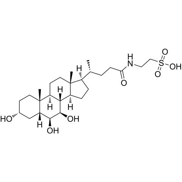
Tauro-β-muricholic acid (TβMCA) is an orally active trihydroxylated bile acid and a competitive, reversible FXR antagonist (IC50=40 μM). Tauro-β-muricholic acid inhibits bile acid-induced hepatocyte apoptosis by maintaining mitochondrial membrane potential, while simultaneously inhibiting intestinal FXR signaling, affecting bile acid synthesis, hepatic lipid metabolism, and insulin sensitivity. Accumulation of tauro-β-muricholic acid disrupts metabolic homeostasis, promoting cancer stem cell proliferation and tumor progression. The mechanisms of tauro-β-muricholic acid involve two aspects: first, inhibiting the translocation of the pro-apoptotic protein Bax to mitochondria and maintaining mitochondrial membrane potential (MMP); and second, blocking the FXR signaling pathway to regulate bile acid metabolism, reduce serum ceramide production, and downregulate the hepatic SREBP1C/CIDEA pathway. Tauro-β-muricholic acid possesses anti-hepatocyte apoptosis, bile acid homeostasis regulation, and liver fat accumulation reduction properties, and also functions as a biomarker, making it useful in the study of diseases such as bile acid metabolism disorders, non-alcoholic fatty liver disease, colorectal cancer, and liver fibrosis.
For research use only. We do not sell to patients.
- Purity : 99.91%
- CAS No.: 25696-60-0
- Formula: C26H45NO7S
- Molecular Weight:515.70
-
Storage:
-20°C, sealed storage, away from moisture
* In solvent : -80°C, 6 months; -20°C, 1 month (sealed storage, away from moisture)

To place orders, for customer services and technical support, please contact: MedChemExpress USA
Tel: 609-228-6898 E-mail: [email protected] [email protected]


-
 Biological Activity
-
Chemical Information
-
Solvent & Solubility
- Protocols
- Purity & Documentation
- References
-
 Help & FAQs


-
HY-133707
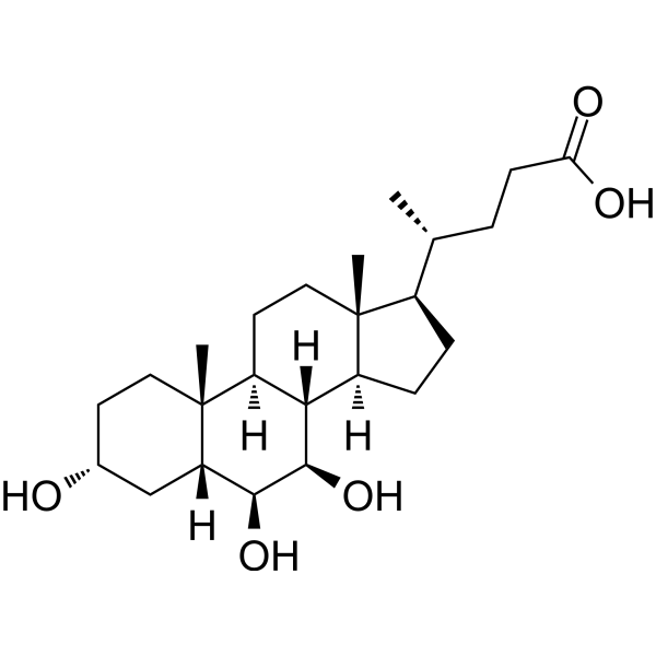
β-Muricholic acidBiliary Cholesterol-Desaturating Agent -
HY-B0351
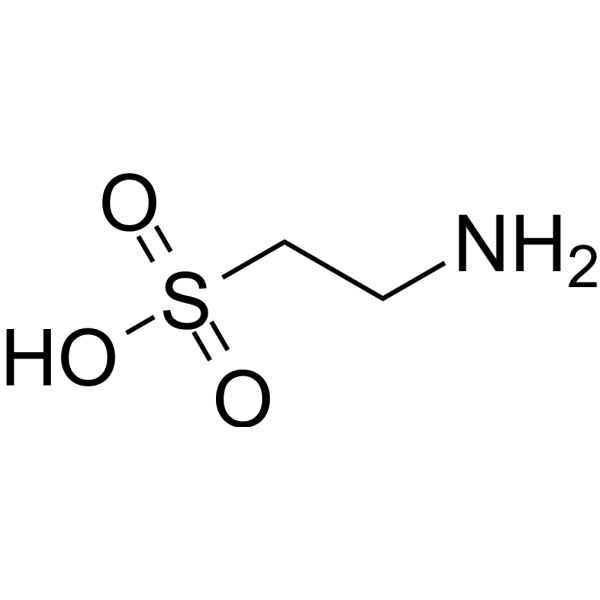
TaurineAutophagy Inducer -
HY-B1899
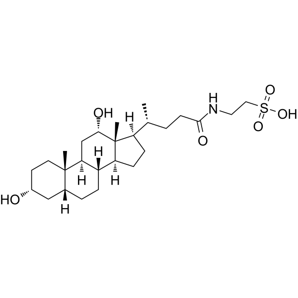
Taurodeoxycholic acidAnti-inflammatory Agent -
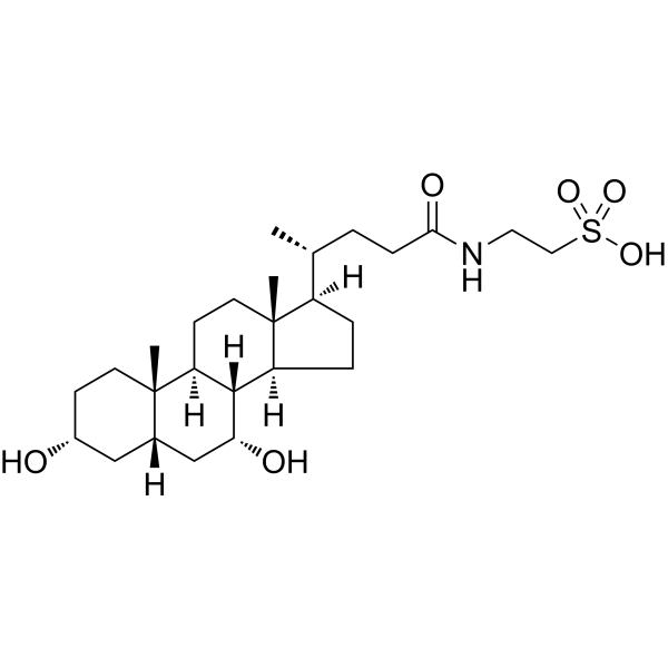
HY-N2027
Taurochenodeoxycholic acidEndogenous Metabolite -

HY-113308
Taurolithocholic acidFerroptosis Suppressor -
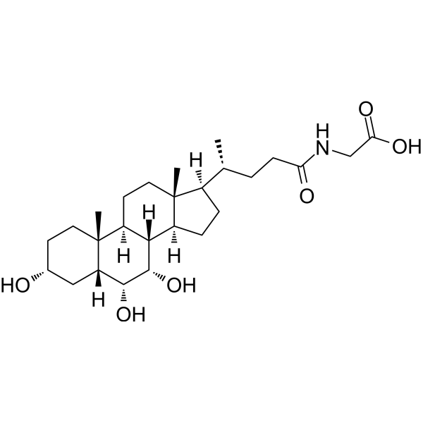
HY-122141
Glycohyocholic acidConjugated Bile Acid -
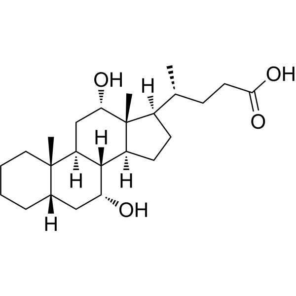
HY-W399297
Isodeoxycholic acidEndogenous Metabolite -
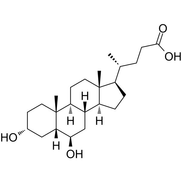
HY-N0169B
Murideoxycholic acid6β-hydroxylated Bile Acid -

HY-133890
Tauro-α-muricholic acidFXR Antagonist
Publications Citing Use of MedChemExpress (MCE) Tauro-β-muricholic acid
More- Cancer Cell. 2026 Aug 10;44(8):1653-1671.e16.
- Cell. 2025 Aug 21;188(17):4530-4548.e25. [Abstract]
- Cell Host Microbe. 2024 Feb 14;32(2):191-208.e9. [Abstract]
- J Pharm Biomed Anal. 2026 Apr 27;278:117541.
- J Pharm Biomed Anal. 2025 Jul 15:259:116760. [Abstract]
- World J Gastrointest Oncol. 2025 Aug 15;17(8):107453. [Abstract]
 Customer Validation & Images

-
Bio/Physico-chemical Assay

-
RT-PCR


Biological Activity
Description
IC50 & Target
40 μM (FXR)[1]
In Vitro
Co-incubation of tauro-β-muricholic acid (25 μM; 4 h) with GCDCA reduced apoptosis in human Ntcp-HepG2 cells, and co-incubation with palmitic acid restored the GCDCA-induced disruption of mitochondrial membrane potential (MMP). Both treatments also inhibited the translocation of the pro-apoptotic protein Bax to the mitochondria[1].
MedChemExpress (MCE) has not independently confirmed the accuracy of these methods. They are for reference only. Further protocols information, click here.
-
Cell Line:Human Ntcp-transfected HepG2 cells, Primary rat hepatocytes, Primary mouse hepatocytes
-
Concentration:25 μmol/L (Ntcp-HepG2 cells, primary rat hepatocytes), 100 μmol/L (primary mouse hepatocytes)
-
Incubation Time:24 h
-
Result:When co-incubated with glycochenodeoxycholic acid (GCDCA), Tauro-β-muricholic acid reduced GCDCA-induced hepatocellular apoptosis to 49% in Ntcp-HepG2 cells, and significantly decreased apoptosis in primary rat and mouse hepatocytes after 4 hours of treatment; nuclear fragmentation induced by GCDCA was also inhibited as visualized by Hoechst 33342 staining.
In Vivo
MedChemExpress (MCE) has not independently confirmed the accuracy of these methods. They are for reference only.
-
Animal Model:Swiss Webster mice (male, 9-16 weeks old), conventional raised (CONV-R) and germ-free (GF) mice; C57BL/6 mice (male, 9-16 weeks old), Fxr-deficient mice (backcrossed 8 generations on C57BL/6 background), CONV-R and GF mice [2]
-
Dosage:Combined with TCA, 400 mg/kg, 200 mg/kg/day
-
Administration:Oral administration
-
Result:Significantly reduced the TCA-induced expression of Fgf15 and Shp in the ileum of GF C57BL/6 mice. Antibiotic treatment increased the levels of TbMCA in the gallbladder of CONV-R C57BL/6 mice, suppressed ileal Fgf15 expression, and increased hepatic Cyp7a1 expression. GF mice had higher levels of TbMCA and a larger bile acid pool compared with CONV-R mice, and TbMCA functioned as a natural FXR antagonist to inhibit bile acid synthesis.


Chemical Information
-
CAS No. 25696-60-0
-
Appearance Solid
-
Molecular Weight 515.70
-
Formula C26H45NO7S
-
Color White to off-white
-
SMILES
C[C@@]12[C@]3([H])[C@]([C@H]([C@H]([C@]1([H])C[C@@H](CC2)O)O)O)([H])[C@@]4([H])[C@](CC3)([C@@](CC4)([H])[C@H](C)CCC(NCCS(=O)(O)=O)=O)C
-
Synonyms
TβMCA
-
Structure Classification
-
Initial Source
-
Shipping
Room temperature in continental US; may vary elsewhere.
-
Storage
-20°C, sealed storage, away from moisture
* In solvent : -80°C, 6 months; -20°C, 1 month (sealed storage, away from moisture)


Publications (6)
-
Journal Impact Factor
-
Most Recent
-
-
Cell
A microbial amino-acid-conjugated bile acid, tryptophan-cholic acid, improves glucose homeostasis via the orphan receptor MRGPRE. [Abstract]2025 Aug 21;188(17):4530-4548.e25. PMID: 40446798 -
Cell Host Microbe
A gut microbiota-bile acid axis promotes intestinal homeostasis upon aspirin-mediated damage. [Abstract]2024 Feb 14;32(2):191-208.e9. PMID: 38237593
Tauro-β-muricholic acid purchased from MedChemExpress. Usage Cited in: Cell Host Microbe. 2024 Feb 14;32(2):191-208.e9. [Abstract]
The expression of two FXR downstream markers, fibroblast growth factor 19 (FGF19) and small heterodimer partner (SHP), were increased by CDCA. But this was abrogated by TβMCA (Tauro-β-muricholic acid sodium, 20 μM), UDCA, and 7-keto-LCA in both intestinal cell lines.
-
-
J Pharm Biomed Anal
Integrating 16S rDNA sequencing analysis and targeted metabolomics to explore the mechanism of Xiexin Tang in treating atherosclerosis mice induced by high-fat diet. [Abstract]2025 Jul 15:259:116760. PMID: 40014894
Tauro-β-muricholic acid purchased from MedChemExpress. Usage Cited in: J Pharm Biomed Anal. 2025 Jul 15:259:116760. [Abstract]
The contents of bile acids such as TCA, T-α-MCA, DCA, TDCA, CA, TCDCA, T-β-MCA, CDCA, and TUDCA were significantly altered when AS occurred (p < 0.01, vs. Ctrl group). However, XXT could only significantly reverse the changes of CDCA, DCA, T-β-MCA, TCA, and TCDCA (p < 0.01, vs. AS group).
-
World J Gastrointest Oncol
Deoxycholic acid induces reactive oxygen species accumulation and promotes colorectal cancer cell apoptosis through the CaMKII-Ca2+ pathway. [Abstract]2025 Aug 15;17(8):107453. PMID: 40837770


Solvent & Solubility
In Vitro:
DMSO : 25 mg/mL (48.48 mM; Need ultrasonic; Hygroscopic DMSO has a significant impact on the solubility of product, please use newly opened DMSO)
Please refer to the solubility information to select the appropriate solvent. Once prepared, please aliquot and store the solution to prevent product inactivation from repeated freeze-thaw cycles.
Storage method and period of stock solution: -80°C, 6 months; -20°C, 1 month (sealed storage, away from moisture). When stored at -80°C, please use it within 6 months. When stored at -20°C, please use it within 1 month.
Please refer to the solubility information to select the appropriate solvent. Once prepared, please aliquot and store the solution to prevent product inactivation from repeated freeze-thaw cycles.
Storage method and period of stock solution: -80°C, 6 months; -20°C, 1 month (sealed storage, away from moisture). When stored at -80°C, please use it within 6 months. When stored at -20°C, please use it within 1 month.
Concentration (start) × Volume (start) = Concentration (final) × Volume (final)

Protocols
-
Collagen: Sirius Red Staining
Sirius Red or picrosirius red staining is a histochemical method for visualizing collagen-rich extracellular matrix in tissue sections, and collagen fibers are detected as red-stained structures under bright-field microscopy with enhanced birefringence under polarized light. Picrosirius red is useful for assessing total collagen organization, distribution, and fibrosis burden, but polarized color should not be interpreted as a definitive collagen type I versus type III readout because color is affected by fiber orientation, thickness, and packing.
-
Somatic Cell Culture
A method of simulating the in vivo environment in vitro to maintain the cell growth, differentation and main functions.
-
Connective Tissue: Masson's Trichrome/Collagen Trichrome Staining
Masson’s Trichrome (collagen/trichrome staining) is a histological technique that differentially stains tissue compartments using sequential acidic dyes to distinguish collagen from muscle and cytoplasmic components based on dye affinity and tissue permeability differences, enabling visualization of fibrosis and connective tissue architecture in histological sections. The classical formulation typically uses Weigert's iron hematoxylin for nuclear staining, Biebrich scarlet-acid fuchsin for cytoplasm and muscle, and aniline blue (or light green variants) for collagen, producing a characteristic blue/green collagen signal contrasted against red cytoplasm and dark nuclei. The staining principle relies on selective displacement of smaller dye molecules by larger anionic dyes in collagen-rich regions under controlled acidified conditions, which enhances collagen-specific dye retention. This property makes the method widely used for fibrosis assessment in organs such as heart, liver, lung, a
-
Apoptosis
Apoptosis, also called programmed cell death, is generally characterized by distinct morphological characteristics.
-
TUNEL staining for apoptotic DNA fragmentation
TUNEL staining detects DNA strand breaks by using terminal deoxynucleotidyl transferase to add labeled nucleotides to exposed 3′-OH DNA termini, generating either microscopic staining in fixed cells or tissue sections, or fluorescence/cytometric signal in cell suspensions. TUNEL positivity reflects DNA fragmentation but should not be interpreted alone as definitive apoptosis, because TUNEL can also label necrotic, autolytic, mechanically damaged, or DNA-repair-associated DNA breaks.
-
Cell Cytotoxicity Assay
Cytotoxicity assays are usually based on the assessment of cell membrane damage, which can also be indirectly detected by measuring cell viability. Detection methods include MTT assay, CKK-8 assay, LDH assay and ATP assay, etc.
-
Research Protocol for Endocrine Diseases
Endocrine diseases often arise from disrupted hormone production, hormone signaling, or target-tissue responsiveness; for diabetes-focused endocrine disease models, insulin signaling regulates glucose uptake, hepatic glucose output, lipid metabolism, and β-cell compensation. Type 2 diabetes develops through interacting defects in insulin resistance, β-cell dysfunction, adipose inflammation, hepatic glucose overproduction, altered incretin signaling, and ectopic lipid metabolism. A major unresolved question is whether endocrine dysfunction is driven primarily by target-tissue insulin resistance, intrinsic β-cell failure, immune/inflammatory stress, or combined multi-organ failure that differs by disease stage.
-
Fluorescent plasma-membrane potential dye assay
Fluorescent plasma-membrane potential dye assays measure changes in cell membrane potential using voltage-sensitive dyes whose fluorescence changes when cells depolarize or hyperpolarize. Anionic bis-oxonol dyes such as DiBAC4(3) enter depolarized cells more readily and show increased fluorescence after intracellular binding, while hyperpolarization reduces dye accumulation and fluorescence. FMP/FLIPR membrane-potential dyes are used for faster, homogeneous microplate assays of ion-channel or receptor-mediated membrane-potential changes.
-
Mitochondrial membrane-potential and mitochondrial mass staining
Mitochondrial membrane potential staining measures the electrochemical polarization across the mitochondrial inner membrane in live cells using lipophilic cationic fluorescent probes; early rhodamine-based work showed that selective mitochondrial dye accumulation is lost when the mitochondrial transmembrane potential is dissipated. JC-1 reports mitochondrial polarization by shifting from green monomer fluorescence to red J-aggregate fluorescence as dye concentration increases within energized mitochondria; therefore, the red/green fluorescence ratio is used as a relative readout of mitochondrial membrane potential. TMRE or TMRM staining provides a single-channel relative readout because these cationic rhodamine esters accumulate in polarized mitochondria, and lower fluorescence indicates reduced mitochondrial polarization when acquisition and dye-loading conditions are controlled. Mitochondrial mass staining is commonly performed with MitoTracker Green FM or related MitoTracker dyes as
-
Apoptosis Solutions
Apoptosis is a regulated, generally non-lytic cell-death pathway that removes unwanted, damaged, infected, or abnormal cells through coordinated morphological changes, caspase activation, DNA fragmentation, and membrane remodeling. The intrinsic apoptosis pathway is controlled mainly by mitochondrial outer membrane permeabilization, BCL-2 family proteins, cytochrome c release, apoptosome formation, caspase-9 activation, and downstream executioner caspase-3/7 activation. The extrinsic apoptosis pathway is initiated by death receptors such as Fas, TNFR, and TRAIL receptors, which recruit adaptor proteins and activate caspase-8 before engaging executioner caspases or mitochondrial amplification through BID cleavage. Apoptosis is linked to many phenotypes, including cancer cell killing, tissue homeostasis, immune regulation, neurodegeneration, infection response, and treatment-induced cytotoxicity; unresolved questions include how apoptosis interacts with necroptosis, pyroptosis, ferroptos
-
MTT Cell Proliferation Assay
The MTT assay is a colorimetric endpoint assay for estimating viable cell number, cell growth, cytotoxicity, or cell activation in cultured mammalian cells. Living cells reduce the yellow tetrazolium salt MTT into purple/blue formazan, while dead cells do not generate the same signal; the resulting color can be quantified with a multiwell spectrophotometer. MTT reduction is commonly interpreted as a readout of metabolic activity that often correlates with viable cell number, but it should not be treated as a direct cell-counting method unless the assay is optimized for the cell type and experimental condition. Studies show that MTT reduction can involve mitochondrial and non-mitochondrial reducing systems, and formazan may accumulate in intracellular lipid droplets rather than simply marking mitochondria.
-
Mitochondrial membrane-potential fluorescent assay
Mitochondrial membrane potential fluorescent assays estimate ΔΨm in living cells using lipophilic cationic dyes such as TMRM, TMRE, rhodamine 123, and JC-1, which accumulate in mitochondria according to membrane polarization; loss of signal after FCCP or CCCP treatment is interpreted as mitochondrial depolarization. TMRM/TMRE and rhodamine 123 are commonly used for semi-quantitative live-cell microscopy or flow cytometry, while JC-1 can report a shift from red aggregate fluorescence to green monomer fluorescence during depolarization; interpretation requires controls because dye concentration, quenching mode, cell type, dye efflux, and mitochondrial mass can affect fluorescence independently of ΔΨm.
-
Annexin V plus membrane-impermeant dye apoptosis staining
Annexin V-based apoptosis assays rely on the detection of phosphatidylserine (PS) externalization from the inner leaflet of the plasma membrane to the outer leaflet, an early biochemical hallmark of apoptosis. Fluorescently labeled Annexin V binds PS in a calcium-dependent manner, enabling identification of early apoptotic cells by flow cytometry or fluorescence microscopy. When combined with a membrane-impermeant DNA-binding dye (e. g. , propidium iodide), this approach allows discrimination between viable (Annexin V−/dye−), early apoptotic (Annexin V+/dye−), and late apoptotic or necrotic (Annexin V+/dye+) cell populations by assessing membrane integrity and PS exposure.
-
BrdU Incorporation Assay
Bromodeoxyuridine (BrdU) incorporation assay is based on the principle that BrdU, a thymidine analog, is incorporated into newly synthesized DNA during the S phase of the cell cycle, thereby serving as a marker of DNA replication and cellular proliferation. Incorporated BrdU can be detected using anti-BrdU antibodies following DNA denaturation, enabling visualization or quantification of proliferating cells through immunochemical detection methods such as immunofluorescence or immunohistochemistry.
-
CFSE Dye Dilution Proliferation Assay
The CFSE (carboxyfluorescein diacetate succinimidyl ester) dye dilution proliferation assay is based on the covalent labeling of intracellular proteins by a cell-permeant fluorescent dye that becomes fluorescent upon intracellular ester cleavage and then is stably retained within cells. As labeled cells divide, the dye is partitioned equally between daughter cells, resulting in a stepwise halving of fluorescence intensity that can be quantified by flow cytometry to determine the number of cell divisions undergone by each cell population. This fluorescence dilution approach enables quantitative tracking of lymphocyte proliferation at the single-cell level over multiple rounds of division. CFSE-based proliferation analysis has been widely applied to measure antigen-driven lymphocyte expansion in vitro, where discrete fluorescence peaks correspond to successive cell divisions and allow reconstruction of proliferative history within heterogeneous populations.
-
Dye-dilution cell tracking and proliferation staining
Dye-dilution cell tracking assays quantify cell proliferation by covalently labeling intracellular proteins with a stable fluorescent dye that is equally partitioned between daughter cells during mitosis, resulting in stepwise halving of fluorescence intensity with each cell division as measured by flow cytometry histograms. Carboxyfluorescein diacetate succinimidyl ester (CFSE) is a prototypical dye that diffuses into cells, is enzymatically converted into a fluorescent compound, and then covalently binds intracellular amine groups, producing long-lived fluorescence suitable for tracking multiple rounds of division in vitro and in vivo. Successive generations of dividing cells form discrete peaks of decreasing fluorescence intensity, enabling estimation of proliferation history, precursor frequency, and division index within heterogeneous populations. Alternative dyes such as CellTrace Violet (CTV) and far-red membrane dyes (e. g. , PKH26) follow the same dilution principle but differ
-
CCK-8/WST-8 Cell Proliferation Assay
The CCK-8/WST-8 assay is based on the reduction of the water-soluble tetrazolium salt WST-8 to a water-soluble formazan product by cellular dehydrogenases in metabolically active cells, where the generated formazan amount is proportional to the number of living cells and is quantified by measuring absorbance in the visible range, providing a colorimetric readout for cell viability and proliferation assessment. This class of tetrazolium-based assays improves upon earlier MTT-based systems by producing a water-soluble formazan, eliminating the need for organic solubilization steps and enabling direct spectrophotometric measurement in culture medium.
-
Protocol for Cell Cycle
Cell-cycle analysis by flow cytometry measures DNA content in single cells to estimate the fraction of cells in G0/G1, S, and G2/M phases. Propidium iodide intercalates into DNA, and after RNA removal with RNase, fluorescence intensity reflects cellular DNA content: 2N cells are assigned to G0/G1, cells between 2N and 4N to S phase, and 4N cells to G2/M. DNA-content analysis alone cannot reliably separate G0 from G1 or G2 from M. Ki-67 can distinguish quiescent G0 cells from cycling cells, EdU or BrdU incorporation marks active DNA synthesis in S phase, and phospho-histone H3 staining identifies mitotic cells within the 4N population.
-
Cell Counting-Based Growth Curve Assay
Cell counting-based growth curve assays quantify cell proliferation by directly measuring changes in viable cell number over time using manual or automated counting methods such as hemocytometer-based counting or instrument-assisted cell enumeration, enabling construction of growth curves that reflect population expansion dynamics in response to culture conditions. A widely used approach is trypan blue exclusion with hemocytometer counting, where membrane-compromised (non-viable) cells take up the dye, allowing discrimination between viable and non-viable cells while simultaneously enabling total cell number quantification. Repeated sampling across time points allows estimation of proliferation rate, growth phases, and comparative growth kinetics between experimental conditions.
-
Colony Formation (Clonogenic) Assay
The clonogenic (colony formation) assay measures the ability of a single cell to retain reproductive viability and form a macroscopic colony, typically defined as a cluster derived from one progenitor cell after a defined growth period. This assay is widely used to evaluate cell survival after exposure to ionizing radiation or cytotoxic treatments and is considered a standard method in radiation biology for generating dose-response relationships of reproductive cell death. Colony formation reflects long-term proliferative capacity rather than short-term metabolic activity, and survival is quantified by comparing treated versus untreated conditions based on colony number and derived survival fractions.
-
Research Protocol for Metabolic Diseases
AMP-activated protein kinase, AMPK, is a conserved cellular energy sensor that responds to reduced cellular energy status and coordinates metabolism by increasing ATP-generating catabolic pathways while suppressing ATP-consuming anabolic processes. In metabolic disease research, the AMPK pathway is experimentally relevant because it regulates hepatic lipid synthesis, fatty acid oxidation, glucose production, skeletal-muscle glucose disposal, mTORC1-linked biosynthesis, autophagy, mitochondrial homeostasis, and whole-body energy balance. The central pathway logic is that energy stress, metformin, exercise-like stimulation, or direct AMPK activators increase AMPKα Thr172 phosphorylation and downstream substrate phosphorylation, including ACC and RAPTOR. Phosphorylation of ACC suppresses lipogenesis and supports fatty acid oxidation, whereas phosphorylation of RAPTOR suppresses mTORC1 signaling and links cellular energy status to growth and protein synthesis control. The pathway is linked
-
Cell Viability Determination by MTT Colorimetric Assay
The following protocol uses the MTT colorimetric assay as a classic literature-established method for assessing cell viability/metabolic activity in cultured mammalian cells. MTT[3-(4,5-dimethylthiazol-2-yl)-2,5-diphenyltetrazolium bromide] is reduced by metabolically active cells to a colored formazan product; the amount of formazan is quantified spectrophotometrically and provides an indirect measure of metabolically active viable cells. Importantly, MTT reduction reflects cellular oxidoreductase/metabolic activity rather than an absolute direct count of living cells, so changes in cellular metabolism can alter the signal independently of cell number.
-
Liver Histomorphometry
Liver histomorphometry is a quantitative histological approach used to measure structural alterations in hepatic tissue, including parenchymal loss, steatosis, fibrosis, and vascular remodeling, by combining stained tissue section analysis with stereological or computerized image-based measurements. Classical morphometric frameworks quantify volume fractions of liver compartments and fibrotic regions using systematic sampling and image analysis, enabling objective comparison of pathological changes across experimental groups. These approaches are widely applied in liver cirrhosis and fibrosis studies to reduce subjectivity in histological scoring and improve reproducibility of tissue evaluation. Recent methodological advances integrate automated image analysis and radiomics-based extraction of histological features from standard liver stains (e. g. , H&E and fibrotic stains), enabling quantitative correlation between morphometric features and fibrosis stages in non-alcoholic fatty live
-
Fibrosis/Collagen Morphometry
Fibrosis and collagen morphometry is based on the quantitative visualization of fibrillar collagen deposition in tissue sections using histochemical stains such as Sirius Red (Picrosirius Red) or Masson's trichrome, followed by image-based or polarization-enhanced analysis to estimate collagen proportional area as a surrogate of extracellular matrix accumulation during fibrotic remodeling. Sirius Red combined with polarized light microscopy enhances detection of collagen fibers due to birefringence properties, enabling more specific visualization of collagen type I and III fibrils compared to conventional bright-field histology, while whole-section or region-restricted digital morphometry reduces field-selection bias in fibrosis assessment. Alternative quantitative approaches include second harmonic generation (SHG) and two-photon excited fluorescence microscopy, which enable label-free detection of fibrillar collagen and have been validated against histological staining and biochemica
-
EdU Incorporation Assay (Click Chemistry-Based DNA Synthesis Measurement)
The EdU incorporation assay measures DNA synthesis by adding the thymidine analog 5-ethynyl-2′-deoxyuridine to cells or tissues, where it is incorporated into newly synthesized DNA during S phase. Incorporated EdU is detected by copper-catalyzed azide-alkyne cycloaddition, in which a fluorescent azide covalently reacts with the ethynyl group on EdU, allowing S-phase cells to be detected by fluorescence microscopy, flow cytometry, or high-content imaging. EdU detection does not require DNA denaturation or anti-BrdU antibody access, which preserves sample structure and improves compatibility with immunostaining and multiparameter cytometry compared with BrdU-based detection. EdU can be cytotoxic in a cell-type- and exposure-dependent manner, so pulse duration, concentration, and continuous-labeling designs should be validated for each cell type.
-
Ki-67 Immunostaining Proliferation Assay
Ki-67 immunostaining measures the growth fraction of a cell population by detecting Ki-67, a nuclear antigen present in proliferating cells and absent in quiescent G0 cells. The readout is the percentage of Ki-67-positive nuclei among total counted cells, commonly called the Ki-67 labeling index or proliferation index.
-
PCNA Immunodetection Proliferation Assay
PCNA immunodetection measures proliferative activity by detecting proliferating cell nuclear antigen, a nuclear protein associated with DNA polymerase δ function and DNA replication. The assay readout is the proportion of PCNA-positive nuclei among total counted cells, but PCNA labeling is not identical to BrdU labeling because PCNA can mark late G1/early S-associated replication competence and may persist beyond active DNA synthesis depending on fixation and extraction conditions.
-
Protocol for Cell Counting and Cell Density Analysis
Cell counting and cell-density analysis estimate the number of cells in a known volume or field area. Manual hemocytometer counting uses a chamber of defined geometry to convert counted cells into cells/mL, while automated counters and image-analysis workflows detect cell objects from optical, brightfield, fluorescence, impedance, or digital-image features. Trypan blue viability counting is based on dye exclusion: viable cells with intact membranes exclude dye, while non-viable cells with compromised membranes stain blue. The readout is total cell density, viable-cell density, dead-cell density, and percent viability. Cell density can also be estimated from microscopy images by counting objects per image area, from flow cytometry using calibrated volume or reference particles, or from in situ microscopy in bioreactors after calibration against reference methods such as hemocytometer or flow cytometry.


Purity & Documentation
-
Data Sheet (280 KB)
-
SDS (252 KB)
- English - EN (252 KB)
- Français - FR (252 KB)
- Deutsch - DE (252 KB)
- Norwegian - NO (252 KB)
- Español - ES (252 KB)
- Swedish - SV (252 KB)
- Italian - IT (252 KB)
- Korean - KR (252 KB)
- Portuguese - PT (252 KB)
-
Handling Instructions (2659 KB)
References
[1]. Denk GU, et al. Tauro-β-muricholic acid restricts bile acid-induced hepatocellular apoptosis by preserving the mitochondrial membrane potential. Biochem Biophys Res Commun. 2012 Aug 10;424(4):758-64. [Content Brief]
[2]. Sayin SI, et al. Gut microbiota regulates bile acid metabolism by reducing the levels of tauro-beta-muricholic acid, a naturally occurring FXR antagonist. Cell Metab. 2013 Feb 5;17(2):225-35. [Content Brief]
[3]. Zhao DS, et al. Identification of urine tauro-β-muricholic acid as a promising biomarker in Polygoni Multiflori Radix-induced hepatotoxicity by targeted metabolomics of bile acids. Food Chem Toxicol. 2017 Oct;108(Pt B):532-542. [Content Brief]
[4]. Jiang C, et al. Intestinal farnesoid X receptor signaling promotes nonalcoholic fatty liver disease. J Clin Invest. 2015 Jan;125(1):386-402. [Content Brief]


Complete Stock Solution Preparation Table
Please refer to the solubility information to select the appropriate solvent. Once prepared, please aliquot and store the solution to prevent product inactivation from repeated freeze-thaw cycles.
Storage method and period of stock solution: -80°C, 6 months; -20°C, 1 month (sealed storage, away from moisture). When stored at -80°C, please use it within 6 months. When stored at -20°C, please use it within 1 month.
| Optional Solvent | Concentration Solvent Mass | 1 mg | 5 mg | 10 mg | 25 mg |
|---|---|---|---|---|---|
| DMSO | 1 mM | 1.9391 mL | 9.6956 mL | 19.3911 mL | 48.4778 mL |
| 5 mM | 0.3878 mL | 1.9391 mL | 3.8782 mL | 9.6956 mL | |
| 10 mM | 0.1939 mL | 0.9696 mL | 1.9391 mL | 4.8478 mL | |
| 15 mM | 0.1293 mL | 0.6464 mL | 1.2927 mL | 3.2319 mL | |
| 20 mM | 0.0970 mL | 0.4848 mL | 0.9696 mL | 2.4239 mL | |
| 25 mM | 0.0776 mL | 0.3878 mL | 0.7756 mL | 1.9391 mL | |
| 30 mM | 0.0646 mL | 0.3232 mL | 0.6464 mL | 1.6159 mL | |
| 40 mM | 0.0485 mL | 0.2424 mL | 0.4848 mL | 1.2119 mL |


Tauro-β-muricholic acid Related Classifications
HY-N9933 Related Classifications
 Powered by Bioz