



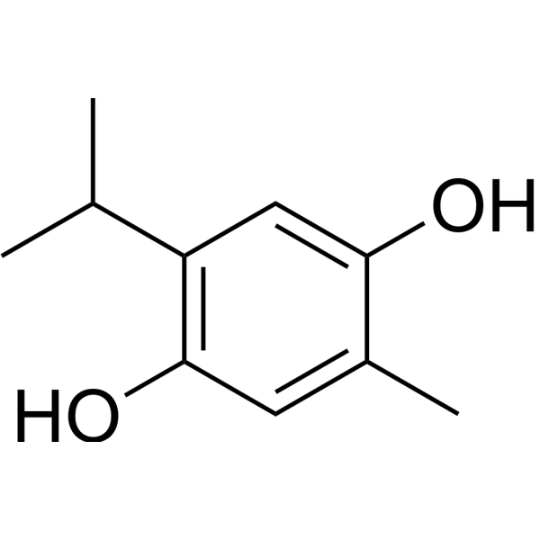
Thymohydroquinone

Based on 1 Customer Validation
Thymohydroquinone is a COX-2 inhibitor and anti-SARS-CoV-2 agent that induces cytotoxicity, antiproliferative effects and inhibits tumor growth. Thymohydroquinone cannot scavenge superoxide radicals via σ (hydrogen atom transfer) and π-π attacks with superoxide anions. Thymohydroquinone can be used in research related to squamous cell carcinoma, fibrosarcoma and coronavirus disease 2019 (SARS-CoV-2 infection).
For research use only. We do not sell to patients.
- Purity : 99.91%
- CAS No.: 2217-60-9
- Formula: C10H14O2
- Molecular Weight:166.22
-
Storage:
4°C, protect from light
* In solvent : -80°C, 6 months; -20°C, 1 month (protect from light)

To place orders, for customer services and technical support, please contact: MedChemExpress USA
Tel: 609-228-6898 E-mail: [email protected] [email protected]


-
 Biological Activity
-
Chemical Information
-
Solvent & Solubility
- Protocols
- Purity & Documentation
- References
-
 Help & FAQs


-
HY-N3300
MelleinNatural Product -
HY-B0932S
Levocarnitine propionate-d3 hydrochlorideStable Isotope -
HY-N1584B
Halofuginone hydrochlorideNatural Product -
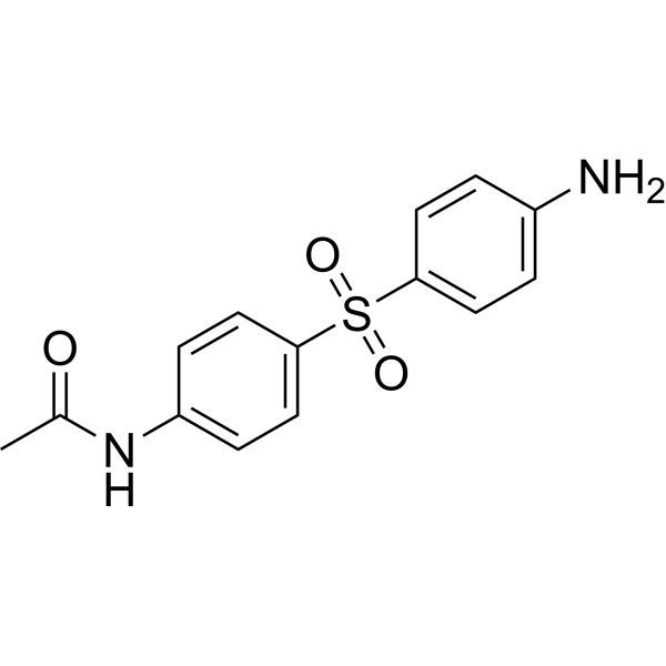
HY-G0016
N-Acetyl dapsoneDapsone Metabolite -

HY-N8280
IKD-8344Macrocyclic Dilactone -

HY-N1584BR
Halofuginone hydrochloride (Standard)Reference Standard -
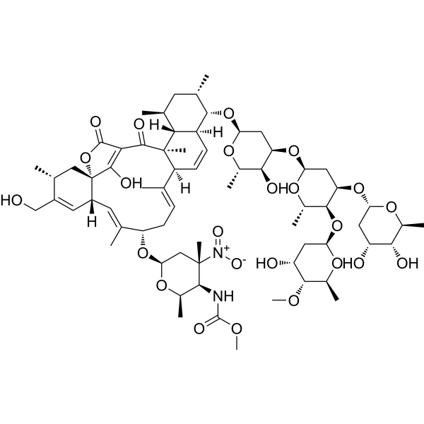
HY-125049
KijanimicinAntibiotic -

HY-N8373
Beauvericin AAntibacterial Agent -

HY-N9475
BetulonaldehydeAnti-inflammatory Agent -
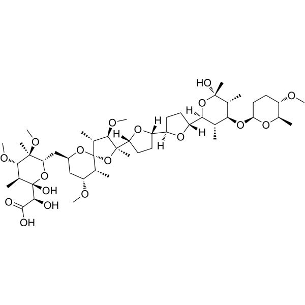
HY-136943
K-41Antimalarial/Antibacterial Agent -
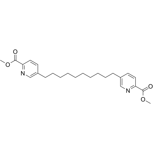
HY-124301
Penicolinate APicolinic Acid Derivative -

HY-N11982
Emeguisin Aantibacterial


Biological Activity
Description
IC50 & Target
|
COX-2 |
Cellular Effect
|
Cell Line

|
Type | Value | Description | References |
|---|---|---|---|---|
| A2780 | IC50 |
3.1 μM
Compound: 18
|
Growth inhibition of human A2780 cells after 72 hrs by SRB assay
Growth inhibition of human A2780 cells after 72 hrs by SRB assay
|
[PMID: 29519737] |
| A2780cis | IC50 |
9.8 μM
Compound: 18
|
Growth inhibition of human A2780cis cells after 72 hrs by SRB assay
Growth inhibition of human A2780cis cells after 72 hrs by SRB assay
|
[PMID: 29519737] |
| OVCAR-8 | IC50 |
8.9 μM
Compound: 18
|
Growth inhibition of human OVCAR8 cells after 72 hrs by SRB assay
Growth inhibition of human OVCAR8 cells after 72 hrs by SRB assay
|
[PMID: 29519737] |
In Vitro
Thymohydroquinone (0.01-0.1 mg/mL; 24 h) exhibits dose-dependent, statistically significant cytotoxic activity against L929 mouse fibroblast cells, SCC VII mouse squamous cell carcinoma cells, and FsaR mouse fibrosarcoma cells[1].
Thymohydroquinone inhibits SARS-CoV-2 in VERO-E6 cells with an IC50 of 23.15 ng/mL[2].
MedChemExpress (MCE) has not independently confirmed the accuracy of these methods. They are for reference only. Further protocols information, click here.
-
Cell Line:L929 mouse fibroblasts, SCC VII mouse squamous cell carcinoma cells, FsaR mouse fibrosarcoma cells
-
Concentration:0.01-0.1 mg/mL
-
Incubation Time:24 h
-
Result:Induced 21% cell growth inhibition in L929 cells, 17% cell growth inhibition in SCC VII cells, and 15% cell growth inhibition in FsaR cells at 0.01 mg/mL.
Induced 63% cell growth inhibition in L929 cells, 87% cell growth inhibition in SCC VII cells, and 92% cell growth inhibition in FsaR cells at 0.1 mg/mL.
Showed statistically significant (p < 0.01) dose-dependent cytotoxic activity, with greater inhibition against tumor cell lines (SCC VII, FsaR) than against normal L929 fibroblasts at the 0.1 mg/mL concentration.
In Vivo
MedChemExpress (MCE) has not independently confirmed the accuracy of these methods. They are for reference only.
-
Animal Model:C3HHf/BuZgr/Hr (male, 3 months old; fibrosarcoma or squamous cell carcinoma cell subcutaneous transplantation)[1]
-
Dosage:5 mg/kg per injection; total cumulative dose of 20 mg/kg
-
Administration:intratumoral; on days 3, 4, 5, 11 post-tumor cell inoculation
-
Result:Achieved a tumor growth inhibition (TGI) of up to 49% against SCC VII tumors and up to 33% against FsaR tumors.
Showed statistically significant differences in tumor growth compared with control mice (p < 0.05) at multiple time points post-inoculation.
Exhibited no statistically significant difference in in vivo antitumor activity compared with thymoquinone.


Chemical Information
-
CAS No. 2217-60-9
-
Appearance Solid
-
Molecular Weight 166.22
-
Formula C10H14O2
-
Color White to off-white
-
SMILES
OC1=CC(C(C)C)=C(O)C=C1C
-
Structure Classification
-
Initial Source
-
Shipping
Room temperature in continental US; may vary elsewhere.
-
Storage
4°C, protect from light
* In solvent : -80°C, 6 months; -20°C, 1 month (protect from light)


Solvent & Solubility
In Vitro:
H2O : 1 mg/mL (6.02 mM; ultrasonic and warming and heat to 60°C)
DMSO : 1 mg/mL (6.02 mM; Need ultrasonic and warming; Hygroscopic DMSO has a significant impact on the solubility of product, please use newly opened DMSO)
Please refer to the solubility information to select the appropriate solvent. Once prepared, please aliquot and store the solution to prevent product inactivation from repeated freeze-thaw cycles.
Storage method and period of stock solution: -80°C, 6 months; -20°C, 1 month (protect from light). When stored at -80°C, please use it within 6 months. When stored at -20°C, please use it within 1 month.
* Note: If you choose water as the stock solution, please dilute it to the working solution, then filter and sterilize it with a 0.22 μm filter before use.
Please refer to the solubility information to select the appropriate solvent. Once prepared, please aliquot and store the solution to prevent product inactivation from repeated freeze-thaw cycles.
Storage method and period of stock solution: -80°C, 6 months; -20°C, 1 month (protect from light). When stored at -80°C, please use it within 6 months. When stored at -20°C, please use it within 1 month.
* Note: If you choose water as the stock solution, please dilute it to the working solution, then filter and sterilize it with a 0.22 μm filter before use.
Concentration (start) × Volume (start) = Concentration (final) × Volume (final)

Protocols
-
Research Protocol for Infectious Diseases
Infectious-disease experiments test how pathogens interact with host barriers, innate immune receptors, inflammatory signaling, pathogen replication, and tissue injury; pattern-recognition receptors such as TLRs, RIG-I-like receptors, NOD-like receptors, and inflammasomes detect microbial molecules and activate NF-κB, interferon, and cytokine responses. The central hypothesis is that infection severity reflects the balance between pathogen burden and host response: protective inflammation restricts pathogen growth, whereas excessive or mislocalized inflammation contributes to tissue damage and disease phenotype. Unresolved questions include which host pathways are protective versus pathogenic, why some infection models fail to translate to human disease, and which combined readouts best predict clinically relevant infection outcomes.
-
Cell Cytotoxicity Assay
Cytotoxicity assays are usually based on the assessment of cell membrane damage, which can also be indirectly detected by measuring cell viability. Detection methods include MTT assay, CKK-8 assay, LDH assay and ATP assay, etc.
-
Mammalian live/dead viability and cytotoxicity staining
Live/dead viability and cytotoxicity staining assays are based on the simultaneous detection of intracellular esterase activity in metabolically active (viable) cells and membrane integrity loss in non-viable cells. In commonly used dual-staining approaches, membrane-permeant fluorogenic substrates are converted by intracellular esterases into fluorescent products in live cells, while impermeant DNA-binding dyes selectively enter cells with compromised plasma membranes and label nucleic acids in dead or dying cells, enabling discrimination between viable and non-viable populations by fluorescence microscopy or flow cytometry.
-
Patient-Derived Xenograft (PDX)
Patient-derived xenograft (PDX) models are generated by engrafting primary human tumor tissue directly into immunodeficient mice, allowing in vivo propagation of patient tumor biology without initial in vitro adaptation. These models are used to preserve key histopathological and molecular characteristics of the original tumor and enable assessment of tumor growth dynamics and therapeutic response in a living organism. The biological readout is tumor engraftment and subsequent growth in the murine host, which reflects the ability of human tumor cells to survive, vascularize, and expand in an immunocompromised microenvironment.
-
Patient-Derived Orthotopic Xenograft (PDOX)
Patient-derived orthotopic xenograft (PDOX) modeling implants fresh patient tumor tissue or patient-derived tumor cells into the anatomically corresponding organ or tissue site of immunodeficient mice, usually by surgical orthotopic implantation, to preserve patient tumor histology, local microenvironmental context, invasion, metastatic behavior, and treatment-response features better than subcutaneous implantation. PDOX readouts include tumor engraftment, orthotopic tumor growth, local invasion, metastasis, recurrence after resection, histologic similarity to the donor tumor, biomarker retention, molecular concordance, survival, and response or resistance to therapy. PDOX models are used for preclinical drug testing and individualized therapy evaluation, but engraftment success varies by tumor type and specimen quality.
-
Subcutaneous Cell-Line-Derived Xenograft
Subcutaneous cell-line-derived xenograft (CDX) models are established by implanting cultured human cancer cell lines into immunodeficient mice, where the injected cells form localized tumors that can be monitored in vivo as a measure of tumorigenic potential, growth kinetics, and treatment response. These models are widely used in oncology research because they allow reproducible tumor formation and enable comparative assessment of tumor growth between different cell lines or genetic manipulations in a controlled in vivo microenvironment. Subcutaneous implantation of cancer cells in immunodeficient mice is a standard approach for evaluating tumor growth behavior and therapeutic response across multiple cancer types, including prostate, esophageal, pancreatic, and colon cancer models.
-
Orthotopic Cell-Line Xenograft
Orthotopic cell-line xenograft models involve implantation of human cancer cell lines into the anatomically corresponding organ of immunodeficient mice to reproduce tumor growth within a native microenvironment, enabling more clinically relevant tumor behavior compared with subcutaneous models. These models are widely used because orthotopic placement better recapitulates tumor progression, including invasion and metastatic spread, which are often underrepresented in heterotopic implantation systems. Compared with conventional xenografts, orthotopic implantation is described as more technically complex but provides improved simulation of tumor-microenvironment interactions and metastatic behavior, making it particularly valuable for translational oncology research. Surgical orthotopic implantation approaches have been emphasized as enabling faithful reproduction of clinical cancer features, including metastasis and disease progression patterns that align with the tumor’s organ of origi
-
Cell Viability Determination by MTT Colorimetric Assay
The following protocol uses the MTT colorimetric assay as a classic literature-established method for assessing cell viability/metabolic activity in cultured mammalian cells. MTT[3-(4,5-dimethylthiazol-2-yl)-2,5-diphenyltetrazolium bromide] is reduced by metabolically active cells to a colored formazan product; the amount of formazan is quantified spectrophotometrically and provides an indirect measure of metabolically active viable cells. Importantly, MTT reduction reflects cellular oxidoreductase/metabolic activity rather than an absolute direct count of living cells, so changes in cellular metabolism can alter the signal independently of cell number.


Purity & Documentation
-
Data Sheet (276 KB)
-
SDS (393 KB)
- English - EN (393 KB)
- Français - FR (393 KB)
- Deutsch - DE (393 KB)
- Norwegian - NO (393 KB)
- Español - ES (393 KB)
- Swedish - SV (393 KB)
- Italian - IT (393 KB)
- Korean - KR (393 KB)
- Portuguese - PT (393 KB)
-
Handling Instructions (2659 KB)
References
[1]. Ivankovic S, et al. The antitumor activity of thymoquinone and thymohydroquinone in vitro and in vivo. Exp Oncol. 2006;28(3):220-224. [Content Brief]
[2]. Esharkawy ER, et al. In vitro potential antiviral SARS-CoV-19- activity of natural product thymohydroquinone and dithymoquinone from Nigella sativa. Bioorg Chem. 2022;120:105587. [Content Brief]
[3]. Sakib R, et al. Antioxidant Properties of Thymoquinone, Thymohydroquinone and Black Cumin (Nigella sativa L.) Seed Oil: Scavenging of Superoxide Radical Studied Using Cyclic Voltammetry, DFT and Single Crystal X-ray Diffraction. Antioxidants (Basel). 2023;12(3):607. Published 2023 Mar 1. [Content Brief]


Complete Stock Solution Preparation Table
Please refer to the solubility information to select the appropriate solvent. Once prepared, please aliquot and store the solution to prevent product inactivation from repeated freeze-thaw cycles.
Storage method and period of stock solution: -80°C, 6 months; -20°C, 1 month (protect from light). When stored at -80°C, please use it within 6 months. When stored at -20°C, please use it within 1 month.
| Optional Solvent | Concentration Solvent Mass | 1 mg | 5 mg | 10 mg | 25 mg |
|---|---|---|---|---|---|
| H2O / DMSO | 1 mM | 6.0161 mL | 30.0806 mL | 60.1612 mL | 150.4031 mL |
| 5 mM | 1.2032 mL | 6.0161 mL | 12.0322 mL | 30.0806 mL |
* Note: If you choose water as the stock solution, please dilute it to the working solution, then filter and sterilize it with a 0.22 μm filter before use.


Thymohydroquinone Related Classifications
HY-W196368 Related Classifications
 Powered by Bioz