



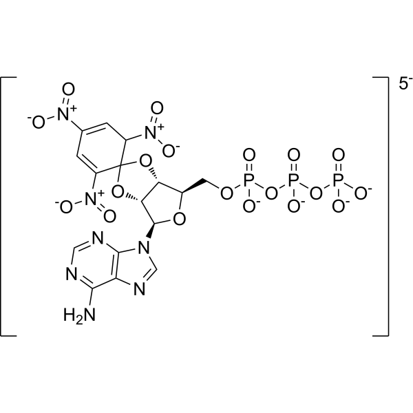
TNP-ATP triethylammonium

Based on 1 Customer Validation
TNP-ATP triethylammonium is a P2X receptor antagonist with an IC50 of 0.010 μM for P2X3 and an IC50 of 0.062 μM for P2X2/3. TNP-ATP triethylammonium acts as an inhibitor of CheA autophosphorylation, with a Ki of 0.7 µM. TNP-ATP triethylammonium blocks the functional activation of P2X1-7 receptors. TNP-ATP triethylammonium attenuates hypoxia-induced IL-1β expression and release. TNP-ATP triethylammonium alleviates visceral pain, and improves hypoxia-induced cognitive impairment, insufficient myelination and neuroinflammation. Binding of TNP-ATP triethylammonium to CheA enhances the fluorescence of the TNP group. TNP-ATP triethylammonium can be used in studies related to visceral pain. NP-ATP triethylammonium can be used in studies related to hypoxia-induced insufficient myelination and cognitive decline.
For research use only. We do not sell to patients.
- Purity : 98.7%
- CAS No.: 61368-63-6
- Formula: C40H77N12O19P3
- Molecular Weight:1123.03
-
Storage:
Solution, -20°C, 2 years

To place orders, for customer services and technical support, please contact: MedChemExpress USA
Tel: 609-228-6898 E-mail: [email protected] [email protected]


-
 Biological Activity
-
Chemical Information
- Protocols
- Purity & Documentation
- References
-
 Help & FAQs


-
HY-150270A
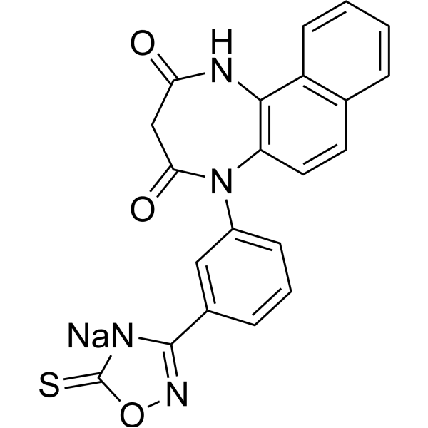
NP-1815-PX sodiumP2X4 Inhibitor -
HY-110237
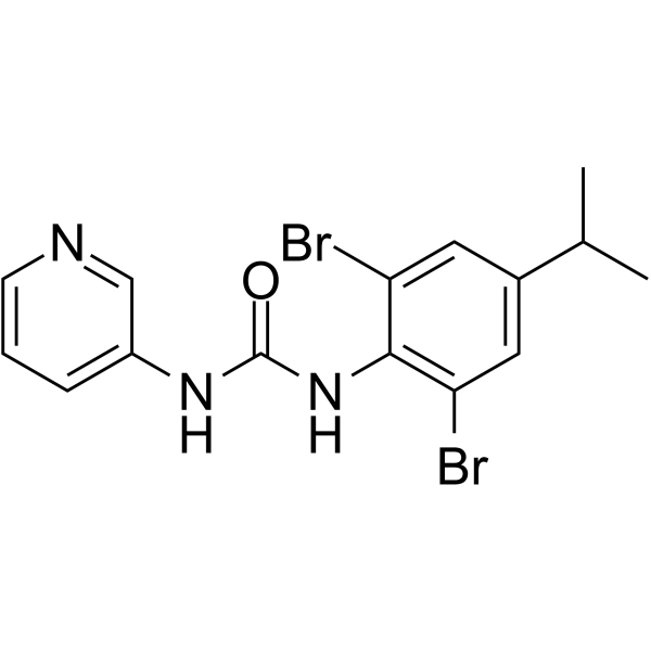
BX430P2X4 Receptor Antagonist -
HY-15488
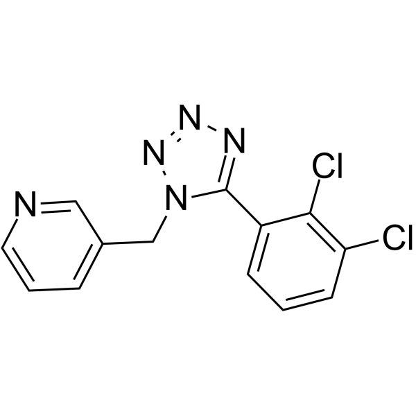
A 438079P2X Receptor Antagonist -
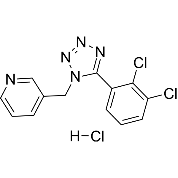
HY-15488A
A 438079 hydrochlorideP2X7 Receptor Antagonist -
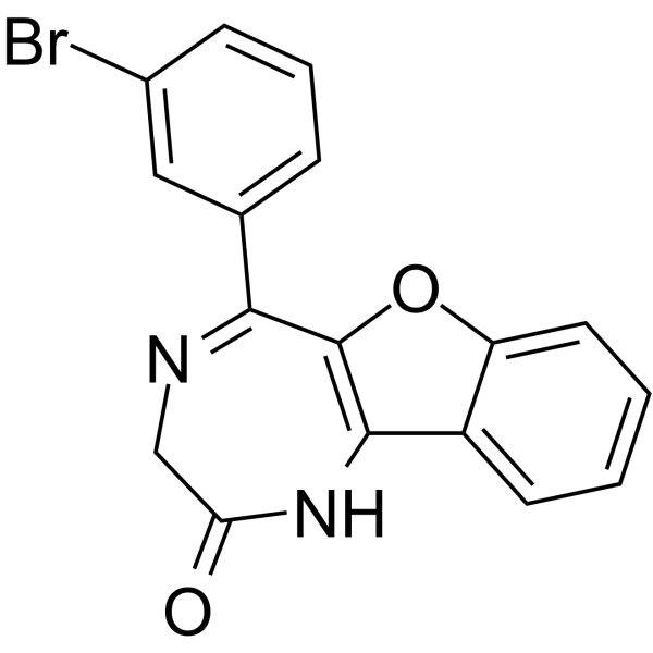
HY-101911
5-BDBDP2X4 Receptor Antagonist -
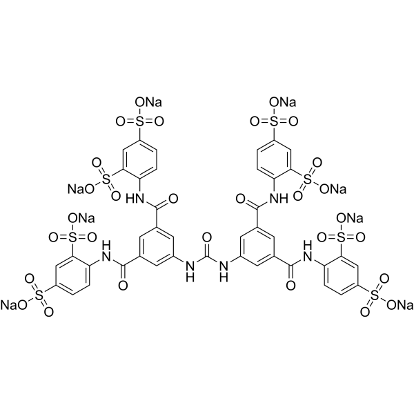
HY-112461A
NF449 octasodiumP2X1 Receptor Antagonist -
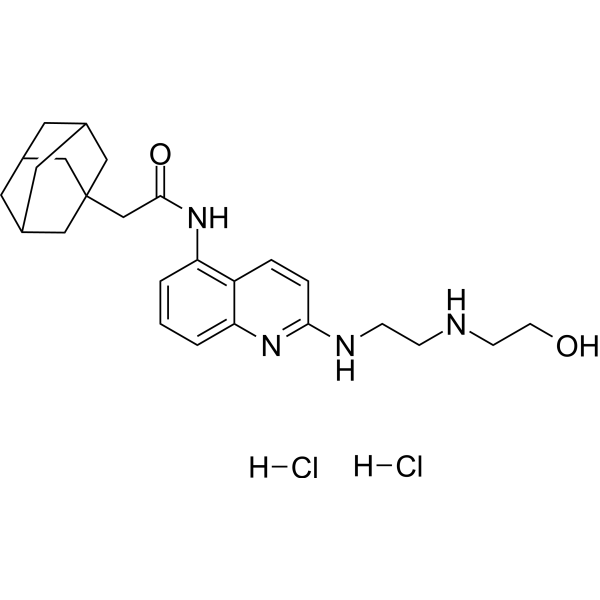
HY-108669
AZ10606120 dihydrochlorideP2X Receptor Antagonist -
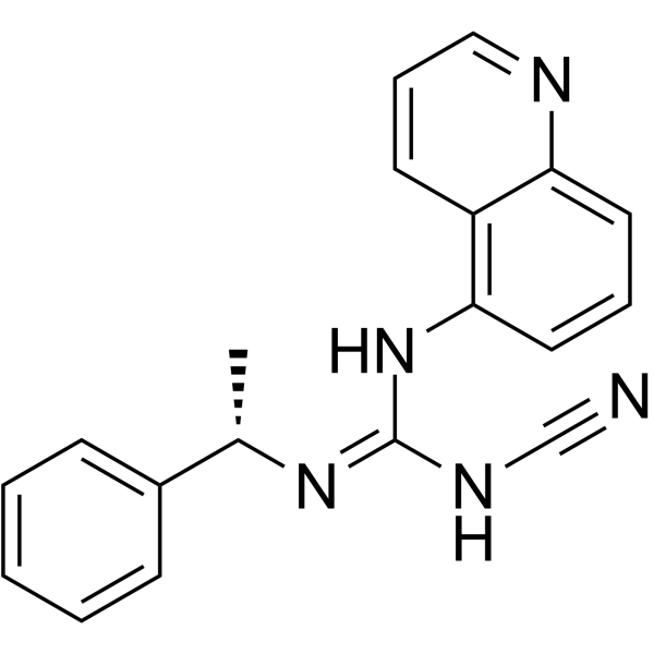
HY-100483
A-804598P2X7 Antagonist -
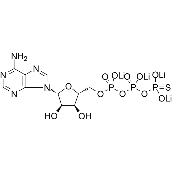
HY-108666
ATPγS tetralithium saltP2Y11 Receptor Agonist -
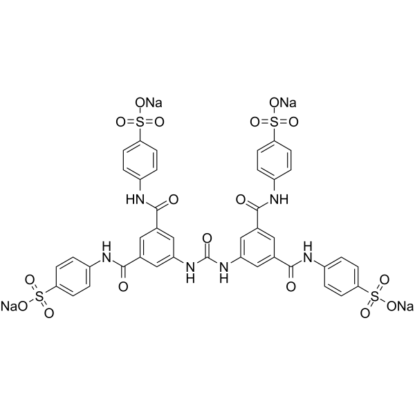
HY-108671
NF110P2X3 Receptor Antagonist -
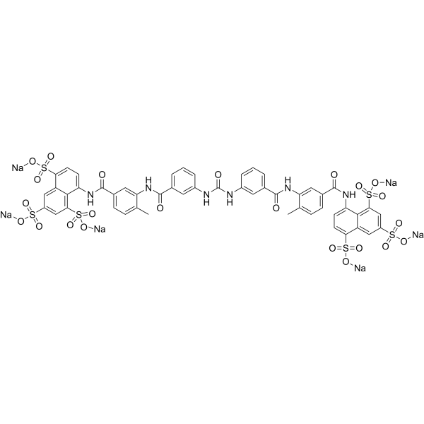
HY-B0879A
Suramin sodium saltPTPases Inhibitor -
HY-15568
A-317491P2X3/P2X2/3 Antagonist
All P2X Receptor Isoforms
More

Biological Activity
Description
|
P2X3 Receptor 0.010 μM (IC50) |
P2X2/3 0.062 μM (IC50) |
IL-1β |
In Vitro
TNP-ATP triethylammonium (3 min) potently blocks agonist-mediated activation of recombinant P2X3 receptors (IC50 = 0.010 μM) and recombinant P2X2/3 receptors (IC50 = 0.062 μM) in 1321N1 cells by inhibiting cytoplasmic calcium influx[1].
TNP-ATP triethylammonium attenuates hypoxia-induced IL-1β expression and release in vitro[2].
TNP-ATP triethylammonium binds to the dimer of E. coli CheA (H48Q mutant), with macroscopic dissociation constants of Kd1 = 0.4 µM and Kd2 = 1.8 µM[3].
TNP-ATP (0.66-9.9 µM) triethylammonium potently inhibits autophosphorylation of wild-type E. coli CheA, with a competitive inhibition constant (Ki) of 0.7 µM[3].
MedChemExpress (MCE) has not independently confirmed the accuracy of these methods. They are for reference only. Further protocols information, click here.
In Vivo
TNP-ATP (1-8 mg/kg; i.p.; single dose; 2 h after hypoxia exposure) triethylammonium significantly ameliorates hypoxia-induced cognitive dysfunction, hypomyelination, neuroinflammation, and glutamate deregulation, with peak efficacy across all measured endpoints at 2 mg/kg[2].
MedChemExpress (MCE) has not independently confirmed the accuracy of these methods. They are for reference only.
-
Animal Model:129J (adult male, 20-25 g)[1]
-
Dosage:1.28 μmol/kg; 12.8 μmol/kg; 38.4 μmol/kg; 128 μmol/kg
-
Administration:i.p.; single dose (3 minutes prior to acetic acid injection)
-
Result:Reduced acetic acid-induced abdominal constrictions dose-dependently, with an ED50 of 6.35 μmol/kg (i.p.).
Completely abolished nociceptive behavior at 128 μmol/kg (i.p.).
Showed antinociceptive potency comparable to morphine (ED50=3 μmol/kg, i.p.), and 6-10 fold greater than suramin and PPADS, and 10 fold greater than TNP-AMP.
-
Animal Model:Sprague-Dawley (postnatal day 0, either sex; male used for P30 Y-maze, P45 Morris water maze, and P60 MRI DTI testing; neonatal hypoxic brain injury model)[2]
-
Dosage:1 mg/kg; 2 mg/kg; 4 mg/kg; 8 mg/kg
-
Administration:i.p.; single dose; 2 h after hypoxia exposure
-
Result:Reduced days needed to reach learning criterion in Y-maze test (P30 male rats) in a dose-dependent manner, with peak efficacy at 2 mg/kg (P < 0.01 vs hypoxia-only group).
Increased time spent in target quadrant and number of platform location crossings in Morris water maze test (P45 male rats) at 2 mg/kg (P < 0.05 vs hypoxia-only group), reversing hypoxia-induced spatial memory deficits.
Attenuated hypoxia-induced expansion of septum and lateral ventricles, restored destroyed myelin structure in corpus callosum (CC), and fully restored fractional anisotropy (FA) values in CC and middle corpus callosum (MCC) to normal control levels at 2 mg/kg (P < 0.01 vs hypoxia-only group) in MRI DTI scanning (P60 male rats).
Reduced hypoxia-induced elevation of IL-1β protein levels in brain tissue at 1 day post-hypoxia at 2 mg/kg (P < 0.05 vs hypoxia-only group).
Prevented hypoxia-induced glutamate elevation in periventricular zone at both 4 h and 1 day post-hypoxia at 2 mg/kg (P < 0.01 at 4 h, P < 0.05 at 1 day vs hypoxia-only group).


Chemical Information
-
CAS No. 61368-63-6
-
Appearance Liquid
-
Molecular Weight 1123.03
-
Formula C40H77N12O19P3
-
Color Orange to red
-
SMILES
CCN(CC)CC.CCN(CC)CC.CCN(CC)CC.CCN(CC)CC.NC1=NC=NC2=C1N=CN2[C@H](O3)[C@H](O4)[C@H](OC54C([N+]([O-])=O)=CC([N+]([O-])=O)=CC5[N+]([O-])=O)[C@H]3COP(O)(OP(OP(O)(O)=O)(O)=O)=O
-
Shipping
Room temperature in continental US; may vary elsewhere.
-
Storage
Solution, -20°C, 2 years

Protocols
-
RNA extraction experimental
By lysing cells, releasing RNA, and removing impurities such as proteins and DNA, high-purity RNA products are finally obtained. The commonly used traditional method is the guanidine isothiocyanate/phenol/chloroform method (Trizol), which is suitable for a variety of animal materials including animal tissues, microorganisms, cultured cells, etc., and most plant materials.
-
Cell Cytotoxicity Assay
Cytotoxicity assays are usually based on the assessment of cell membrane damage, which can also be indirectly detected by measuring cell viability. Detection methods include MTT assay, CKK-8 assay, LDH assay and ATP assay, etc.
-
Research Protocol for Neurological Diseases
PINK1/Parkin-mediated mitophagy pathway is a mitochondrial quality-control signaling axis in which mitochondrial depolarization stabilizes PINK1 on damaged mitochondria, activates Parkin recruitment and E3 ubiquitin ligase activity, promotes ubiquitination of outer mitochondrial membrane proteins, recruits selective autophagy adaptors, and drives lysosomal degradation of damaged mitochondria. In neurological disease research, this pathway is experimentally important because neurons, especially dopaminergic neurons, are highly dependent on mitochondrial integrity, and defective mitochondrial turnover can lead to mitochondrial dysfunction, oxidative stress, impaired neuronal survival, α-synuclein accumulation, and neuroinflammatory damage-associated signals. The genetic disease link is strongest in Parkinson’s disease because mutations in PRKN/parkin cause autosomal recessive juvenile parkinsonism, mutations in PINK1 cause hereditary early-onset Parkinson’s disease, and Drosophila studie
-
Pyroptosis Solutions
Pyroptosis is a lytic inflammatory cell-death pathway executed by gasdermin pores, most classically through inflammasome-mediated activation of caspase-1, cleavage of gasdermin D, membrane pore formation, LDH release, and secretion of IL-1β and IL-18. The canonical pathway is commonly modeled by priming cells with an inflammatory signal such as LPS to induce pro-IL-1β and inflammasome components, followed by an activation signal such as ATP or nigericin to activate NLRP3, ASC speck formation, caspase-1 cleavage, GSDMD cleavage, cytokine release, and pyroptotic membrane rupture. The non-canonical pathway is triggered when cytosolic LPS activates mouse caspase-11 or human caspase-4/5, leading to GSDMD cleavage and pyroptosis, and this can secondarily activate NLRP3-dependent IL-1β release. Pyroptosis is linked to inflammatory injury, infection, cancer, liver disease, ocular disease, placental inflammation, and other disease phenotypes, but unresolved questions include which gasdermin fam
-
Alzheimer’s Disease Modeling
Alzheimer’s Disease (AD) is a neurodegenerative disorder characterized by a progressive decline in cognitive functions and loss of specific types of neurons and synapses. Alzheimer's symptoms can be simulated in mice by injecting drugs (such as Aβ) or genetically modified.


Purity & Documentation
-
Data Sheet (276 KB)
-
SDS (251 KB)
- English - EN (251 KB)
- Français - FR (251 KB)
- Deutsch - DE (251 KB)
- Norwegian - NO (251 KB)
- Español - ES (251 KB)
- Swedish - SV (251 KB)
- Italian - IT (251 KB)
- Korean - KR (251 KB)
- Portuguese - PT (251 KB)
-
Handling Instructions (2659 KB)
References
[1]. Honore P, et al. TNP-ATP, a potent P2X3 receptor antagonist, blocks acetic acid-induced abdominal constriction in mice: comparison with reference analgesics. Pain. 2002 Mar;96(1-2):99-105. [Content Brief]
[2]. Xiao J, et al. TNP-ATP is Beneficial for Treatment of Neonatal Hypoxia-Induced Hypomyelination and Cognitive Decline. Neurosci Bull. 2016;32(1):99-107. [Content Brief]
[3]. Stewart RC, et al. TNP-ATP and TNP-ADP as probes of the nucleotide binding site of CheA, the histidine protein kinase in the chemotaxis signal transduction pathway of Escherichia coli. Biochemistry. 1998;37(35):12269-12279. [Content Brief]

Calculators
Concentration (start) × Volume (start) = Concentration (final) × Volume (final)


TNP-ATP triethylammonium Related Classifications
HY-108667 Related Classifications
 Powered by Bioz