



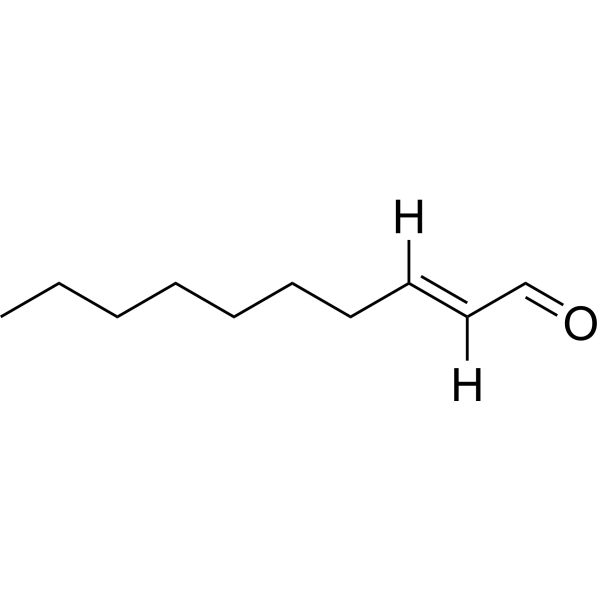
trans-2-Decenal

Based on 1 Customer Validation
trans-2-Decenal ((E)-Dec-2-enal) acts as a urease inhibitor and antibacterial agent against Helicobacter pylori, with an IC50 of 9.484 μg/mL against Helicobacter pylori urease. trans-2-Decenal reduces the urease activity of Helicobacter pylori, and possesses antibacterial, bactericidal, anti-biofilm and anti-migratory activities. It alters the morphology of Helicobacter pylori, induces bacterial rupture, inhibits biofilm formation, reduces the number of mature biofilms and impairs the migratory capacity of Helicobacter pylori. trans-2-Decenal disrupts the cell wall integrity of Phytophthora capsici, damages membrane integrity and permeability, triggers intracellular reactive oxygen species (ROS) accumulation, decreases glutathione levels and disrupts the mitochondrial membrane potential of Phytophthora capsici. trans-2-Decenal is applicable to studies related to Helicobacter pylori and plant diseases induced by and Phytophthora capsici.
For research use only. We do not sell to patients.
- Purity : 98.33%
- CAS No.: 3913-81-3
- Formula: C10H18O
- Molecular Weight:154.25
-
Storage:Pure form -20°C, 3 years , 4°C, 2 years ; In solvent -80°C, 6 months , -20°C, 1 month

To place orders, for customer services and technical support, please contact: MedChemExpress USA
Tel: 609-228-6898 E-mail: [email protected] [email protected]


-
 Biological Activity
-
Chemical Information
-
Solvent & Solubility
- Protocols
- Purity & Documentation
- References
-
 Help & FAQs


-

HY-W127521
trans,trans-2,4-NonadienalBiochemical Assay Reagent -

HY-W015551R
trans-2-Decenal (Standard)Standard -

HY-W089800R
trans-2-Nonenal (Standard)Biochemical Assay Reagent -

HY-W089800
trans-2-NonenalCOX Inhibitor -

HY-W134163
(E)-2-OctenalAntifungal Agent -
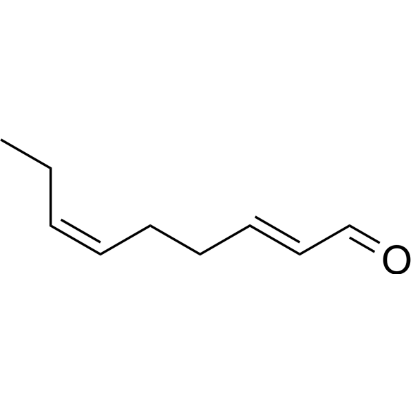
HY-W127515
Trans-2,cis-6-NonadienalBiochemical Assay Reagent -
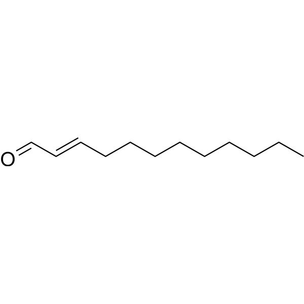
HY-W111720
Trans-2-dodecenalBiochemical Assay Reagent -

HY-W127515R
trans-2,cis-6-Nonadienal (Standard)Biochemical Assay Reagent


Biological Activity
Description
In Vitro
trans-2-Decenal (4-512 μg/mL; 3-5 d) exhibits in vitro bacteriostatic and bactericidal activities against clinically drug-resistant strains of Helicobacter pylori (C907, C101, R1, R2, R4, R10, R16, R24, R36, R40) and the standard strain ATCC43504, with MIC values ranging from 8 to 16 μg/mL and MBC values ranging from 16 to 32 μg/mL[1].
trans-2-Decenal (4-16 μg/mL; 72 h) inhibits the growth of the standard Helicobacter pylori strain ATCC43504 in a concentration-dependent manner within 72 h, with complete growth inhibition achieved at 16 μg/mL[1].
trans-2-Decenal (16-64 μg/mL; 24 h) exhibits rapid, concentration-dependent bactericidal activity against the standard Helicobacter pylori strain ATCC43504: 16 μg/mL eliminates all viable bacteria within 4 h, while complete bactericidal effect is achieved within 1 h at 32 μg/mL or 64 μg/mL[1].
trans-2-Decenal (16 μg/mL; 4 h) alters the morphology of the standard Helicobacter pylori strain ATCC43504 and causes damage to its ultrastructure after 4 h of incubation[1].
trans-2-Decenal (8-64 μg/mL; 1-3 d) inhibits biofilm formation and mature biofilm growth of the standard Helicobacter pylori strain ATCC43504 in a concentration-dependent manner, with the maximum inhibitory effect achieved at 16 μg/mL[1].
trans-2-Decenal (0-16 μg/mL; 20 min) inhibits Helicobacter pylori urease activity in a concentration-dependent manner, with an IC50 of 9.484 μg/mL[1].
trans-2-Decenal (4-32 μg/mL; 4 d) inhibits the migration ability of the standard Helicobacter pylori strain ATCC43504 in a concentration-dependent manner, and complete growth inhibition is observed at the concentration of 32 μg/mL[1].
Trans-2-decenal (16-32 μg/mL; 12-48 h) induces dose-dependent intracellular ROS accumulation and depletes glutathione levels in Phytophthora capsici strain LT263: a strong DCFH-DA fluorescence signal is detected at the concentration of 32 μg/mL after 12 h of treatment; after 48 h of treatment, the GSH content decreases to 44.39% of that in the control group at the concentration of 32 μg/mL[2].
Trans-2-decenal (16-32 μg/mL; 12 h) dose-dependently disrupts the mitochondrial membrane potential of Phytophthora capsici strain LT263[2].
MedChemExpress (MCE) has not independently confirmed the accuracy of these methods. They are for reference only. Further protocols information, click here.


Chemical Information
-
CAS No. 3913-81-3
-
Appearance Liquid (Density: 0.841 g/cm3)
-
Molecular Weight 154.25
-
Formula C10H18O
-
Color Colorless to light yellow
-
SMILES
CCCCCCC/C=C/C=O
-
Synonyms
(E)-Dec-2-enal
-
Initial Source
-
Shipping
Room temperature in continental US; may vary elsewhere.
-
Storage
Pure form -20°C 3 years 4°C 2 years In solvent -80°C 6 months -20°C 1 month


Solvent & Solubility
In Vitro:
DMSO : 200 mg/mL (1296.60 mM; Need ultrasonic; Hygroscopic DMSO has a significant impact on the solubility of product, please use newly opened DMSO)
Please refer to the solubility information to select the appropriate solvent. Once prepared, please aliquot and store the solution to prevent product inactivation from repeated freeze-thaw cycles.
Storage method and period of stock solution: -80°C, 6 months; -20°C, 1 month. When stored at -80°C, please use it within 6 months. When stored at -20°C, please use it within 1 month.
Please refer to the solubility information to select the appropriate solvent. Once prepared, please aliquot and store the solution to prevent product inactivation from repeated freeze-thaw cycles.
Storage method and period of stock solution: -80°C, 6 months; -20°C, 1 month. When stored at -80°C, please use it within 6 months. When stored at -20°C, please use it within 1 month.
Concentration (start) × Volume (start) = Concentration (final) × Volume (final)
In Vivo:
Select the appropriate dissolution method based on your experimental animal and administration route.
- For the following dissolution methods, please ensure to first prepare a clear stock solution using an In Vitro approach and then sequentially add co-solvents:
- To ensure reliable experimental results, the clarified stock solution can be appropriately stored based on storage conditions. As for the working solution for In Vivo experiments, it is recommended to prepare freshly and use it on the same day.
- The percentages shown for the solvents indicate their volumetric ratio in the final prepared solution. If precipitation or phase separation occurs during preparation, heat and/or sonication can be used to aid dissolution.
Add each solvent one by one: 10% DMSO 90% (20% SBE-β-CD in Saline)
Solubility: ≥ 5 mg/mL (32.41 mM); Clear solution
This protocol yields a clear solution of ≥ 5 mg/mL (saturation unknown).
Taking 1 mL working solution as an example, add 100 μL DMSO stock solution (50.0 mg/mL) to 900 μL 20% SBE-β-CD in Saline, and mix evenly.
Preparation of 20% SBE-β-CD in Saline (4°C, storage for one week): 2 g SBE-β-CD powder is dissolved in 10 mL Saline, completely dissolve until clear.
In Vivo Dissolution Calculator
Please enter the basic information of animal experiments:
-
-
-
-
Recommended: Prepare an additional quantity of animals to account for potential losses during experiments.
Please enter your animal formula composition:
-
%DMSO +
Recommended: Keep the proportion of DMSO in working solution below 2% if your animal is weak.
-
%+
-
+%Tween-80 + +
-
%Saline +
The co-solvents required include: DMSO, . All of co-solvents are available by MedChemExpress (MCE). , Tween 80. All of co-solvents are available by MedChemExpress (MCE).
Working solution concentration: 0.22 mg/mL
Method for preparing stock solution: mg drug dissolved in μL DMSO. Stock solution concentration: mg/mL.
1. Take μL DMSO stock solution;
2. Add μL .
μL , mix evenly;
3. Then add μL Tween 80, mix evenly;
4. Then add μL
Please ensure that the stock solution in the first step is dissolved to a clear state, and add co-solvents in sequence. You can use ultrasonic heating (ultrasonic cleaner, recommended frequency 20-40 kHz), vortexing, etc. to assist dissolution.

Protocols
-
Research Protocol for Infectious Diseases
Infectious-disease experiments test how pathogens interact with host barriers, innate immune receptors, inflammatory signaling, pathogen replication, and tissue injury; pattern-recognition receptors such as TLRs, RIG-I-like receptors, NOD-like receptors, and inflammasomes detect microbial molecules and activate NF-κB, interferon, and cytokine responses. The central hypothesis is that infection severity reflects the balance between pathogen burden and host response: protective inflammation restricts pathogen growth, whereas excessive or mislocalized inflammation contributes to tissue damage and disease phenotype. Unresolved questions include which host pathways are protective versus pathogenic, why some infection models fail to translate to human disease, and which combined readouts best predict clinically relevant infection outcomes.
-
Mitochondrial membrane-potential fluorescent assay
Mitochondrial membrane potential fluorescent assays estimate ΔΨm in living cells using lipophilic cationic dyes such as TMRM, TMRE, rhodamine 123, and JC-1, which accumulate in mitochondria according to membrane polarization; loss of signal after FCCP or CCCP treatment is interpreted as mitochondrial depolarization. TMRM/TMRE and rhodamine 123 are commonly used for semi-quantitative live-cell microscopy or flow cytometry, while JC-1 can report a shift from red aggregate fluorescence to green monomer fluorescence during depolarization; interpretation requires controls because dye concentration, quenching mode, cell type, dye efflux, and mitochondrial mass can affect fluorescence independently of ΔΨm.
-
Cell Cytotoxicity Assay
Cytotoxicity assays are usually based on the assessment of cell membrane damage, which can also be indirectly detected by measuring cell viability. Detection methods include MTT assay, CKK-8 assay, LDH assay and ATP assay, etc.
-
Cell migration
Cell migration is a method that plays an important role in wound healing, cell differentiation, embryonic development, etc.
-
ROS/oxidative-stress fluorescent staining
ROS/oxidative-stress fluorescent staining uses cell-permeant fluorogenic probes that become fluorescent after oxidation inside cells or tissues; commonly used examples include DCFH-DA/DCFDA for broad cellular oxidant detection, DHE for superoxide-related signal detection, MitoSOX for mitochondrial superoxide-related signal detection, and CellROX probes for oxidative-stress-associated fluorescence readouts. The assay detects probe oxidation rather than a single ROS species unless the probe and analysis method have been chemically validated for that species. DCFH-DA enters cells, is deacetylated by intracellular esterases to DCFH, and produces fluorescent DCF after oxidation, so the readout is used as an operational measure of total cellular oxidative stress rather than a species-specific ROS measurement. DHE and MitoSOX can report superoxide-related oxidation, but red fluorescence alone can include non-specific ethidium-like oxidation products; HPLC or optimized spectral approaches are
-
Fluorescent plasma-membrane potential dye assay
Fluorescent plasma-membrane potential dye assays measure changes in cell membrane potential using voltage-sensitive dyes whose fluorescence changes when cells depolarize or hyperpolarize. Anionic bis-oxonol dyes such as DiBAC4(3) enter depolarized cells more readily and show increased fluorescence after intracellular binding, while hyperpolarization reduces dye accumulation and fluorescence. FMP/FLIPR membrane-potential dyes are used for faster, homogeneous microplate assays of ion-channel or receptor-mediated membrane-potential changes.
-
Mitochondrial membrane-potential and mitochondrial mass staining
Mitochondrial membrane potential staining measures the electrochemical polarization across the mitochondrial inner membrane in live cells using lipophilic cationic fluorescent probes; early rhodamine-based work showed that selective mitochondrial dye accumulation is lost when the mitochondrial transmembrane potential is dissipated. JC-1 reports mitochondrial polarization by shifting from green monomer fluorescence to red J-aggregate fluorescence as dye concentration increases within energized mitochondria; therefore, the red/green fluorescence ratio is used as a relative readout of mitochondrial membrane potential. TMRE or TMRM staining provides a single-channel relative readout because these cationic rhodamine esters accumulate in polarized mitochondria, and lower fluorescence indicates reduced mitochondrial polarization when acquisition and dye-loading conditions are controlled. Mitochondrial mass staining is commonly performed with MitoTracker Green FM or related MitoTracker dyes as
-
Transepithelial/transendothelial electrical resistance assay
TEER measures electrical resistance across epithelial or endothelial monolayers cultured on permeable supports, and the readout reflects ionic conductance through the cell barrier, especially the paracellular pathway regulated by junctional integrity. TEER can be measured without destroying the monolayer and is commonly used before or during transport, permeability, barrier-disruption, and barrier-maturation experiments. TEER values are influenced by biological maturation and technical conditions; reported factors include temperature, medium formulation, passage number, electrode geometry, membrane properties, and junctional length during early monolayer maturation. Therefore, TEER should be interpreted with blank-insert subtraction, area normalization, repeated readings, and, when possible, orthogonal barrier readouts such as FITC-dextran flux or tight-junction staining.
-
Fungal Biofilm Culture
Fungal biofilm culture is an in vitro method for growing surface-attached fungal communities, most commonly Candida albicans, on abiotic substrates such as polystyrene wells, silicone elastomer, or polymethylmethacrylate; the assay models adhesion, proliferation, filamentation, extracellular-matrix-associated maturation, and dispersion. Biofilm output can be read by optical density at 600 nm for adherent biomass, XTT reduction for metabolic activity, CFU recovery for viable attached or dispersed cells, and microscopy for architecture.


Purity & Documentation
-
Data Sheet (270 KB)
-
SDS (537 KB)
- English - EN (537 KB)
- Français - FR (537 KB)
- Deutsch - DE (537 KB)
- Norwegian - NO (537 KB)
- Español - ES (537 KB)
- Swedish - SV (537 KB)
- Italian - IT (537 KB)
- Korean - KR (537 KB)
- Portuguese - PT (537 KB)
-
Handling Instructions (2659 KB)
References


Complete Stock Solution Preparation Table
Please refer to the solubility information to select the appropriate solvent. Once prepared, please aliquot and store the solution to prevent product inactivation from repeated freeze-thaw cycles.
Storage method and period of stock solution: -80°C, 6 months; -20°C, 1 month. When stored at -80°C, please use it within 6 months. When stored at -20°C, please use it within 1 month.
| Optional Solvent | Concentration Solvent Mass | 1 mg | 5 mg | 10 mg | 25 mg |
|---|---|---|---|---|---|
| DMSO | 1 mM | 6.4830 mL | 32.4149 mL | 64.8298 mL | 162.0746 mL |
| 5 mM | 1.2966 mL | 6.4830 mL | 12.9660 mL | 32.4149 mL | |
| 10 mM | 0.6483 mL | 3.2415 mL | 6.4830 mL | 16.2075 mL | |
| 15 mM | 0.4322 mL | 2.1610 mL | 4.3220 mL | 10.8050 mL | |
| 20 mM | 0.3241 mL | 1.6207 mL | 3.2415 mL | 8.1037 mL | |
| 25 mM | 0.2593 mL | 1.2966 mL | 2.5932 mL | 6.4830 mL | |
| 30 mM | 0.2161 mL | 1.0805 mL | 2.1610 mL | 5.4025 mL | |
| 40 mM | 0.1621 mL | 0.8104 mL | 1.6207 mL | 4.0519 mL | |
| 50 mM | 0.1297 mL | 0.6483 mL | 1.2966 mL | 3.2415 mL | |
| 60 mM | 0.1080 mL | 0.5402 mL | 1.0805 mL | 2.7012 mL | |
| 80 mM | 0.0810 mL | 0.4052 mL | 0.8104 mL | 2.0259 mL | |
| 100 mM | 0.0648 mL | 0.3241 mL | 0.6483 mL | 1.6207 mL |


trans-2-Decenal Related Classifications
HY-W015551 Related Classifications
 Powered by Bioz