



Trierucin

Based on 1 Customer Validation
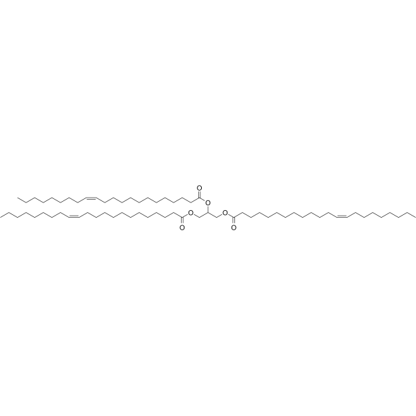
Trierucin (Trierucoylglycerol) is a triester formed by glycerol and erucic acid, which serves as an occlusive skin conditioning agent and non-aqueous thickener in cosmetics. Long-term feeding of Trierucin to rats induces chronic toxicities such as cardiac lipid deposition and fibrosis.
For research use only. We do not sell to patients.
- Purity : 99.89%
- CAS No.: 2752-99-0
- Formula: C69H128O6
- Molecular Weight:1053.75
-
Storage:Pure form -20°C, 3 years , 4°C, 2 years
* The compound is unstable in solutions, freshly prepared is recommended.

To place orders, for customer services and technical support, please contact: MedChemExpress USA
Tel: 609-228-6898 E-mail: [email protected] [email protected]


-
 Biological Activity
-
Chemical Information
-
Solvent & Solubility
- Protocols
- Purity & Documentation
- References
-
 Help & FAQs


-
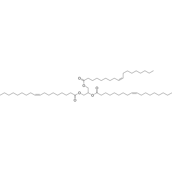
HY-N1981
TrioleinMonoacid Triglyceride -

HY-N1981R
Triolein (Standard)Monoacid Triglyceride -
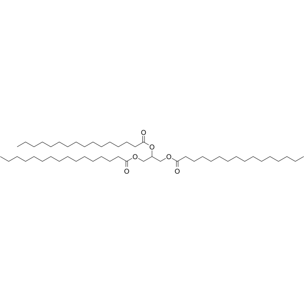
HY-W013061
Propane-1,2,3-triyl tripalmitateGlycerol Tristearate Lymphatic Transport Promoter -

HY-W923355
1,2,3-Trieicosapentaenoyl glycerolGlyceride -
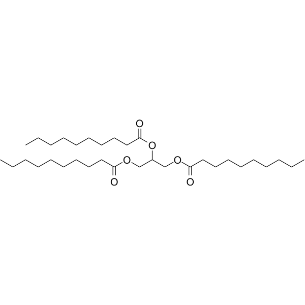
HY-N6660
TrisdecanoinDA Precursor -
HY-126573
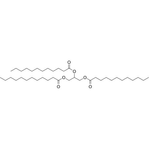
TrilaurinNeoplasm Inhibitor -
HY-W127408
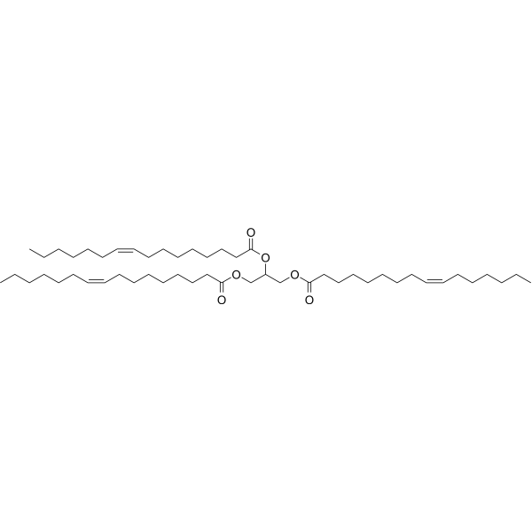
TripalmitoleinBiochemical Assay Reagent -
HY-B1804
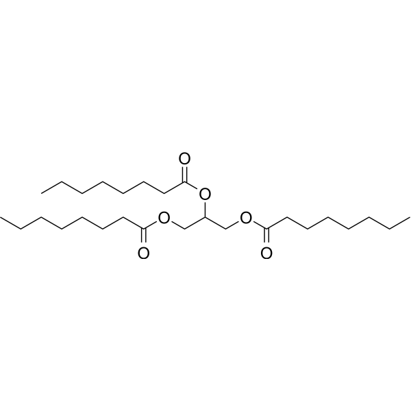
TricaprilinKetogenic Agent -

HY-Y1039
1-Stearoyl-rac-glycerolBiochemical Assay Reagent -
HY-W127506
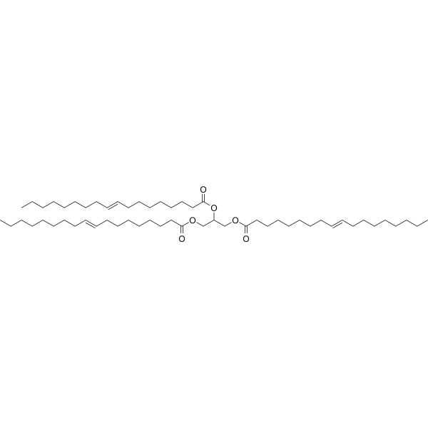
TrielaidinBiochemical Assay Reagent -
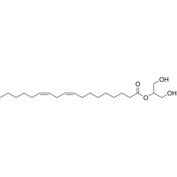
HY-130311
2-Linoleoyl glycerolCB1 Receptor Agonist -
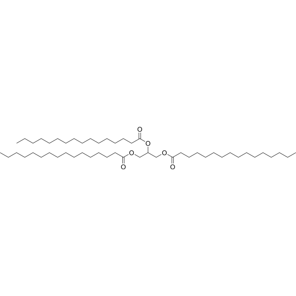
HY-W013061R
Propane-1,2,3-triyl tripalmitate (Standard)Standard of Propane-1,2,3-triyl Tripalmitate


Biological Activity
Description
IC50 & Target
|
Human Endogenous Metabolite |
In Vivo
MedChemExpress (MCE) has not independently confirmed the accuracy of these methods. They are for reference only.
-
Animal Model:Wistar rats (male, 3 weeks old)[1]
-
Dosage:30 cal %
-
Administration:dietary administration; daily; up to 24 weeks
-
Result:Induced severe cardiac lipidosis after 1 week of dosing.
Induced cardiac lesions (lipidosis and/or focal fibrosis) after 24 weeks, with less severe cardiac lipidosis than in rats killed after 1 week.
Induced cardiac fibrosis in rats.
Induced tubular dilatation, proteinaceous casts, or interstitial foci of fibrosis in kidneys rats killed after 24 weeks.


Chemical Information
-
CAS No. 2752-99-0
-
Appearance <31.5°C Solid,>32°C Liquid
-
Molecular Weight 1053.75
-
Formula C69H128O6
-
Color White to off-white
-
SMILES
CCCCCCCC/C=C\CCCCCCCCCCCC(OCC(OC(CCCCCCCCCCC/C=C\CCCCCCCC)=O)COC(CCCCCCCCCCC/C=C\CCCCCCCC)=O)=O
-
Synonyms
Trierucoylglycerol
-
Structure Classification
-
Initial Source
-
Shipping
Room temperature in continental US; may vary elsewhere.
-
Storage
Pure form -20°C 3 years 4°C 2 years * The compound is unstable in solutions, freshly prepared is recommended.


Solvent & Solubility
In Vitro:
DMSO : 32.5 mg/mL (30.84 mM; Need ultrasonic; Hygroscopic DMSO has a significant impact on the solubility of product, please use newly opened DMSO)
Please refer to the solubility information to select the appropriate solvent. The compound is unstable in solutions, freshly prepared is recommended.
Please refer to the solubility information to select the appropriate solvent. The compound is unstable in solutions, freshly prepared is recommended.
Concentration (start) × Volume (start) = Concentration (final) × Volume (final)
In Vivo:
Select the appropriate dissolution method based on your experimental animal and administration route.
- For the following dissolution methods, please ensure to first prepare a clear stock solution using an In Vitro approach and then sequentially add co-solvents:
- To ensure reliable experimental results, the clarified stock solution can be appropriately stored based on storage conditions. As for the working solution for In Vivo experiments, it is recommended to prepare freshly and use it on the same day.
- The percentages shown for the solvents indicate their volumetric ratio in the final prepared solution. If precipitation or phase separation occurs during preparation, heat and/or sonication can be used to aid dissolution.
Add each solvent one by one: 10% DMSO 40% PEG300 5% Tween-80 45% Saline
Solubility: ≥ 1.08 mg/mL (1.02 mM); Clear solution
This protocol yields a clear solution of ≥ 1.08 mg/mL (saturation unknown).
Taking 1 mL working solution as an example, add 100 μL DMSO stock solution (10.8 mg/mL) to 400 μL PEG300, and mix evenly; then add 50 μL Tween-80 and mix evenly; then add 450 μL Saline to adjust the volume to 1 mL.
Preparation of Saline: Dissolve 0.9 g sodium chloride in ddH₂O and dilute to 100 mL to obtain a clear Saline solution.
Add each solvent one by one: 10% DMSO 90% (20% SBE-β-CD in Saline)
Solubility: ≥ 1.08 mg/mL (1.02 mM); Clear solution
This protocol yields a clear solution of ≥ 1.08 mg/mL (saturation unknown).
Taking 1 mL working solution as an example, add 100 μL DMSO stock solution (10.8 mg/mL) to 900 μL 20% SBE-β-CD in Saline, and mix evenly.
Preparation of 20% SBE-β-CD in Saline (4°C, storage for one week): 2 g SBE-β-CD powder is dissolved in 10 mL Saline, completely dissolve until clear.
In Vivo Dissolution Calculator
Please enter the basic information of animal experiments:
-
-
-
-
Recommended: Prepare an additional quantity of animals to account for potential losses during experiments.
Please enter your animal formula composition:
-
%DMSO +
Recommended: Keep the proportion of DMSO in working solution below 2% if your animal is weak.
-
%+
-
+%Tween-80 + +
-
%Saline +
The co-solvents required include: DMSO, . All of co-solvents are available by MedChemExpress (MCE). , Tween 80. All of co-solvents are available by MedChemExpress (MCE).
Working solution concentration: 0.22 mg/mL
Method for preparing stock solution: mg drug dissolved in μL DMSO. Stock solution concentration: mg/mL. * The compound is unstable in solutions, freshly prepared is recommended.
1. Take μL DMSO stock solution;
2. Add μL .
μL , mix evenly;
3. Then add μL Tween 80, mix evenly;
4. Then add μL
Please ensure that the stock solution in the first step is dissolved to a clear state, and add co-solvents in sequence. You can use ultrasonic heating (ultrasonic cleaner, recommended frequency 20-40 kHz), vortexing, etc. to assist dissolution.


Purity & Documentation
-
Data Sheet (266 KB)
-
SDS (254 KB)
- English - EN (254 KB)
- Français - FR (254 KB)
- Deutsch - DE (254 KB)
- Norwegian - NO (254 KB)
- Español - ES (254 KB)
- Swedish - SV (254 KB)
- Italian - IT (254 KB)
- Korean - KR (254 KB)
- Portuguese - PT (254 KB)
-
Handling Instructions (2659 KB)
References
[1]. Johnson W Jr, et al. Final report on the safety assessment of trilaurin, triarachidin, tribehenin, tricaprin, tricaprylin, trierucin, triheptanoin, triheptylundecanoin, triisononanoin, triisopalmitin, triisostearin, trilinolein, trimyristin, trioctanoin, triolein, tripalmitin, tripalmitolein, triricinolein, tristearin, triundecanoin, glyceryl triacetyl hydroxystearate, glyceryl triacetyl ricinoleate, and glyceryl stearate diacetate. Int J Toxicol. 2001;20 Suppl 4:61-94. [Content Brief]


Complete Stock Solution Preparation Table
Please refer to the solubility information to select the appropriate solvent. The compound is unstable in solutions, freshly prepared is recommended.
| Optional Solvent | Concentration Solvent Mass | 1 mg | 5 mg | 10 mg | 25 mg |
|---|---|---|---|---|---|
| DMSO | 1 mM | 0.9490 mL | 4.7450 mL | 9.4899 mL | 23.7248 mL |
| 5 mM | 0.1898 mL | 0.9490 mL | 1.8980 mL | 4.7450 mL | |
| 10 mM | 0.0949 mL | 0.4745 mL | 0.9490 mL | 2.3725 mL | |
| 15 mM | 0.0633 mL | 0.3163 mL | 0.6327 mL | 1.5817 mL | |
| 20 mM | 0.0474 mL | 0.2372 mL | 0.4745 mL | 1.1862 mL | |
| 25 mM | 0.0380 mL | 0.1898 mL | 0.3796 mL | 0.9490 mL | |
| 30 mM | 0.0316 mL | 0.1582 mL | 0.3163 mL | 0.7908 mL |


Trierucin Related Classifications
HY-N7055 Related Classifications
 Powered by Bioz