



TrkB agonist-1

Based on 1 Customer Validation
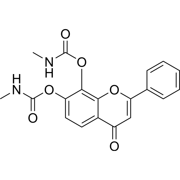
TrkB agonist-1 is an orally active, blood-brain barrier-permeable TrkB agonist. TrkB agonist-1 activates the TrkB signaling pathway and its downstream AKT and ERK/MAPK pathways via its released parent compound, reduces AEP enzyme activity and inhibits its activation, and simultaneously suppresses the production of pro-inflammatory cytokines. TrkB agonist-1 is used in the research of Alzheimer's disease.
For research use only. We do not sell to patients.
- Purity : 99.2%
- CAS No.: 1609067-49-3
- Formula: C19H16N2O6
- Molecular Weight:368.34
-
Storage:Powder -20°C, 3 years ; In solvent -80°C, 6 months , -20°C, 1 month

To place orders, for customer services and technical support, please contact: MedChemExpress USA
Tel: 609-228-6898 E-mail: [email protected] [email protected]


-
 Biological Activity
-
Chemical Information
- Protocols
- Purity & Documentation
- References
-
 Help & FAQs


-
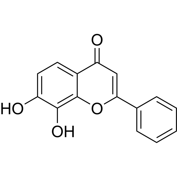
HY-W013372
7,8-Dihydroxyflavone -
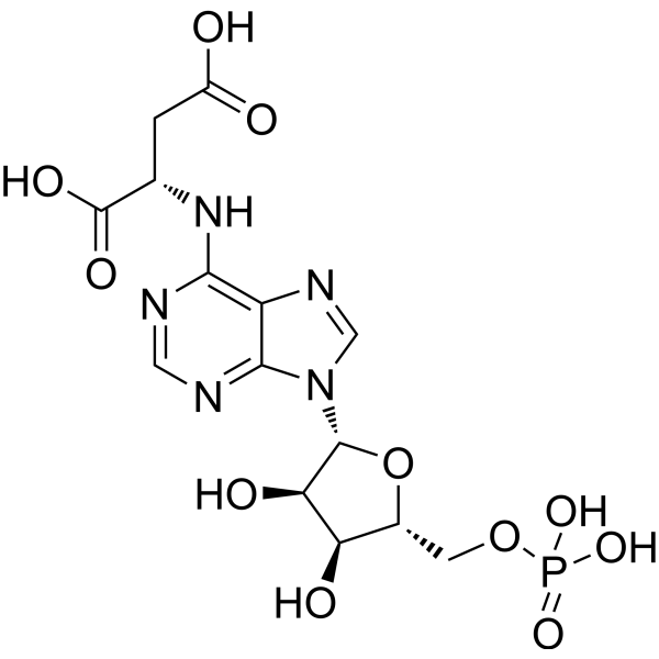
HY-127137
Adenylosuccinic acidNucleotide Cycle Metabolite -
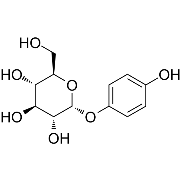
HY-N3002
α-ArbutinTyrosinase Inhibitor -

HY-N3002R
α-Arbutin (Standard)Tyrosinase Inhibitor -

HY-W013372R
7,8-Dihydroxyflavone (Standard) -
HY-W011641
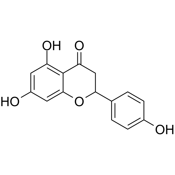
(±)-NaringeninAnti-inflammatory Agents -
HY-W011641R
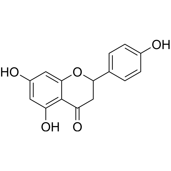
(±)-Naringenin (Standard) -
HY-115434
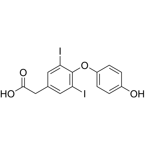
3,5-Diiodothyroacetic acidThyroid Hormone Analog


Biological Activity
Description
IC50 & Target
[1]|
TrkB |
In Vitro
TrkB agonist-1 (R13) exhibits favorable in vitro ADME properties, with adequate intestinal stability and efficient conversion to 7,8-DHF in liver microsomes and plasma[1].
TrkB agonist-1 displays strong stability in acidic pH matching gastric conditions, and stepwise hydrolysis into active 7,8-DHF under neutral pH conditions[1].
MedChemExpress (MCE) has not independently confirmed the accuracy of these methods. They are for reference only. Further protocols information, click here.
Parmacokinetics
| Species | Dose | Route | Cmax | Tmax | AUC0-t | T1/2 | Bioavailability |
|---|---|---|---|---|---|---|---|
| Mice[1] | 36 mg/kg | p.o. | 129 ng/mL | 30 min | 11880 min·ng/mL | 219.6 min | 10.5 % |
In Vivo
TrkB agonist-1 (21.8-72.5 mg/kg; p.o.; single dose) a single oral dose dose-dependently releases 7,8-DHF into both systemic circulation and brain tissue, with the intermediate hydrolysis product T1 restricted to plasma and not penetrating the brain parenchyma[1].
TrkB agonist-1 (36-72.5 mg/kg; p.o.; single dose) single oral administration provides sustained release of 7,8-DHF with markedly improved T1/2, oral bioavailability, and extended brain exposure in mice[1].
MedChemExpress (MCE) has not independently confirmed the accuracy of these methods. They are for reference only.
-
Animal Model:5XFAD transgenic mice (bred in a pathogen-free environment, starting treatment at 2 months of age)[1]
-
Dosage:7.25 mg/kg/d; 21.8 mg/kg/d; 43.6 mg/kg/d
-
Administration:p.o.; daily; 3 months
-
Result:Increased TrkB phosphorylation levels in a dose-dependent manner, with phosphorylated AKT and phosphorylated ERK downstream of TrkB also elevated in a dose-dependent manner.
Reversed pathological reduction of hippocampal CA1 region synaptic density in a dose-dependent manner, and increased long-term potentiation in a dose-dependent fashion.
Reduced Aβ deposition in both hippocampus and frontal cortex in a dose-dependent manner, and reduced brain Aβ40 concentrations at all tested doses.
Attenuated spatial memory acquisition deficit measured by Morris water maze latency AUC and swim path distance AUC, and significantly improved spatial memory recall in the probe trial.
Reduced mature active AEP enzymatic activity at 21.8 mg/kg and 43.6 mg/kg doses, and inhibited AEP-mediated pathological cleavage of APP and Tau.
Significantly reduced brain concentrations of the proinflammatory cytokines IL-1β, IL-6, and TNFα across all tested doses.
-
Animal Model:Male CD1 mice[1]
-
Dosage:21.8 mg/kg; 43.6 mg/kg; 72.5 mg/kg
-
Administration:p.o.; single dose
-
Result:Reached mean plasma 7,8-DHF concentrations of 2.1 ng/mL at 21.8 mg/kg, 4.8 ng/mL at 43.6 mg/kg, and 6.9 ng/mL at 72.5 mg/kg at 4 hours post-administration.
Reached mean brain 7,8-DHF concentration of 3.4 ng/g at 4 hours post-administration for the 72.5 mg/kg dose.
Detected the intermediate T1 in plasma at mean concentrations of 10.2 ng/mL (21.8 mg/kg), 34.0 ng/mL (43.6 mg/kg), and 42.3 ng/mL (72.5 mg/kg), with T1 undetectable in brain across all doses.
-
Animal Model:Mice[1]
-
Dosage:36 mg/kg; 72.5 mg/kg
-
Administration:p.o.; single dose
-
Result:Achieved mean Cmax of released 7,8-DHF of 129 ng/mL, Tmax of 30 min, AUC (0-t) value of 11880 min·ng/mL, and 7,8-DHF T1/2 of 219.6 min after a single 36 mg/kg oral dose, resulting in 10.5% oral bioavailability for R13.
Reached mean Cmax of 7,8-DHF in plasma of 56 ng/mL at 2 hours post-dose, and mean Cmax in brain tissue of 46 ng/g at 4 hours post-dose after a single 72.5 mg/kg oral dose, with 7,8-DHF concentrations remaining above 5 ng/g in brain for at least 240 minutes and 7,8-DHF still detectable in plasma at 8 hours after administration.


Chemical Information
-
CAS No. 1609067-49-3
-
Appearance Solid
-
Molecular Weight 368.34
-
Formula C19H16N2O6
-
Color White to off-white
-
SMILES
O=C1C=C(C2=CC=CC=C2)OC3=C(OC(NC)=O)C(OC(NC)=O)=CC=C13
-
Shipping
Room temperature in continental US; may vary elsewhere.
-
Storage
Powder -20°C 3 years In solvent -80°C 6 months -20°C 1 month

Protocols
-
LPS-Induced Endotoxemia/Systemic Inflammation
Lipopolysaccharide (LPS)-induced endotoxemia is a widely used in vivo model of acute systemic inflammation in which LPS, a Gram-negative bacterial endotoxin, activates innate immune signaling primarily through TLR4, leading to rapid and transient induction of pro-inflammatory cytokines such as TNF-α, IL-6, and IL-1β in circulation and tissues. This cytokine surge is commonly used as a measurable readout of systemic inflammatory activation and immune dysregulation, and is typically assessed within hours after intraperitoneal LPS administration in mouse models of endotoxemia. The model captures key features of systemic inflammatory response syndrome, including cytokine release, immune cell activation, and downstream tissue responses, and has been used to evaluate anti-inflammatory interventions such as cytokine modulation, lipid mediators, and immune cell-targeting therapies.
-
Research Protocol for Inflammation-related Diseases
The NLRP3 inflammasome is a cytosolic innate immune signaling platform that integrates priming signals and danger-signal activation to promote caspase-1 activation, maturation of IL-1β and IL-18, and gasdermin D-mediated pyroptotic cell death. The core experimental logic is to determine whether inflammatory disease phenotypes are driven by increased NLRP3 expression, ASC-containing inflammasome assembly, caspase-1 cleavage, GSDMD cleavage, and extracellular release of IL-1β/IL-18 rather than by nonspecific cell injury alone. The pathway is strongly linked to inflammation-related disease phenotypes because monosodium urate crystals activate NALP3/NLRP3 inflammasome signaling in gout-like crystal inflammation, cholesterol crystals activate NLRP3 inflammasomes in atherogenesis models, and DSS-induced intestinal inflammation has been reported to involve NLRP3 inflammasome activity. However, experimental colitis studies also show context-dependent protective effects of NLRP3 inflammasome co
-
Alzheimer’s Disease Modeling
Alzheimer’s Disease (AD) is a neurodegenerative disorder characterized by a progressive decline in cognitive functions and loss of specific types of neurons and synapses. Alzheimer's symptoms can be simulated in mice by injecting drugs (such as Aβ) or genetically modified.


Purity & Documentation
-
Data Sheet (287 KB)
-
SDS (393 KB)
- English - EN (393 KB)
- Français - FR (393 KB)
- Deutsch - DE (393 KB)
- Norwegian - NO (393 KB)
- Español - ES (393 KB)
- Swedish - SV (393 KB)
- Italian - IT (393 KB)
- Korean - KR (393 KB)
- Portuguese - PT (393 KB)
-
Handling Instructions (2659 KB)
References

Calculators
Concentration (start) × Volume (start) = Concentration (final) × Volume (final)


TrkB agonist-1 Related Classifications
HY-153189 Related Classifications
 Powered by Bioz