



Typhaneoside
Based on 4 publication(s) in Google Scholar
Typhaneoside is an orally active activator of PI3K/Akt/mTOR and farnesoid X receptor. Typhaneoside promotes the activation of AMPK and Caspase-3, induces apoptosis, ferroptosis, autophagy, ROS accumulation, cell cycle arrest at the G2/M phase, and reduces cancer cell viability. Typhaneoside improves glucose and lipid metabolism, alleviates inflammatory responses, oxidative stress and hepatic lipid accumulation, and exerts hepatoprotective effects. Typhaneoside can be used in research related to heart failure after myocardial infarction, acute myeloid leukemia, non-alcoholic fatty liver disease and neurological disorders.
For research use only. We do not sell to patients.
- Purity : 99.74%
- CAS No.: 104472-68-6
- Formula: C34H42O20
- Molecular Weight:770.69
-
Storage:
4°C, protect from light
* In solvent : -80°C, 6 months; -20°C, 1 month (protect from light)

To place orders, for customer services and technical support, please contact: MedChemExpress USA
Tel: 609-228-6898 E-mail: [email protected] [email protected]


-
 Biological Activity
-
Chemical Information
-
Solvent & Solubility
- Protocols
- Purity & Documentation
- References
-
 Help & FAQs


-
HY-N0527
PentagalloylglucoseAntioxidant -
HY-13653
(-)-Epigallocatechin GallateAnticancer Agent -
HY-N1362
Salvianolic acid BAntioxidant -
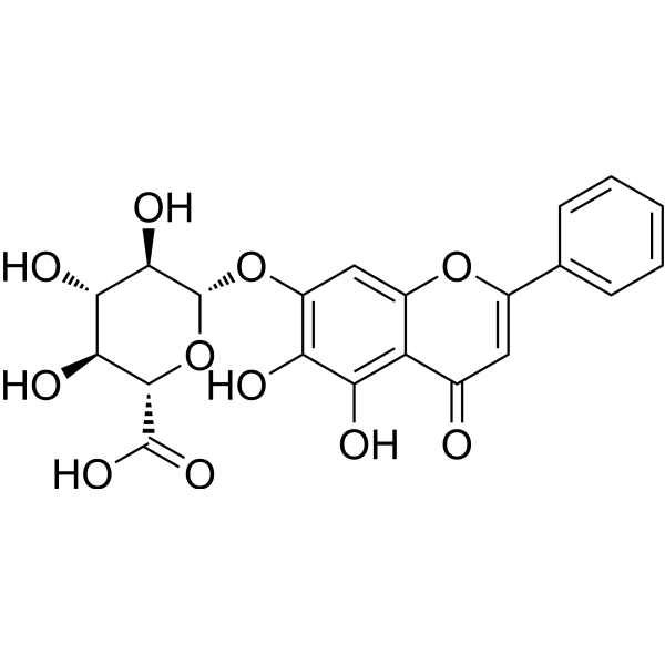
HY-N0197
BaicalinAutophagy Inducer -
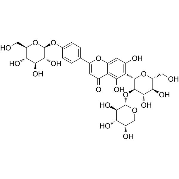
HY-N1419
VaccarinAKT/ERK/AMPK Agonists -
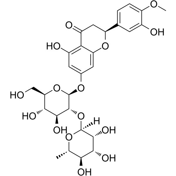
HY-N0101
NeohesperidinWnt/β-Catenin Activator -
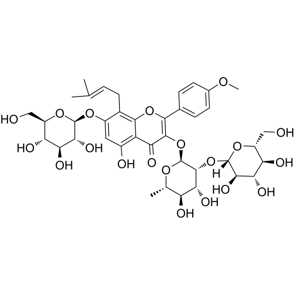
HY-N0257
Epimedin ANatural Product -
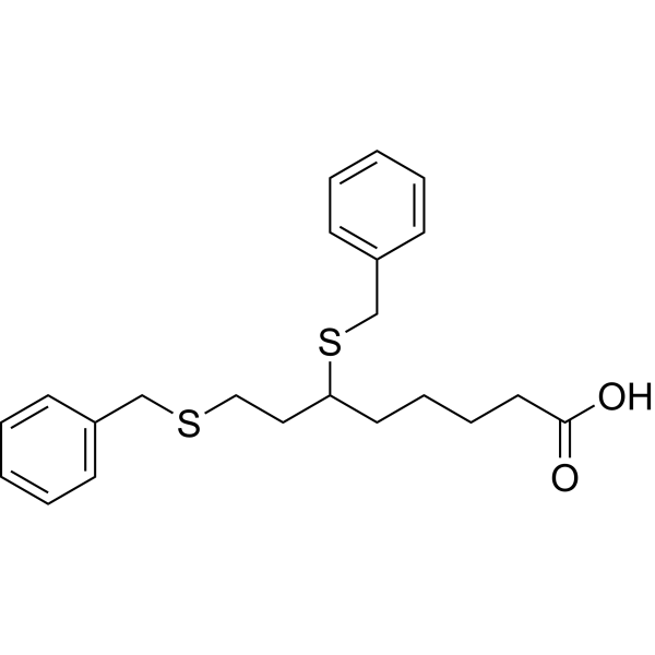
HY-15453
DevimistatMitochondrial Metabolism Inhibitor -

HY-N0712R
Typhaneoside (Standard)Autophagy Inhibitor -
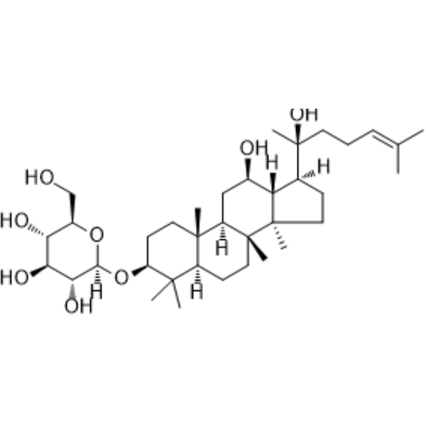
HY-N0605
Ginsenoside Rh2Caspase Activator -
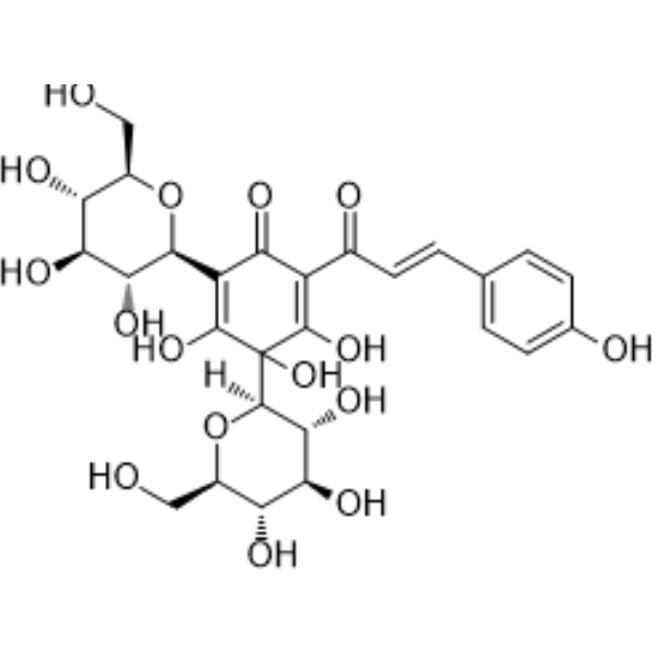
HY-N0567
Hydroxysafflor yellow AAnticancer Agent -
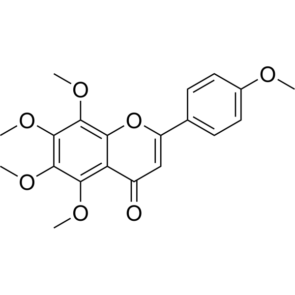
HY-N0133
TangeretinApoptosis Inducer
Publications Citing Use of MedChemExpress (MCE) Typhaneoside
MoreAll Calcium Channel Isoforms
More

Biological Activity
Description
Cellular Effect
|
Cell Line

|
Type | Value | Description | References |
|---|---|---|---|---|
| N9 | IC50 |
>100 μM
Compound: 10
|
Antineuroinflammatory activity in mouse N9 cells assessed as inhibition of LPS-induced nitric oxide production after 24 hrs by Griess assay
Antineuroinflammatory activity in mouse N9 cells assessed as inhibition of LPS-induced nitric oxide production after 24 hrs by Griess assay
|
[PMID: 28073678] |
| Raji | IC50 |
592 molar ratio
Compound: 12
|
Inhibition of TPA-induced EBV-early antigen activation in human Raji cells relative to TPA
Inhibition of TPA-induced EBV-early antigen activation in human Raji cells relative to TPA
|
[PMID: 17190444] |
In Vitro
Typhaneoside (0-50 μM; 12-48 h) significantly reduces the viability of Kas-1, HL60 and NB4 acute myeloid leukemia cells in a time- and dose-dependent manner, while exerts no significant effect on K562 acute myeloid leukemia cells or normal 293T cells[2].
Typhaneoside (20-40 μM; 24 h) induces apoptosis in Kas-1, HL60 and NB4 acute myeloid leukemia cells in a dose-dependent manner, with the highest apoptosis rate induced by treatment at 40 μM[2].
Typhaneoside (20-40 μM; 24 h) dose-dependently reduces the mRNA expression levels of genes associated with mitochondrial dysfunction (NDUFS3, SDHB, UQCRFS1, TFAM, ClpP) in Kas-1, HL60 and NB4 acute myeloid leukemia cells[2].
The effects of typhaneoside (40 μM; 24 h) on inducing reactive oxygen species production and apoptosis in Kas-1, HL60 and NB4 acute myeloid leukemia cells are significantly reversed by pre-treatment with NAC or DFO[2].
Typhaneoside (40 μM; 24 h) upregulates autophagy-related proteins (ATG5, ATG7, Beclin 1, LC3) in Kas-1, HL60, and NB4 acute myeloid leukemia cells, and this effect is reversed by pre-treatment with 5 mM NAC for 2 h[2].
Typhaneoside (12.5-50 μM; 24 h) activates FXR-dependent BSEP promoter activity in HEK293T cells, with significant activation observed at the concentration of 50 μM after 24 h[3].
Typhaneoside (12.5-50 μM; 24 h) alleviates OAPA-induced lipid accumulation, oxidative stress, inflammatory response and glucose metabolism disorder in HepG2 cells by activating the AKT/GSK3β signaling pathway[3].
Typhaneoside (5-50 μM; 10 min) concentration-dependently inhibits 4-aminopyridine (HY-B0604)-induced glutamate release from rat cerebral cortical synaptosomes, with an IC50 of 20 μM[4].
Typhaneoside (20 μM; 10 min)-mediated inhibition of 4-aminopyridine-induced glutamate release from rat cerebral cortex synaptosomes targets Ca2+-dependent vesicular exocytosis, rather than reverse transport via Ca2+-independent glutamate transporters[4].
Typhaneoside (20 μM) inhibits KCl-induced glutamate release from rat cerebral cortex synaptosomes[4].
Typhaneoside (20 μM; 10 min)-mediated inhibition of 4-aminopyridine-induced glutamate release from rat cerebral cortex synaptosomes depends on N-type (Cav2.2) voltage-dependent Ca2+ channels, and does not involve intracellular Ca2+ release or mitochondrial Na+/Ca2+ exchange[4].
The inhibitory effect of Typhaneoside (20 μM; 10 min) on 4-aminopyridine-induced glutamate release from rat cerebral cortex synaptosomes depends on the MAPK/ERK signaling pathway, and does not involve PKA or PKC[4].
Typhaneoside (20 μM; 10 min) reduces the phosphorylation levels of 4-aminopyridine-induced ERK1/2 and its presynaptic target synapsin I in rat cerebral cortex synaptosomes, but exerts no such effect on JNK or p38[4].
MedChemExpress (MCE) has not independently confirmed the accuracy of these methods. They are for reference only.
-
Cell Line:Kas-1, HL60, NB4, K562, 293T
-
Concentration:0, 10, 20, 30, 40 and 50 μM
-
Incubation Time:12 h, 24 h, 48 h
-
Result:Reduced cell viability of Kas-1, HL60, and NB4 cells in a time- and dose-dependent manner.
Showed no significant effect on cell viability of K562 or 293T cells.
-
Cell Line:Kas-1, HL60, NB4
-
Concentration:20, 30 and 40 μM
-
Incubation Time:24 h
-
Result:Dose-dependently arrested Kas-1, HL60, and NB4 cells at the G2/M phase.
Decreased G1/G0 phase cell percentage in a dose-dependent manner for all three cell lines.
Decreased S phase cell percentage in a dose-dependent manner for all three cell lines.
-
Cell Line:Kas-1, HL60, NB4
-
Concentration:20, 30 and 40 μM
-
Incubation Time:24 h
-
Result:Dose-dependently reduced protein expression of Cyclin B1 and p-Cdc2 in Kas-1, HL60, and NB4 cells.
Dose-dependently enhanced protein expression of p53 and p27 in Kas-1, HL60, and NB4 cells.\nDose-dependently reduced protein expression of anti-apoptotic Bcl-2 in Kas-1, HL60, and NB4 cells.
Dose-dependently increased protein expression of pro-apoptotic Bax and cleaved Caspase-3 in Kas-1, HL60, and NB4 cells.\nDose-dependently increased protein expression of p-AMPK in Kas-1, HL60, and NB4 cells.
Dose-dependently decreased protein expression of p-mTOR in Kas-1, HL60, and NB4 cells.\nDose-dependently increased protein expression of ATG5, ATG7, Beclin 1, and LC3 in Kas-1, HL60, and NB4 cells.
-
Cell Line:Kas-1, HL60, NB4
-
Concentration:40 μM
-
Incubation Time:24 h (with 2 h pre-treatment of 5 mM NAC)
-
Result:Induced upregulation of ATG5, ATG7, Beclin 1, and LC3 protein expression that was reversed by pretreatment with 5 mM NAC in Kas-1, HL60, and NB4 cells.
In Vivo
Typhaneoside (10-30 mg/kg; i.p.; daily; 30 days) dose-dependently suppresses AML tumor growth in BALB/c nude mice, with the 30 mg/kg dose achieving the greatest tumor volume reduction and the highest 30-day survival rate of ~50%, while showing no overt tissue toxicity[2].
Typhaneoside (30 mg/kg; i.p.; daily; 30 days) causes no overt tissue toxicity in healthy BALB/c nude mice[2].
Typhaneoside (15-60 mg/kg/day; p.o.; daily; 8 weeks) dose-dependently alleviates HFD-induced NAFLD in male C57BL/6 mice by activating FXR signaling, reducing body and adipose tissue weight, improving lipid and glucose homeostasis, mitigating liver injury, oxidative stress and inflammation, and enhancing BAT thermogenesis and energy expenditure[3].
MedChemExpress (MCE) has not independently confirmed the accuracy of these methods. They are for reference only.
-
Animal Model:BALB/c nude (4-week-old male; AML model via subcutaneous injection of 1×10⁷ HL60 cells, tumor grown to 50 mm3 before treatment)[2]
-
Dosage:10 mg/kg; 20 mg/kg; 30 mg/kg
-
Administration:i.p.; daily; 30 days
-
Result:Significantly reduced mean tumor volume to ~1000 mm3 (10 mg/kg), ~800 mm3 (20 mg/kg), and ~500 mm3 (30 mg/kg) at day 30 compared to controls.
Improved 30-day survival rate to ~20% (10 mg/kg), ~30% (20 mg/kg), and ~50% (30 mg/kg) in a dose-dependent manner.
Significantly reduced white blood cell counts in a dose-dependent manner compared to controls.
-
Animal Model:C57BL/6 (male, 8-week-old, 20-22 g, SPF, HFD-induced NAFLD)[3]
-
Dosage:15 mg/kg/day; 30 mg/kg/day; 60 mg/kg/day
-
Administration:p.o.; daily; 8 weeks
-
Result:Reduced HFD-induced body weight gain in a dose-dependent manner, with a significant decrease in area under the curve for body weight across all doses.
Dose-dependently reduced liver weight,
Dose-dependently reversed HFD-induced serum and hepatic lipid abnormalities: decreased serum triglyceride (TG), low-density lipoprotein cholesterol (LDL-C), and free fatty acid (FFA) levels; increased serum high-density lipoprotein cholesterol (HDL-C) levels; decreased hepatic TG levels.
Dose-dependently reversed HFD-induced liver injury, oxidative stress, and inflammation: decreased serum and hepatic AST, ALT, and MDA levels; increased serum and hepatic SOD levels; suppressed HFD-induced upregulation of proinflammatory mRNA (IL-6, IL-1β, NF-κB, TNF-α) in liver and WAT.
Dose-dependently upregulated mRNA expression of FXR, SHP, BSEP, and TGR5 in liver, and increased hepatic FXR protein expression detected by immunofluorescence.
- Mammalian live/dead viability and cytotoxicity staining
- Annexin V plus membrane-impermeant dye apoptosis staining
- ROS/oxidative-stress fluorescent staining
- BrdU Incorporation Assay
- Protocol for Cell Cycle
- CCK-8/WST-8 Cell Proliferation Assay
- Research Protocol for Cardiovascular Diseases
- Research Protocol for Metabolic Diseases
- Cell Viability Determination by MTT Colorimetric Assay
- 3T3-L1 preadipocyte-to-adipocyte differentiation
- Detection of Apoptosis
- TUNEL staining for apoptotic DNA fragmentation
- Flow cytometric DNA-content cell-cycle staining
- Cell Cytotoxicity Assay
- Autophagy
- Apoptosis Solutions
- Ferroptosis Solutions
- Lysosome and acidic-vesicle live-cell staining
- MTT Cell Proliferation Assay
- Macroautophagy Solutions
- Lipid Droplets: Oil Red O/Sudan Dye Lipid Staining


Chemical Information
-
CAS No. 104472-68-6
-
Appearance Solid
-
Molecular Weight 770.69
-
Formula C34H42O20
-
Color Light yellow to yellow
-
SMILES
O=C1C(O[C@H](O[C@H](CO[C@H](O[C@@H](C)[C@H](O)[C@H]2O)[C@@H]2O)[C@@H](O)[C@@H]3O)[C@@H]3O[C@@](O[C@@H](C)[C@H](O)[C@H]4O)([H])[C@@H]4O)=C(C5=CC(OC)=C(O)C=C5)OC6=CC(O)=CC(O)=C16
-
Structure Classification
-
Initial Source
-
Shipping
Room temperature in continental US; may vary elsewhere.
-
Storage
4°C, protect from light
* In solvent : -80°C, 6 months; -20°C, 1 month (protect from light)


Publications (4)
-
Journal Impact Factor
-
Most Recent
-
Microbiome
Desulfovibrio vulgaris interacts with novel gut epithelial immune receptor LRRC19 and exacerbates colitis. [Abstract]2024 Jan 3;12(1):4. PMID: 38172943 -
Food Chem
Effects of sun drying combined with baking processes on the flavor quality of Chongqing Tuocha raw tea. [Abstract]2025 Dec 30:497:146992. PMID: 41285060 -
Food Chem
Flavonoid-mediated metabolic underpinning quality variation in red bud-sport pear mutants. [Abstract]2025 Oct 15:489:144992. PMID: 40466530 -


Solvent & Solubility
In Vitro:
DMSO : 250 mg/mL (324.38 mM; Need ultrasonic; Hygroscopic DMSO has a significant impact on the solubility of product, please use newly opened DMSO)
Please refer to the solubility information to select the appropriate solvent. Once prepared, please aliquot and store the solution to prevent product inactivation from repeated freeze-thaw cycles.
Storage method and period of stock solution: -80°C, 6 months; -20°C, 1 month (protect from light). When stored at -80°C, please use it within 6 months. When stored at -20°C, please use it within 1 month.
Please refer to the solubility information to select the appropriate solvent. Once prepared, please aliquot and store the solution to prevent product inactivation from repeated freeze-thaw cycles.
Storage method and period of stock solution: -80°C, 6 months; -20°C, 1 month (protect from light). When stored at -80°C, please use it within 6 months. When stored at -20°C, please use it within 1 month.
Concentration (start) × Volume (start) = Concentration (final) × Volume (final)
In Vivo:
Select the appropriate dissolution method based on your experimental animal and administration route.
- For the following dissolution methods, please ensure to first prepare a clear stock solution using an In Vitro approach and then sequentially add co-solvents:
- To ensure reliable experimental results, the clarified stock solution can be appropriately stored based on storage conditions. As for the working solution for In Vivo experiments, it is recommended to prepare freshly and use it on the same day.
- The percentages shown for the solvents indicate their volumetric ratio in the final prepared solution. If precipitation or phase separation occurs during preparation, heat and/or sonication can be used to aid dissolution.
Add each solvent one by one: 10% DMSO 40% PEG300 5% Tween-80 45% Saline
Solubility: ≥ 2.08 mg/mL (2.70 mM); Clear solution
This protocol yields a clear solution of ≥ 2.08 mg/mL (saturation unknown).
Taking 1 mL working solution as an example, add 100 μL DMSO stock solution (20.8 mg/mL) to 400 μL PEG300, and mix evenly; then add 50 μL Tween-80 and mix evenly; then add 450 μL Saline to adjust the volume to 1 mL.
Preparation of Saline: Dissolve 0.9 g sodium chloride in ddH₂O and dilute to 100 mL to obtain a clear Saline solution.
Add each solvent one by one: 10% DMSO 90% (20% SBE-β-CD in Saline)
Solubility: ≥ 2.08 mg/mL (2.70 mM); Clear solution
This protocol yields a clear solution of ≥ 2.08 mg/mL (saturation unknown).
Taking 1 mL working solution as an example, add 100 μL DMSO stock solution (20.8 mg/mL) to 900 μL 20% SBE-β-CD in Saline, and mix evenly.
Preparation of 20% SBE-β-CD in Saline (4°C, storage for one week): 2 g SBE-β-CD powder is dissolved in 10 mL Saline, completely dissolve until clear.
In Vivo Dissolution Calculator
Please enter the basic information of animal experiments:
-
-
-
-
Recommended: Prepare an additional quantity of animals to account for potential losses during experiments.
Please enter your animal formula composition:
-
%DMSO +
Recommended: Keep the proportion of DMSO in working solution below 2% if your animal is weak.
-
%+
-
+%Tween-80 + +
-
%Saline +
The co-solvents required include: DMSO, . All of co-solvents are available by MedChemExpress (MCE). , Tween 80. All of co-solvents are available by MedChemExpress (MCE).
Working solution concentration: 0.22 mg/mL
Method for preparing stock solution: mg drug dissolved in μL DMSO. Stock solution concentration: mg/mL. * In solvent : -80°C, 6 months; -20°C, 1 month (protect from light)
1. Take μL DMSO stock solution;
2. Add μL .
μL , mix evenly;
3. Then add μL Tween 80, mix evenly;
4. Then add μL
Please ensure that the stock solution in the first step is dissolved to a clear state, and add co-solvents in sequence. You can use ultrasonic heating (ultrasonic cleaner, recommended frequency 20-40 kHz), vortexing, etc. to assist dissolution.


Purity & Documentation
-
Data Sheet (296 KB)
-
SDS (393 KB)
- English - EN (393 KB)
- Français - FR (393 KB)
- Deutsch - DE (393 KB)
- Norwegian - NO (393 KB)
- Español - ES (393 KB)
- Swedish - SV (393 KB)
- Italian - IT (393 KB)
- Korean - KR (393 KB)
- Portuguese - PT (393 KB)
-
Handling Instructions (2659 KB)
References
[1]. Zhang X, et al. Effect of typhaneoside on ventricular remodeling and regulation of PI3K/Akt/mTOR pathway. Wirkung von Typhaneosid auf ventrikuläres Remodeling und Regulierung des PI3K/Akt/mTOR-Signalwegs. Herz. 2020;45(Suppl 1):113-122. [Content Brief]
[2]. Zhu HY, et al. Typhaneoside prevents acute myeloid leukemia (AML) through suppressing proliferation and inducing ferroptosis associated with autophagy. Biochem Biophys Res Commun. 2019;516(4):1265-1271. [Content Brief]
[3]. Zheng Y, et al. Hepatoprotective effect of Typhaneoside on non-alcoholic fatty liver disease via farnesoid X receptor in vivo and in vitro. Biomed Pharmacother. 2023;164:114957. [Content Brief]
[4]. Chiu KM, et al. Typhaneoside Suppresses Glutamate Release Through Inhibition of Voltage-Dependent Calcium Entry in Rat Cerebrocortical Nerve Terminals. Chem Res Toxicol. 2021;34(5):1286-1295. [Content Brief]


Complete Stock Solution Preparation Table
Please refer to the solubility information to select the appropriate solvent. Once prepared, please aliquot and store the solution to prevent product inactivation from repeated freeze-thaw cycles.
Storage method and period of stock solution: -80°C, 6 months; -20°C, 1 month (protect from light). When stored at -80°C, please use it within 6 months. When stored at -20°C, please use it within 1 month.
| Optional Solvent | Concentration Solvent Mass | 1 mg | 5 mg | 10 mg | 25 mg |
|---|---|---|---|---|---|
| DMSO | 1 mM | 1.2975 mL | 6.4877 mL | 12.9754 mL | 32.4385 mL |
| 5 mM | 0.2595 mL | 1.2975 mL | 2.5951 mL | 6.4877 mL | |
| 10 mM | 0.1298 mL | 0.6488 mL | 1.2975 mL | 3.2438 mL | |
| 15 mM | 0.0865 mL | 0.4325 mL | 0.8650 mL | 2.1626 mL | |
| 20 mM | 0.0649 mL | 0.3244 mL | 0.6488 mL | 1.6219 mL | |
| 25 mM | 0.0519 mL | 0.2595 mL | 0.5190 mL | 1.2975 mL | |
| 30 mM | 0.0433 mL | 0.2163 mL | 0.4325 mL | 1.0813 mL | |
| 40 mM | 0.0324 mL | 0.1622 mL | 0.3244 mL | 0.8110 mL | |
| 50 mM | 0.0260 mL | 0.1298 mL | 0.2595 mL | 0.6488 mL | |
| 60 mM | 0.0216 mL | 0.1081 mL | 0.2163 mL | 0.5406 mL | |
| 80 mM | 0.0162 mL | 0.0811 mL | 0.1622 mL | 0.4055 mL | |
| 100 mM | 0.0130 mL | 0.0649 mL | 0.1298 mL | 0.3244 mL |


Typhaneoside Related Classifications
HY-N0712 Related Classifications
 Powered by Bioz
Keywords
- Typhaneoside
- 104472-68-6
- mTOR
- Akt
- FXR
- PI3K
- Autophagy
- Ferroptosis
- Apoptosis
- Reactive Oxygen Species (ROS)
- Calcium Channel
- PI3K/Akt/mTOR autophagy transduction pathway
- acute myeloid leukemia
- farnesoid X receptor
- tumor necrosis factor alpha
- matrix metalloproteinase 2
- interleukin-6
- matrix metalloproteinase 9
- cardiomyocyte
- Inhibitor
- inhibitor
- inhibit