



Telavancin
Based on 1 publication(s) in Google Scholar
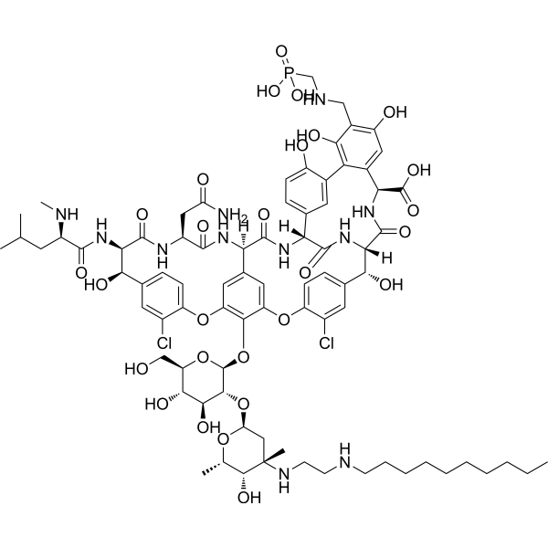
Telavancin (TD-6424) is a semisynthetic lipoglycopeptide vancomycin-derivative, is a novel antimicrobial agent developed by Theravance for overcoming resistant Gram-positive bacterial infections, specifically methicillin-resistant Staphylococcus aureus (MRSA). Telavancin disrupts cell membrane integrity, can be used for research of complicated skin and skin structure infections (cSSSIs) caused by Gram-positive bacteria.
For research use only. We do not sell to patients.
- CAS No.: 372151-71-8
- Formula: C80H106Cl2N11O27P
- Molecular Weight:1755.63
-
Storage:
Please store the product under the recommended conditions in the Certificate of Analysis.

To place orders, for customer services and technical support, please contact: MedChemExpress USA
Tel: 609-228-6898 E-mail: [email protected] [email protected]


-
 Biological Activity
-
Chemical Information
- Protocols
- Purity & Documentation
- References
-
 Help & FAQs


Publications Citing Use of MedChemExpress (MCE) Telavancin
MoreAll Antibiotic Isoforms
More

Biological Activity
Description
IC50 & Target
|
Glycopeptide |
In Vitro
Telavancin has a rapid, concentration-dependent bactericidal effect, due to disruption of cell membrane integrity[1].
Telavancin (5 μg/mL) exerts bacteriocidal effect against a MRSA strain (COL) and a VISA strain (HIP5836) at a concentration of 5 µg/mL[1].
MedChemExpress (MCE) has not independently confirmed the accuracy of these methods. They are for reference only. Further protocols information, click here.
In Vivo
MedChemExpress (MCE) has not independently confirmed the accuracy of these methods. They are for reference only.
-
Animal Model:MRSA bacteremia model in neutropenic mice[1]
-
Dosage:40 mg/kg
-
Administration:Subcutaneous injection; twice every day spaced 12 h apart
-
Result:Resulted a significantly higher 14-day survival compared with vancomycin-treated animals.
-
Animal Model:Rabbit model of S. aureus endocarditis[1]
-
Dosage:30 mg/kg
-
Administration:Intravenous injection; twice every day, spaced 12 h for 4 days
-
Result:Significantly reduced MRSA densities in all target tissues and increased the percentage of these organs rendered culture negative.
Clinical Trial
| NCT Number | Sponsor | Condition | Start Date |
Phase

|
|---|---|---|---|---|
| NCT01329991 | Plexxikon| | 2011-05 | PHASE1 |


Chemical Information
-
CAS No. 372151-71-8
-
Appearance Solid
-
Molecular Weight 1755.63
-
Formula C80H106Cl2N11O27P
-
Color White to off-white
-
SMILES
ClC1=CC([C@H]([C@](C(N[C@@H]2C(O)=O)=O)([H])N3)O)=CC=C1OC4=CC([C@](NC([C@@H](NC5=O)CC(N)=O)=O)([H])C(N[C@](C3=O)([H])C(C=C6C(C2=CC(O)=C7CNC[P](O)(O)=O)=C7O)=CC=C6O)=O)=CC(OC8=CC=C([C@H]([C@H]5NC([C@H](NC)CC(C)C)=O)O)C=C8Cl)=C4O[C@H](O[C@@H]9CO)[C@@H]([C@H]([C@@H]9O)O)O[C@H](O[C@H]%10C)C[C@@](C)([C@@H]%10O)NCCNCCCCCCCCCC
-
Synonyms
TD-6424
-
Shipping
Room temperature in continental US; may vary elsewhere.
-
Storage
Please store the product under the recommended conditions in the Certificate of Analysis.


Publications (1)
-
Journal Impact Factor
-
Most Recent
-
Adv Sci (Weinh)
SRSF5-Mediated Alternative Splicing of M Gene is Essential for Influenza A Virus Replication: A Host-Directed Target Against Influenza Virus. [Abstract]2022 Dec;9(34):e2203088. PMID: 36257906

Protocols
-
Research Protocol for Infectious Diseases
Infectious-disease experiments test how pathogens interact with host barriers, innate immune receptors, inflammatory signaling, pathogen replication, and tissue injury; pattern-recognition receptors such as TLRs, RIG-I-like receptors, NOD-like receptors, and inflammasomes detect microbial molecules and activate NF-κB, interferon, and cytokine responses. The central hypothesis is that infection severity reflects the balance between pathogen burden and host response: protective inflammation restricts pathogen growth, whereas excessive or mislocalized inflammation contributes to tissue damage and disease phenotype. Unresolved questions include which host pathways are protective versus pathogenic, why some infection models fail to translate to human disease, and which combined readouts best predict clinically relevant infection outcomes.
-
Gram Staining of Tissue Sections
Gram staining of tissue sections is a histochemical technique used to differentiate Gram-positive and Gram-negative bacteria within histological specimens based on differences in bacterial cell wall structure and dye retention, adapted from classical bacteriological Gram staining into tissue-compatible “histological Gram stain” variants. In tissue applications, modifications of the Brown-Hopps and Brown-Brenn methods are commonly used to improve differentiation of microorganisms embedded within host connective tissue and to reduce overstaining or loss of Gram-negative signal, which are known limitations of earlier approaches. The principle relies on crystal violet-iodine complex retention in Gram-positive organisms and subsequent decolorization and counterstaining steps that allow contrast visualization of Gram-negative organisms against tissue background.


Purity & Documentation
-
Data Sheet (280 KB)
-
SDS (252 KB)
- Français - FR (252 KB)
- Deutsch - DE (252 KB)
- Norwegian - NO (252 KB)
- Español - ES (252 KB)
- Swedish - SV (252 KB)
- Italian - IT (252 KB)
- Korean - KR (252 KB)
- Portuguese - PT (252 KB)
-
Handling Instructions (2659 KB)
References

Calculators
Concentration (start) × Volume (start) = Concentration (final) × Volume (final)


Telavancin Related Classifications
HY-112959 Related Classifications
 Powered by Bioz