



α-L-Fucosidase, Microorganism
Based on 1 publication(s) in Google Scholar
α-L-Fucosidase, Microorganism (EC 3.2.1.51) is an enzyme that catalyzes the chemical reaction. Serum activity of α-L-Fucosidase, Microorganism, a lysosomal enzyme present in all mammalian cells, has been proposed as a marker of hepatocellular carcinoma (HCC).
For research use only. We do not sell to patients.
- CAS No.: 9037-65-4
-
Storage:
Please store the product under the recommended conditions in the Certificate of Analysis.

To place orders, for customer services and technical support, please contact: MedChemExpress USA
Tel: 609-228-6898 E-mail: [email protected] [email protected]


-
 Biological Activity
-
Chemical Information
- Protocols
- Purity & Documentation
- References
-
 Help & FAQs


-
HY-100944
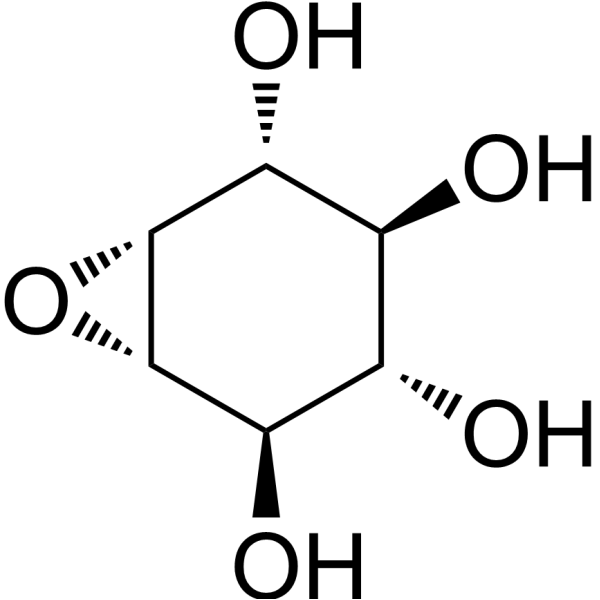
Conduritol B epoxideGcase Inhibitor -

HY-P2929
PNGase FGlycosidase -
HY-17020
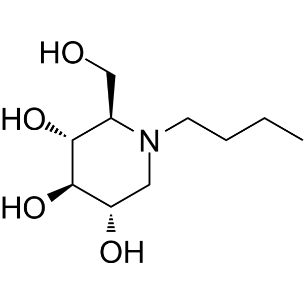
MiglustatCeramide Glucosyltransferase Inhibitor -

HY-P2929A
PNGase F-FastGlycosidase -
HY-P2840
Dextranase, Trichoderma reeseiDextranase -

HY-P2929C
PNGase F (Lyophilized)Glycosidase -
HY-132177B
α1-2,4,6 Fucosidase O, Omnitrophica bacteriumExoglycosidase -
HY-132177C
α1-3,4 Fucosidase, Bifidobacterium bifidumExoglycosidase -
HY-132177A
α-1-2 Fucosidase, bifidobacterium longumExoglycosidase -

HY-P2929D
PNGase F (MS grade)Glycosidase -

HY-P2929B
PNGase F (Immobilized, Microspin)N-glycosidic bond Cleaver -

HY-P2872
ThioglucosidaseS-glycosidic Bond Hydrolase
Publications Citing Use of MedChemExpress (MCE) α-L-Fucosidase, Microorganism
More

Biological Activity
Description
EC Number
3.2.1.51
Enzyme Activity
≥25 U/g
Unit Definition
One unit is defined as the amount of enzyme that will releas 1 μmol of methyl umbelliferone from 4- methyl umbelerone α-l-fucosides per min at 37°C and pH 6.0


Chemical Information
-
CAS No. 9037-65-4
-
Appearance Solid
-
Color White to off-white
-
SMILES
[a-L-Fucosidase, Microorganism]
-
Synonyms
EC 3.2.1.51; FUC
-
Shipping
Room temperature in continental US; may vary elsewhere.
-
Storage
Please store the product under the recommended conditions in the Certificate of Analysis.


Publications (1)
-
Journal Impact Factor
-
Most Recent
-
Aging Cell
A rationally designed fluorescence probe achieves highly specific and long-term detection of senescence in vitro and in vivo. [Abstract]2023 Aug;22(8):e13896. PMID: 37312431

Protocols
-
Lysosome and acidic-vesicle live-cell staining
Lysosome and acidic-vesicle live-cell staining detects acidic intracellular compartments by using membrane-permeant acidotropic probes that accumulate in low-pH vesicles, including lysosomes, late endosomes, autolysosomes, and acidic phagosomes. LysoTracker staining is commonly used as an intensity-based readout of acidic lysosomal compartment abundance or enlargement, while acridine orange produces green fluorescence in less concentrated compartments and red fluorescence after concentration-dependent accumulation in acidic vesicular organelles. Loss or reduction of acridine-orange red signal can be used as a readout of lysosomal membrane permeabilization or reduced acidic-vesicle integrity. This protocol is designed for live cultured cells and can be adapted for fluorescence microscopy, high-content imaging, plate-reader readout, or flow cytometry when the selected literature supports the readout. Because these dyes report acidotropic accumulation rather than lysosome identity alone,
-
Liver Cancer Modeling
Liver cancer can be classified into primary liver cancer and secondary liver cancer. Secondary liver cancer is the metastatic liver cancer. Primary liver cancer includes hepatocellular carcinoma (HCC), intrahepatic cholangiocarcinoma (ICC) and fibrolamellar HCC, of which HCC is the most common form, accounting for approximately 90% of primary liver cancers[1]. HCC mouse models include chemical agent-induced models, transplanted tumor models, and genetic engineered models.


Purity & Documentation
-
Data Sheet (264 KB)
-
SDS (251 KB)
- English - EN (251 KB)
- Français - FR (251 KB)
- Deutsch - DE (251 KB)
- Norwegian - NO (251 KB)
- Español - ES (251 KB)
- Swedish - SV (251 KB)
- Italian - IT (251 KB)
- Korean - KR (251 KB)
- Portuguese - PT (251 KB)
-
Handling Instructions (2659 KB)
References

Calculators
Concentration (start) × Volume (start) = Concentration (final) × Volume (final)


α-L-Fucosidase, Microorganism Related Classifications
HY-132177 Related Classifications
 Powered by Bioz