



κ-Carrageenan
Based on 1 publication(s) in Google Scholar
κ-Carrageenan is a natural polymer which predominantly available in red seaweeds. κ-Carrageenan is an effective agent carrier to deliver curcumin in cancer cells and to induce apoptosis. κ-carrageenan serves as a potential inflammatory agent that magnifies existing intestinal inflammation.
For research use only. We do not sell to patients.
- CAS No.: 11114-20-8
-
Storage:Powder -20°C, 3 years , 4°C, 2 years ; In solvent -80°C, 6 months , -20°C, 1 month


To place orders, for customer services and technical support, please contact: MedChemExpress USA
Tel: 609-228-6898 E-mail: [email protected] [email protected]


-
 Biological Activity
-
Chemical Information
-
Solvent & Solubility
- Protocols
- Purity & Documentation
- References
-
 Help & FAQs


-

HY-W145523
ι-CarrageenanBiochemical Assay Reagents -
HY-119109
LaminaranImmunotherapy Agent -

HY-132179
FucoidanAnticoagulant/Cancer Agent -

HY-B2220
CellulaseEnzyme -

HY-N6664
Gum arabicComplex Branched Polysaccharide -

HY-W145521
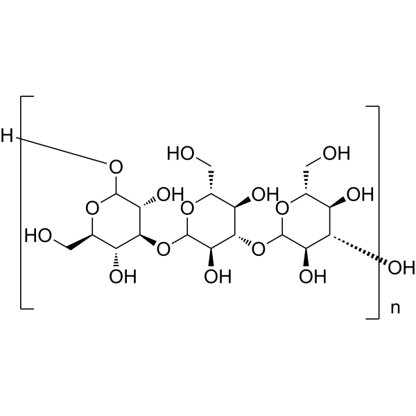
β-1,3-GlucanPolysaccharide -
HY-B2144A
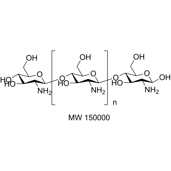
Chitosan (MW 150000) (≥85% deacetylated)Bacterial Inhibitor -
HY-B2144B
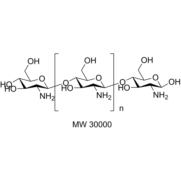
Chitosan (MW 30000) (≥85% deacetylated)Bacterial Inhibitor -

HY-B2144K
Chitosan (≥85% deacetylated, viscosity>90 mPa.s, MW 15000)Polysaccharid -
HY-B2144E
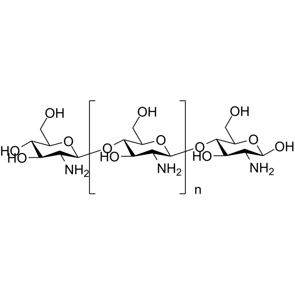
Chitosan (≥90% deacetylated, Low viscosity,<200mPa.s)Biochemical Assay Reagent -
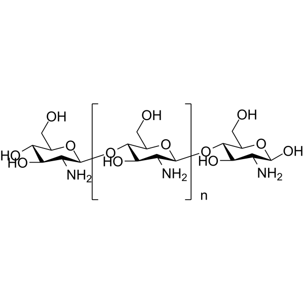
HY-B2144F
Chitosan (≥80% deacetylated, Medium viscosity,200-400mPa.s)Biochemical Assay Reagent -
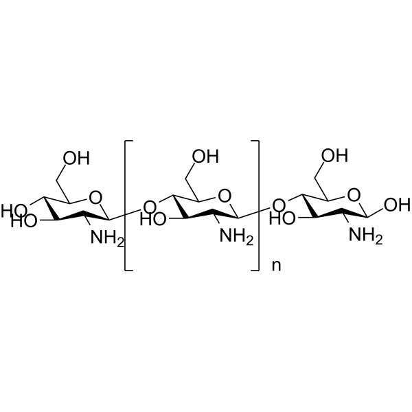
HY-B2144C
Chitosan (≥95% deacetylated,viscosity 100-200 mPa.s)Biochemical Assay Reagent
Publications Citing Use of MedChemExpress (MCE) κ-Carrageenan
More

Biological Activity
Description
In Vitro
κ-Car- Curcumin (Cur) (0-500 μg/mL; 24-72 hours) effectively involves in cancer cell growth inhibition at lower concentrations of 40 μg/mL[1].
The cytotoxicity of the Cur loaded κ-Car has a significantly high apoptotic activity in selected lung cancer cells of A549[1].
κ-Carrageenan (1-60 μg/mL; 0.5-24 hours) enhances LPS-induced IL-8 secretion in HT-29 cells[2].
MedChemExpress (MCE) has not independently confirmed the accuracy of these methods. They are for reference only. Further protocols information, click here.
-
Cell Line:A549 cells
-
Concentration:0-500 μg/mL
-
Incubation Time:24, 48 and 72 hours
-
Result:The dose response effects of cells treated with Cur loaded κ-Car after incubation of 24, 48 and 72 h exhibited a significant IC50 values of 65, 50 and 40 μg/mL respectively, for 24, 48, 72 h ours.
In Vivo
-
Animal Model:Male and female NIH (s) mice[2]
-
Dosage:1.7 mg/kg, LOW; 8.3 mg/kg, MED; or 41.7 mg/kg, HIG
-
Administration:Orally administered for 1 week prior to C. freundii DBS100 treatment
-
Result:Enhanced the C. freundii DBS100-dependent induction of TLR4 and NF-κB in the intestinal mucosa of infected mice.
Induced Disease Models
Please do not refer to only one article to determine the experimental conditions. It is recommended to determine the optimal experimental conditions (animal strain, age, dosage, frequency and cycle, detection time and indicators, etc.) through preliminary experiments before the formal experiment.
κ-Carrageenan can be used in animal modeling to create paw edema, colitis, and thrombosis models. κ-Carrageenan has oral activity; after oral administration, its concentration in BALB/c mice peaks at 3 hours, with an average residence time of 36.6 hours and a clearance rate of 0.1 L/h/kg. Most of the κ-Carrageenan is excreted via feces, while 9.2% is excreted through urine, indicating rapid absorption, slow elimination, and prolonged retention in the body. Additionally, κ-Carrageenan accumulates in the liver and kidneys. After 14 days of oral administration of κ-Carrageenan, hepatocyte necrosis, renal tubular vacuolation, and incomplete colonic epithelial cells are observed. Four hours after intraperitoneal injection of κ-Carrageenan, minor infarcts and erythema appear at the tips of rat tails, with infarct length increasing in a time-dependent manner and stabilizing 24 hours post-injection. Twenty-four hours is determined as the time point for successful establishment of the thrombosis model[4][5][6][7][8][9].
Administration: 0.1 mL • sc • single dose
Molecular changes: Neutrophil migration to the foot area of rats injected with κ-carrageenan, along with elevated expression levels of pro-inflammatory factors such as bradykinin, histamine, tachykinins, complement, and reactive oxygen species.
Administration: 1.7, 8.3, 41.7 mg/kg • po • single dose
Molecular changes: Significant upregulation of IL-2, TNF-α, and IL-6 expression, with a notable decrease in IL-10 expression. The enzymatic activity of SOD and GSH-px in the colonic mucosa is reduced, while the expression of TLR4, NF-κB, p-ERK, p-JNK, p-Jun, IL-8, and MDA is upregulated.
Coagulation parameter changes: In the tails of rats with thrombus formation, prothrombin time (PT) and fibrinogen (FIB) significantly increased, while thrombin time (TT) significantly decreased, as determined by classical coagulation testing methods.
MedChemExpress (MCE) has not independently confirmed the accuracy of these methods. They are for reference only.


Chemical Information
-
CAS No. 11114-20-8
-
Appearance Solid
-
Color Off-white to light yellow
-
SMILES
[K-Carrageenan]
-
Synonyms
K-Carrageenan Karra Type
-
Structure Classification
-
Initial Source
-
Shipping
Room temperature in continental US; may vary elsewhere.
-
Storage
Powder -20°C 3 years 4°C 2 years In solvent -80°C 6 months -20°C 1 month


Publications (1)
-
Journal Impact Factor
-
Most Recent


Solvent & Solubility
In Vitro:
DMSO : 8.33 mg/mL (ultrasonic and warming and heat to 80°C; Hygroscopic DMSO has a significant impact on the solubility of product, please use newly opened DMSO)
H2O : 8 mg/mL (ultrasonic and warming and heat to 80°C)
In Vivo:
Select the appropriate dissolution method based on your experimental animal and administration route.
- For the following dissolution methods, please ensure to first prepare a clear stock solution using an In Vitro approach and then sequentially add co-solvents:
- To ensure reliable experimental results, the clarified stock solution can be appropriately stored based on storage conditions. As for the working solution for In Vivo experiments, it is recommended to prepare freshly and use it on the same day.
- The percentages shown for the solvents indicate their volumetric ratio in the final prepared solution. If precipitation or phase separation occurs during preparation, heat and/or sonication can be used to aid dissolution.
Add each solvent one by one: 10% DMSO 40% PEG300 5% Tween-80 45% Saline
Solubility: ≥ 0.83 mg/mL; Clear solution
This protocol yields a clear solution of ≥ 0.83 mg/mL (saturation unknown).
Taking 1 mL working solution as an example, add 100 μL DMSO stock solution (8.3 mg/mL) to 400 μL PEG300, and mix evenly; then add 50 μL Tween-80 and mix evenly; then add 450 μL Saline to adjust the volume to 1 mL.
Preparation of Saline: Dissolve 0.9 g sodium chloride in ddH₂O and dilute to 100 mL to obtain a clear Saline solution.
Add each solvent one by one: 10% DMSO 90% (20% SBE-β-CD in Saline)
Solubility: ≥ 0.83 mg/mL; Clear solution
This protocol yields a clear solution of ≥ 0.83 mg/mL (saturation unknown).
Taking 1 mL working solution as an example, add 100 μL DMSO stock solution (8.3 mg/mL) to 900 μL 20% SBE-β-CD in Saline, and mix evenly.
Preparation of 20% SBE-β-CD in Saline (4°C, storage for one week): 2 g SBE-β-CD powder is dissolved in 10 mL Saline, completely dissolve until clear.
For the following dissolution methods, please prepare the working solution directly:
It is recommended to prepare fresh solutions and use them promptly within a short period of time.
The percentages shown for the solvents indicate their volumetric ratio in the final prepared solution. If precipitation or phase separation occurs during preparation, heat and/or sonication can be used to aid dissolution.
Add each solvent one by one: PBS
Solubility: 8.33 mg/mL; Clear solution; Need ultrasonic and warming and heat to 60°C
In Vivo Dissolution Calculator
Please enter the basic information of animal experiments:
-
-
-
-
Recommended: Prepare an additional quantity of animals to account for potential losses during experiments.
Please enter your animal formula composition:
-
%DMSO +
Recommended: Keep the proportion of DMSO in working solution below 2% if your animal is weak.
-
%+
-
+%Tween-80 + +
-
%Saline +
The co-solvents required include: DMSO, . All of co-solvents are available by MedChemExpress (MCE). , Tween 80. All of co-solvents are available by MedChemExpress (MCE).
Working solution concentration: 0.22 mg/mL
Method for preparing stock solution: mg drug dissolved in μL DMSO. Stock solution concentration: mg/mL.
1. Take μL DMSO stock solution;
2. Add μL .
μL , mix evenly;
3. Then add μL Tween 80, mix evenly;
4. Then add μL
Please ensure that the stock solution in the first step is dissolved to a clear state, and add co-solvents in sequence. You can use ultrasonic heating (ultrasonic cleaner, recommended frequency 20-40 kHz), vortexing, etc. to assist dissolution.

Protocols
-
Apoptosis
Apoptosis, also called programmed cell death, is generally characterized by distinct morphological characteristics.
-
TUNEL staining for apoptotic DNA fragmentation
TUNEL staining detects DNA strand breaks by using terminal deoxynucleotidyl transferase to add labeled nucleotides to exposed 3′-OH DNA termini, generating either microscopic staining in fixed cells or tissue sections, or fluorescence/cytometric signal in cell suspensions. TUNEL positivity reflects DNA fragmentation but should not be interpreted alone as definitive apoptosis, because TUNEL can also label necrotic, autolytic, mechanically damaged, or DNA-repair-associated DNA breaks.
-
Annexin V plus membrane-impermeant dye apoptosis staining
Annexin V-based apoptosis assays rely on the detection of phosphatidylserine (PS) externalization from the inner leaflet of the plasma membrane to the outer leaflet, an early biochemical hallmark of apoptosis. Fluorescently labeled Annexin V binds PS in a calcium-dependent manner, enabling identification of early apoptotic cells by flow cytometry or fluorescence microscopy. When combined with a membrane-impermeant DNA-binding dye (e. g. , propidium iodide), this approach allows discrimination between viable (Annexin V−/dye−), early apoptotic (Annexin V+/dye−), and late apoptotic or necrotic (Annexin V+/dye+) cell populations by assessing membrane integrity and PS exposure.
-
DSS-Induced Colitis
Dextran sulfate sodium (DSS)-induced colitis is generated by administering DSS in mouse drinking water, producing epithelial injury, barrier disruption, weight loss, diarrhea, fecal blood, colon shortening, histologic mucosal damage, and inflammatory mediator changes; the model is mainly used to study acute or chronic intestinal inflammation resembling selected features of ulcerative colitis. DSS injury is interpreted through clinical and tissue readouts rather than a single molecular endpoint: daily body weight, stool consistency, and bleeding are combined into a disease activity index, while colon length, histology, cytokines, myeloperoxidase activity, intestinal permeability, and tight-junction markers provide complementary measures of inflammation and barrier damage.
-
TNBS-Induced Colitis
TNBS-induced colitis is produced by intrarectal delivery of 2,4,6-trinitrobenzene sulfonic acid in ethanol, where ethanol disrupts the mucosal barrier and TNBS haptenates colonic proteins, generating immune-mediated colonic inflammation with weight loss, diarrhea, ulceration, transmural injury, inflammatory-cell infiltration, and cytokine responses. The model is used as an experimental intestinal inflammation model with Crohn’s disease–like features, especially when Th1-type responses, IL-12–dependent inflammation, chronic relapsing inflammation, or fibrosis-related endpoints are studied.
-
Apoptosis Solutions
Apoptosis is a regulated, generally non-lytic cell-death pathway that removes unwanted, damaged, infected, or abnormal cells through coordinated morphological changes, caspase activation, DNA fragmentation, and membrane remodeling. The intrinsic apoptosis pathway is controlled mainly by mitochondrial outer membrane permeabilization, BCL-2 family proteins, cytochrome c release, apoptosome formation, caspase-9 activation, and downstream executioner caspase-3/7 activation. The extrinsic apoptosis pathway is initiated by death receptors such as Fas, TNFR, and TRAIL receptors, which recruit adaptor proteins and activate caspase-8 before engaging executioner caspases or mitochondrial amplification through BID cleavage. Apoptosis is linked to many phenotypes, including cancer cell killing, tissue homeostasis, immune regulation, neurodegeneration, infection response, and treatment-induced cytotoxicity; unresolved questions include how apoptosis interacts with necroptosis, pyroptosis, ferroptos
-
Research Protocol for Inflammation-related Diseases
The NLRP3 inflammasome is a cytosolic innate immune signaling platform that integrates priming signals and danger-signal activation to promote caspase-1 activation, maturation of IL-1β and IL-18, and gasdermin D-mediated pyroptotic cell death. The core experimental logic is to determine whether inflammatory disease phenotypes are driven by increased NLRP3 expression, ASC-containing inflammasome assembly, caspase-1 cleavage, GSDMD cleavage, and extracellular release of IL-1β/IL-18 rather than by nonspecific cell injury alone. The pathway is strongly linked to inflammation-related disease phenotypes because monosodium urate crystals activate NALP3/NLRP3 inflammasome signaling in gout-like crystal inflammation, cholesterol crystals activate NLRP3 inflammasomes in atherogenesis models, and DSS-induced intestinal inflammation has been reported to involve NLRP3 inflammasome activity. However, experimental colitis studies also show context-dependent protective effects of NLRP3 inflammasome co


Purity & Documentation
-
Data Sheet (284 KB)
-
SDS (252 KB)
- English - EN (252 KB)
- Français - FR (252 KB)
- Deutsch - DE (252 KB)
- Norwegian - NO (252 KB)
- Español - ES (252 KB)
- Swedish - SV (252 KB)
- Italian - IT (252 KB)
- Korean - KR (252 KB)
- Portuguese - PT (252 KB)
-
Handling Instructions (2659 KB)
References
[1]. Sathuvan M, et al. κ-Carrageenan: An effective drug carrier to deliver curcumin in cancer cells and to induce apoptosis. Carbohydr Polym. 2017;160:184-193. [Content Brief]
[2]. Wu W, et al. κ-Carrageenan Enhances Lipopolysaccharide-Induced Interleukin-8 Secretion by Stimulating the Bcl10-NF-κB Pathway in HT-29 Cells and Aggravates C. freundii-Induced Inflammation in Mice. Mediators Inflamm. 2017;2017:8634865. [Content Brief]
[3]. Wei W, et al. Enhanced effect of κ-carrageenan on TNBS-induced inflammation in mice. Int Immunopharmacol. 2016;39:218-228. [Content Brief]
[4]. Wang J, et al. Pharmacokinetics, tissue distribution, and subacute toxicity of oral carrageenan in mice. Int J Biol Macromol. 2024 May;266(Pt 1):130725. [Content Brief]
[5]. Morris CJ. Carrageenan-induced paw edema in the rat and mouse. Methods Mol Biol. 2003;225:115-21. [Content Brief]
[6]. Ochoa-Amaya JE, et al. Dual effects of hyperprolactinemia on carrageenan-induced inflammatory paw edema in rats. Neuroimmunomodulation. 2011;18(4):245-53. [Content Brief]
[7]. Jiang HY, et al. κ-carrageenan induces the disruption of intestinal epithelial Caco-2 monolayers by promoting the interaction between intestinal epithelial cells and immune cells. Mol Med Rep. 2013 Dec;8(6):1635-42. [Content Brief]
[8]. Wei W, et al. Enhanced effect of κ-carrageenan on TNBS-induced inflammation in mice. Int Immunopharmacol. 2016 Oct;39:218-228. [Content Brief]
[9]. Ma N, et al. Preventive Effect of Aspirin Eugenol Ester on Thrombosis in κ-Carrageenan-Induced Rat Tail Thrombosis Model. PLoS One. 2015 Jul 20;10(7):e0133125. [Content Brief]

Calculators
Concentration (start) × Volume (start) = Concentration (final) × Volume (final)


κ-Carrageenan Related Classifications
HY-138962 Related Classifications
 Powered by Bioz
Keywords