



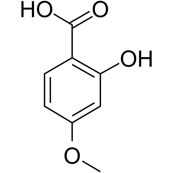
2-Hydroxy-4-methoxybenzoic acid

Based on 1 Customer Validation
2-Hydroxy-4-methoxybenzoic acid is an orally active inhibitor of MCT-1 and MCT-4, as well as a plant biomarker. 2-Hydroxy-4-methoxybenzoic acid can be isolated from roots. 2-Hydroxy-4-methoxybenzoic acid induces Apoptosis and loss of mitochondrial membrane potential. 2-Hydroxy-4-methoxybenzoic acid exhibits anticancer activity against breast cancer. 2-Hydroxy-4-methoxybenzoic acid normalizes lactic acid levels. 2-Hydroxy-4-methoxybenzoic acid neutralizes viper venom and attenuates its lethal, hemorrhagic, coagulant and anticoagulant activities in male albino mice. 2-Hydroxy-4-methoxybenzoic acid possesses antihyperlipidemic, antidiabetic and hepatoprotective activities.
For research use only. We do not sell to patients.
- Purity : 99.64%
- CAS No.: 2237-36-7
- Formula: C8H8O4
- Molecular Weight:168.15
-
Storage:
4°C, stored under nitrogen
* In solvent : -80°C, 6 months; -20°C, 1 month (stored under nitrogen)

To place orders, for customer services and technical support, please contact: MedChemExpress USA
Tel: 609-228-6898 E-mail: [email protected] [email protected]


-
 Biological Activity
-
Chemical Information
-
Solvent & Solubility
- Protocols
- Purity & Documentation
- References
-
 Help & FAQs


-
HY-N0708
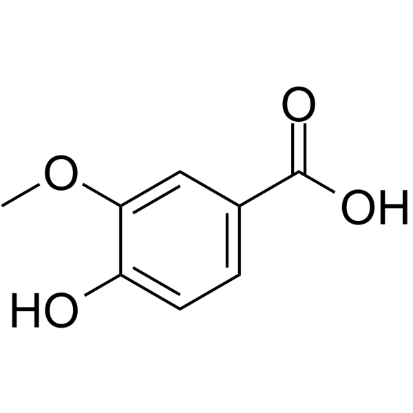
Vanillic acidFlavouring Agent -

HY-N0708R
Vanillic acid (Standard)Flavouring Agent -
HY-Y0760
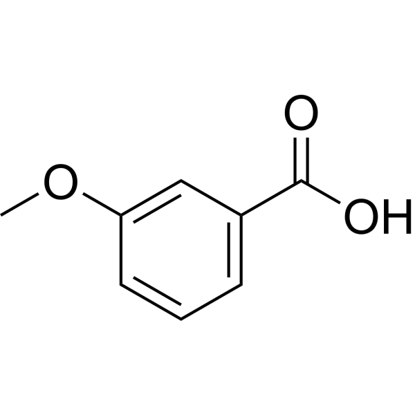
3-Methoxybenzoic acidDrug Intermediate -
HY-N1393
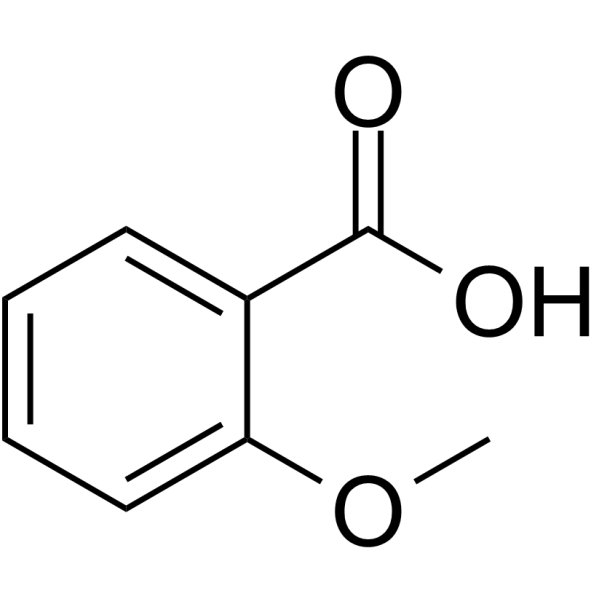
2-Methoxybenzoic acidCarbonic Anhydrase Inhibitor -
HY-W021293
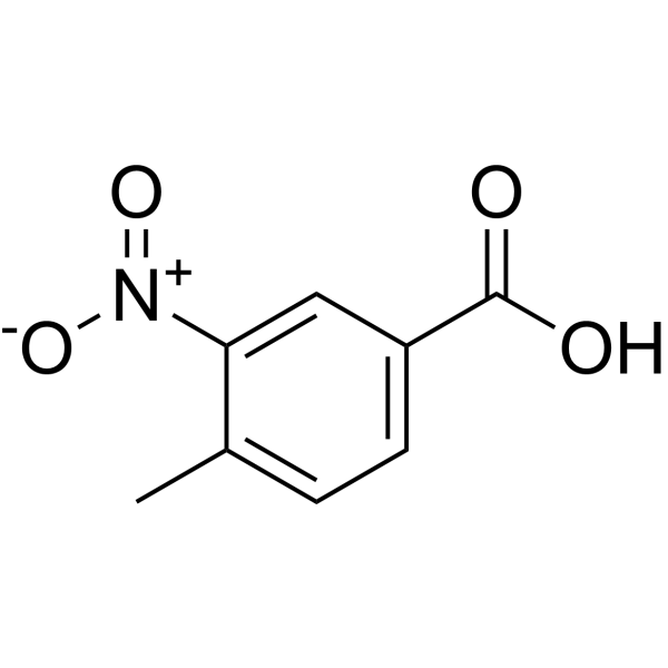
4-Methyl-3-nitrobenzoic acidBiochemical Reagent -

HY-Y0760R
3-Methoxybenzoic acid (Standard) -

HY-N1393R
2-Methoxybenzoic acid (Standard)Endogenous Metabolite


Biological Activity
Description
IC50 & Target
[1]|
MCT1 |
MCT4 |
Cellular Effect
|
Cell Line

|
Type | Value | Description | References |
|---|---|---|---|---|
| RAW264.7 | CC50 |
>40 μM
Compound: 2
|
Cytotoxicity against mouse RAW264.7 cells assessed as reduction in survival rate after 24 hrs by MTT assay
Cytotoxicity against mouse RAW264.7 cells assessed as reduction in survival rate after 24 hrs by MTT assay
|
[PMID: 25200306] |
In Vitro
2-Hydroxy-4-methoxybenzoic acid (1-25 μM; 24 h) potently inhibits the viability of MCF-7 breast cancer cells, with an IC50 of 4.8 μM after 24 h of treatment[2].
2-Hydroxy-4-methoxybenzoic acid (IC25, IC50; 24 h) induces nuclear apoptotic changes in breast cancer MCF-7 cells, with 78% and 87.33% of apoptotic nuclei observed after treatment at IC25 and IC50 concentrations, respectively[2].
2-Hydroxy-4-methoxybenzoic acid (IC25, IC50; 24 h) induces dose-dependent morphological changes of early and late apoptosis, including membrane blebbing and apoptotic body formation, in MCF-7 breast cancer cells following 24 h of treatment at IC25 and IC50 concentrations[2].
2-Hydroxy-4-methoxybenzoic acid (IC25, IC50; 24 h) induces loss of mitochondrial membrane potential in breast cancer MCF-7 cells following 24 h of treatment at IC25 and IC50 concentrations[2].
MedChemExpress (MCE) has not independently confirmed the accuracy of these methods. They are for reference only. Further protocols information, click here.
-
Cell Line:MCF-7 human mammary gland carcinoma cells
-
Concentration:1-25 μM
-
Incubation Time:24 h
-
Result:Reduced MCF-7 cell viability in a concentration-dependent manner, with an IC50 value of 4.8 μM.
-
Cell Line:MCF-7 human mammary gland carcinoma cells
-
Concentration:IC25, IC50
-
Incubation Time:24 h
-
Result:Induced bright nuclear condensation and fragmentation compared to untreated cells with uniform round nuclei and faint blue fluorescence.
Reached 78% apoptotic nuclei in the IC25 HMBA group and 87.33% apoptotic nuclei in the IC50 HMBA group, compared to 42% in untreated control cells.\nInduced yellow (early apoptosis) and orange/red (late apoptosis) staining, along with membrane blebbing and apoptotic body formation in a dose-dependent manner compared to untreated cells with uniformly green-stained viable cells and normal large nuclei.\nCaused decreased red fluorescence and increased green fluorescence, indicating loss of MMP, with a reduced red-to-green fluorescence ratio compared to untreated controls with strong red fluorescence indicating high MMP.
In Vivo
2-Hydroxy-4-methoxybenzoic acid (50 mg/20 g mouse) enhances the positive effects of adjuvants and protects mice against venom-induced death and hemorrhage[1].
2-Hydroxy-4-methoxybenzoic acid (200 μg/kg, p.o.; once daily for 30 days) reduces ethanol-induced hyperlipidemia in male Wistar rats[1].
MedChemExpress (MCE) has not independently confirmed the accuracy of these methods. They are for reference only.
-
Animal Model:Wistar (female, 100-120 g, DMBA-induced mammary gland carcinoma)[2]
-
Dosage:200 μg/kg; 400 μg/kg
-
Administration:i.p.
-
Result:Reduced terminal end buds count from 95.25 to 73.5 (200 μg/kg) and to 57.25 (400 μg/kg).
Reduced alveolar buds count from 118.75 to 70.25, lobules count from 206 to 154.4, and differentiation score from 420 to 282 (400 μg/kg).
Reduced TBARs levels from 233.97 to 185.90, protein carbonyl levels from 90.98 to 70.45, and increased superoxide dismutase levels from 0.06 to 0.10 (200 μg/kg).
Reduced TBARs levels from 233.97 to 134.62, protein carbonyl levels from 90.98 to 64.55, increased superoxide dismutase levels from 0.06 to 0.11, and increased catalase levels from 5.75 to 7.22 (400 μg/kg).
Normalized elevated serum lactate levels induced by DMBA.
Reduced DMBA-induced overexpression of MCT-1, MCT-4, GLUT-1, HIF-1α, and VEGF; downregulated anti-apoptotic Bcl-2 and Apaf-1, and upregulated pro-apoptotic BAX and Caspase-3.
Improved mammary gland architecture, restoring ordered distribution of luminal epithelial cells, myoepithelial cells, and adipocytes toward normal.


Chemical Information
-
CAS No. 2237-36-7
-
Appearance Solid
-
Molecular Weight 168.15
-
Formula C8H8O4
-
Color White to off-white
-
SMILES
O=C(O)C1=C(O)C=C(OC)C=C1
-
Synonyms
4-Methoxysalicylic Acid
-
Structure Classification
-
Initial Source
-
Shipping
Room temperature in continental US; may vary elsewhere.
-
Storage
4°C, stored under nitrogen
* In solvent : -80°C, 6 months; -20°C, 1 month (stored under nitrogen)


Solvent & Solubility
In Vitro:
DMSO : 50 mg/mL (297.35 mM; Need ultrasonic; Hygroscopic DMSO has a significant impact on the solubility of product, please use newly opened DMSO)
Please refer to the solubility information to select the appropriate solvent. Once prepared, please aliquot and store the solution to prevent product inactivation from repeated freeze-thaw cycles.
Storage method and period of stock solution: -80°C, 6 months; -20°C, 1 month (stored under nitrogen). When stored at -80°C, please use it within 6 months. When stored at -20°C, please use it within 1 month.
Please refer to the solubility information to select the appropriate solvent. Once prepared, please aliquot and store the solution to prevent product inactivation from repeated freeze-thaw cycles.
Storage method and period of stock solution: -80°C, 6 months; -20°C, 1 month (stored under nitrogen). When stored at -80°C, please use it within 6 months. When stored at -20°C, please use it within 1 month.
Concentration (start) × Volume (start) = Concentration (final) × Volume (final)
In Vivo:
Select the appropriate dissolution method based on your experimental animal and administration route.
- For the following dissolution methods, please ensure to first prepare a clear stock solution using an In Vitro approach and then sequentially add co-solvents:
- To ensure reliable experimental results, the clarified stock solution can be appropriately stored based on storage conditions. As for the working solution for In Vivo experiments, it is recommended to prepare freshly and use it on the same day.
- The percentages shown for the solvents indicate their volumetric ratio in the final prepared solution. If precipitation or phase separation occurs during preparation, heat and/or sonication can be used to aid dissolution.
Add each solvent one by one: 10% DMSO 40% PEG300 5% Tween-80 45% Saline
Solubility: ≥ 2.5 mg/mL (14.87 mM); Clear solution
This protocol yields a clear solution of ≥ 2.5 mg/mL (saturation unknown).
Taking 1 mL working solution as an example, add 100 μL DMSO stock solution (25.0 mg/mL) to 400 μL PEG300, and mix evenly; then add 50 μL Tween-80 and mix evenly; then add 450 μL Saline to adjust the volume to 1 mL.
Preparation of Saline: Dissolve 0.9 g sodium chloride in ddH₂O and dilute to 100 mL to obtain a clear Saline solution.
Add each solvent one by one: 10% DMSO 90% (20% SBE-β-CD in Saline)
Solubility: ≥ 2.5 mg/mL (14.87 mM); Clear solution
This protocol yields a clear solution of ≥ 2.5 mg/mL (saturation unknown).
Taking 1 mL working solution as an example, add 100 μL DMSO stock solution (25.0 mg/mL) to 900 μL 20% SBE-β-CD in Saline, and mix evenly.
Preparation of 20% SBE-β-CD in Saline (4°C, storage for one week): 2 g SBE-β-CD powder is dissolved in 10 mL Saline, completely dissolve until clear.
In Vivo Dissolution Calculator
Please enter the basic information of animal experiments:
-
-
-
-
Recommended: Prepare an additional quantity of animals to account for potential losses during experiments.
Please enter your animal formula composition:
-
%DMSO +
Recommended: Keep the proportion of DMSO in working solution below 2% if your animal is weak.
-
%+
-
+%Tween-80 + +
-
%Saline +
The co-solvents required include: DMSO, . All of co-solvents are available by MedChemExpress (MCE). , Tween 80. All of co-solvents are available by MedChemExpress (MCE).
Working solution concentration: 0.22 mg/mL
Method for preparing stock solution: mg drug dissolved in μL DMSO. Stock solution concentration: mg/mL. * In solvent : -80°C, 6 months; -20°C, 1 month (stored under nitrogen)
1. Take μL DMSO stock solution;
2. Add μL .
μL , mix evenly;
3. Then add μL Tween 80, mix evenly;
4. Then add μL
Please ensure that the stock solution in the first step is dissolved to a clear state, and add co-solvents in sequence. You can use ultrasonic heating (ultrasonic cleaner, recommended frequency 20-40 kHz), vortexing, etc. to assist dissolution.

Protocols
-
Apoptosis
Apoptosis, also called programmed cell death, is generally characterized by distinct morphological characteristics.
-
TUNEL staining for apoptotic DNA fragmentation
TUNEL staining detects DNA strand breaks by using terminal deoxynucleotidyl transferase to add labeled nucleotides to exposed 3′-OH DNA termini, generating either microscopic staining in fixed cells or tissue sections, or fluorescence/cytometric signal in cell suspensions. TUNEL positivity reflects DNA fragmentation but should not be interpreted alone as definitive apoptosis, because TUNEL can also label necrotic, autolytic, mechanically damaged, or DNA-repair-associated DNA breaks.
-
Mitochondrial membrane-potential fluorescent assay
Mitochondrial membrane potential fluorescent assays estimate ΔΨm in living cells using lipophilic cationic dyes such as TMRM, TMRE, rhodamine 123, and JC-1, which accumulate in mitochondria according to membrane polarization; loss of signal after FCCP or CCCP treatment is interpreted as mitochondrial depolarization. TMRM/TMRE and rhodamine 123 are commonly used for semi-quantitative live-cell microscopy or flow cytometry, while JC-1 can report a shift from red aggregate fluorescence to green monomer fluorescence during depolarization; interpretation requires controls because dye concentration, quenching mode, cell type, dye efflux, and mitochondrial mass can affect fluorescence independently of ΔΨm.
-
Annexin V plus membrane-impermeant dye apoptosis staining
Annexin V-based apoptosis assays rely on the detection of phosphatidylserine (PS) externalization from the inner leaflet of the plasma membrane to the outer leaflet, an early biochemical hallmark of apoptosis. Fluorescently labeled Annexin V binds PS in a calcium-dependent manner, enabling identification of early apoptotic cells by flow cytometry or fluorescence microscopy. When combined with a membrane-impermeant DNA-binding dye (e. g. , propidium iodide), this approach allows discrimination between viable (Annexin V−/dye−), early apoptotic (Annexin V+/dye−), and late apoptotic or necrotic (Annexin V+/dye+) cell populations by assessing membrane integrity and PS exposure.
-
Fluorescent plasma-membrane potential dye assay
Fluorescent plasma-membrane potential dye assays measure changes in cell membrane potential using voltage-sensitive dyes whose fluorescence changes when cells depolarize or hyperpolarize. Anionic bis-oxonol dyes such as DiBAC4(3) enter depolarized cells more readily and show increased fluorescence after intracellular binding, while hyperpolarization reduces dye accumulation and fluorescence. FMP/FLIPR membrane-potential dyes are used for faster, homogeneous microplate assays of ion-channel or receptor-mediated membrane-potential changes.
-
Mitochondrial membrane-potential and mitochondrial mass staining
Mitochondrial membrane potential staining measures the electrochemical polarization across the mitochondrial inner membrane in live cells using lipophilic cationic fluorescent probes; early rhodamine-based work showed that selective mitochondrial dye accumulation is lost when the mitochondrial transmembrane potential is dissipated. JC-1 reports mitochondrial polarization by shifting from green monomer fluorescence to red J-aggregate fluorescence as dye concentration increases within energized mitochondria; therefore, the red/green fluorescence ratio is used as a relative readout of mitochondrial membrane potential. TMRE or TMRM staining provides a single-channel relative readout because these cationic rhodamine esters accumulate in polarized mitochondria, and lower fluorescence indicates reduced mitochondrial polarization when acquisition and dye-loading conditions are controlled. Mitochondrial mass staining is commonly performed with MitoTracker Green FM or related MitoTracker dyes as
-
Apoptosis Solutions
Apoptosis is a regulated, generally non-lytic cell-death pathway that removes unwanted, damaged, infected, or abnormal cells through coordinated morphological changes, caspase activation, DNA fragmentation, and membrane remodeling. The intrinsic apoptosis pathway is controlled mainly by mitochondrial outer membrane permeabilization, BCL-2 family proteins, cytochrome c release, apoptosome formation, caspase-9 activation, and downstream executioner caspase-3/7 activation. The extrinsic apoptosis pathway is initiated by death receptors such as Fas, TNFR, and TRAIL receptors, which recruit adaptor proteins and activate caspase-8 before engaging executioner caspases or mitochondrial amplification through BID cleavage. Apoptosis is linked to many phenotypes, including cancer cell killing, tissue homeostasis, immune regulation, neurodegeneration, infection response, and treatment-induced cytotoxicity; unresolved questions include how apoptosis interacts with necroptosis, pyroptosis, ferroptos
-
Breast Cancer Modeling
Breast cancer is a heterogeneous cancer, and it has been distinguished into four subtypes: luminal A, luminal B, HER2-positive and basal-like. Molecular mutations, epigenetic alterations, hormone exposure and immune microenvironment are related to the progression of breast cancer.


Purity & Documentation
-
Data Sheet (292 KB)
-
SDS (394 KB)
- English - EN (394 KB)
- Français - FR (394 KB)
- Deutsch - DE (394 KB)
- Norwegian - NO (394 KB)
- Español - ES (394 KB)
- Swedish - SV (394 KB)
- Italian - IT (394 KB)
- Korean - KR (394 KB)
- Portuguese - PT (394 KB)
-
Handling Instructions (2659 KB)
References


Complete Stock Solution Preparation Table
Please refer to the solubility information to select the appropriate solvent. Once prepared, please aliquot and store the solution to prevent product inactivation from repeated freeze-thaw cycles.
Storage method and period of stock solution: -80°C, 6 months; -20°C, 1 month (stored under nitrogen). When stored at -80°C, please use it within 6 months. When stored at -20°C, please use it within 1 month.
| Optional Solvent | Concentration Solvent Mass | 1 mg | 5 mg | 10 mg | 25 mg |
|---|---|---|---|---|---|
| DMSO | 1 mM | 5.9471 mL | 29.7354 mL | 59.4707 mL | 148.6768 mL |
| 5 mM | 1.1894 mL | 5.9471 mL | 11.8941 mL | 29.7354 mL | |
| 10 mM | 0.5947 mL | 2.9735 mL | 5.9471 mL | 14.8677 mL | |
| 15 mM | 0.3965 mL | 1.9824 mL | 3.9647 mL | 9.9118 mL | |
| 20 mM | 0.2974 mL | 1.4868 mL | 2.9735 mL | 7.4338 mL | |
| 25 mM | 0.2379 mL | 1.1894 mL | 2.3788 mL | 5.9471 mL | |
| 30 mM | 0.1982 mL | 0.9912 mL | 1.9824 mL | 4.9559 mL | |
| 40 mM | 0.1487 mL | 0.7434 mL | 1.4868 mL | 3.7169 mL | |
| 50 mM | 0.1189 mL | 0.5947 mL | 1.1894 mL | 2.9735 mL | |
| 60 mM | 0.0991 mL | 0.4956 mL | 0.9912 mL | 2.4779 mL | |
| 80 mM | 0.0743 mL | 0.3717 mL | 0.7434 mL | 1.8585 mL | |
| 100 mM | 0.0595 mL | 0.2974 mL | 0.5947 mL | 1.4868 mL |


2-Hydroxy-4-methoxybenzoic acid Related Classifications
HY-75625 Related Classifications
 Powered by Bioz
Keywords
- 2-Hydroxy-4-methoxybenzoic acid
- 2237-36-7
- 4-Methoxysalicylic Acid
- Monocarboxylate Transporter
- Apoptosis
- Mitochondrial Metabolism
- lactate transport
- monocarboxylate transporter 1
- monocarboxylate transporter 4
- MCF-7 human mammary gland carcinoma cells
- female albino Wistar rats
- cancer cells
- mitochondrial apoptotic pathway
- angiogenesis
- DMBA-induced mammary gland carcinoma
- mammary gland carcinoma
- Inhibitor
- inhibitor
- inhibit