



4-Hydroperoxy cyclophosphamide
Based on 13 publication(s) in Google Scholar
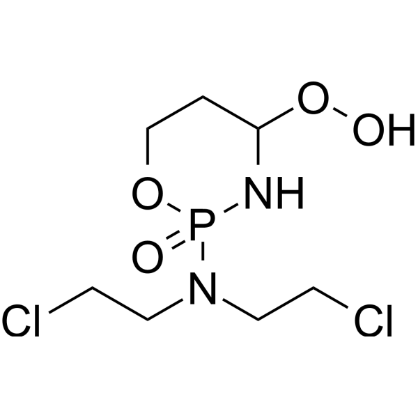
4-Hydroperoxy cyclophosphamide is the active metabolite form of the precursor Cyclophosphamide. 4-Hydroperoxy cyclophosphamide cross-links DNA to induce T cell apoptosis independent of caspase receptor activation, and can activate the mitochondrial death pathway by producing reactive oxygen species (ROS). 4-Hydroperoxy cyclophosphamide can be used in the study of rheumatoid arthritis and autoimmune diseases.
For research use only. We do not sell to patients.
- Purity : 99.92%
- CAS No.: 39800-16-3
- Formula: C7H15Cl2N2O4P
- Molecular Weight:293.08
-
Storage:
-20°C, stored under nitrogen
* In solvent : -80°C, 6 months; -20°C, 1 month (stored under nitrogen)

To place orders, for customer services and technical support, please contact: MedChemExpress USA
Tel: 609-228-6898 E-mail: [email protected] [email protected]


-
 Biological Activity
-
Chemical Information
-
Solvent & Solubility
- Protocols
- Purity & Documentation
- References
-
 Help & FAQs


-
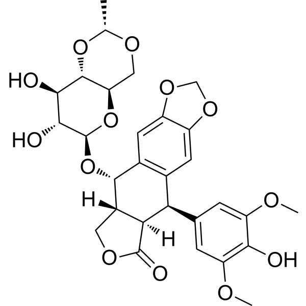
HY-13629
EtoposideTopoisomerase II Inhibitor -
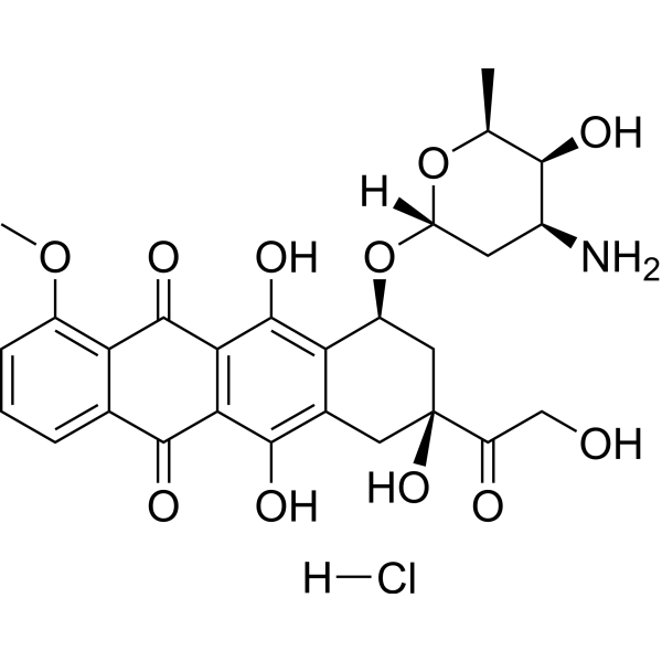
HY-15142
Doxorubicin hydrochlorideTopoisomerase Inhibitor -
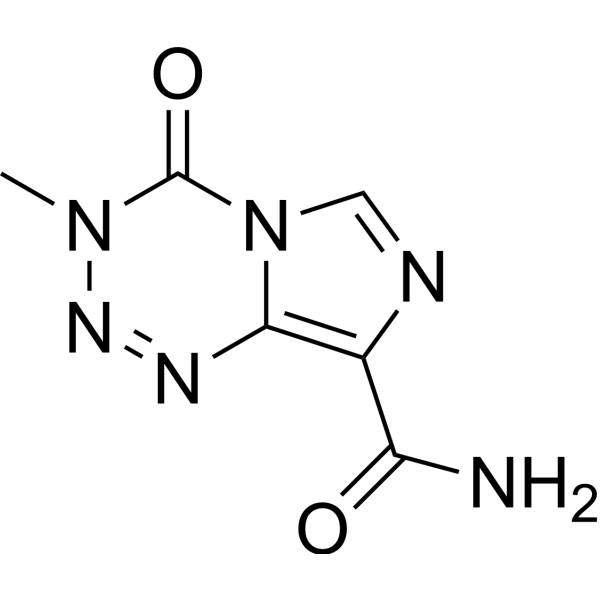
HY-17364
TemozolomideDNA Alkylator -
HY-17393
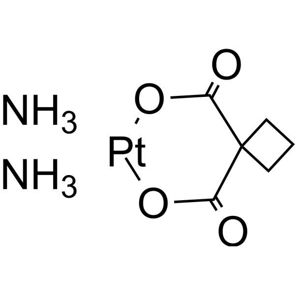
CarboplatinDNA Synthesis Inhibitor -
HY-17420
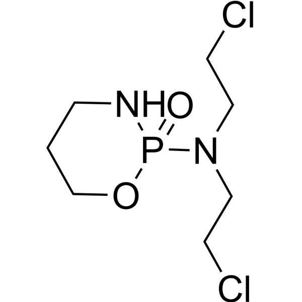
CyclophosphamideAlkylating Agent -
HY-90006
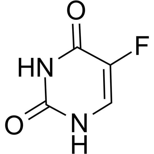
5-FluorouracilThymidylate Synthase Inhibitor -
HY-N0488
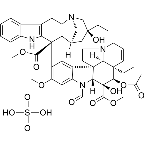
Vincristine sulfateAntineoplastic Agent -
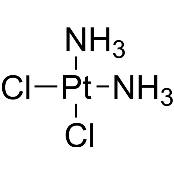
HY-17394
CisplatinDNA Alkylator -
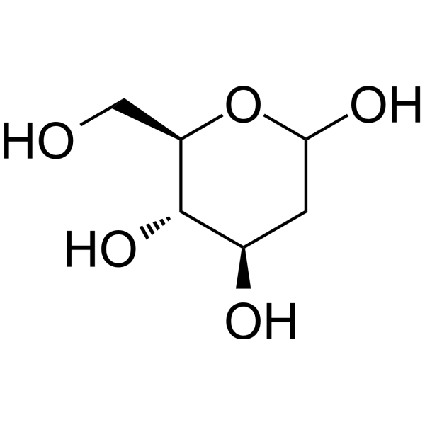
HY-13966
2-Deoxy-D-glucoseGlycolysis Inhibitor -
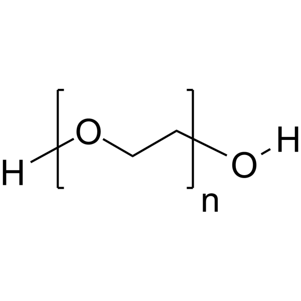
HY-Y0873
PEG300Neutral Polymer -

HY-15141
StaurosporinePKC/PKA Inhibitor -
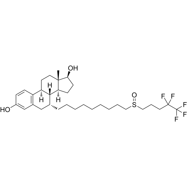
HY-13636
FulvestrantEstrogen Receptor Antagonist
Publications Citing Use of MedChemExpress (MCE) 4-Hydroperoxy cyclophosphamide
More- Cell Death Discov. 2023 Nov 14;9(1):413. [Abstract]
- Apoptosis. 2025 Jun;30(5-6):1331-1350. [Abstract]
- Free Radic Biol Med. 2025 Mar 16:230:112-126. [Abstract]
- Anal Chem. 2025 Jun 3;97(21):11099-11109. [Abstract]
- J Ethnopharmacol. 2026 Mar 1:358:120979. [Abstract]
- J Ethnopharmacol. 2024 Oct 28:333:118405. [Abstract]
- J Agric Food Chem. 2026 Jun 16. [Abstract]
- Int J Cancer. 2024 Jul 15;155(2):324-338. [Abstract]
- Biology (Basel). 2025 Jul 8;14(7):829. [Abstract]
- Eng Comput. 2024;40(3):1469-1499. [Abstract]
- Res Sq. 2026 Jan 9.
- bioRxiv. 2025 Jan 19:2025.01.15.633227. [Abstract]
- AfricArXiv Preprints. 2212.
 Customer Validation & Images

-
Cell Proliferation/Viability Assay
-
Cell Imaging/Staining
-
RT-PCR
-
WB


Biological Activity
Description
Cellular Effect
|
Cell Line

|
Type | Value | Description | References |
|---|---|---|---|---|
| L1210 | IC50 |
90 μM
Compound: 4-HC
|
Concentration required to reduce the viability of L1210 cells by 50% after 1-h incubation at 37 degrees C
Concentration required to reduce the viability of L1210 cells by 50% after 1-h incubation at 37 degrees C
|
[PMID: 1992116] |
| V79 | IC50 |
2.7 μM
Compound: 4-hydroperoxycyclophosphamide
|
Antiproliferative activity against NTR expressing chinese hamster V79 cells after 72 hrs
Antiproliferative activity against NTR expressing chinese hamster V79 cells after 72 hrs
|
[PMID: 16821793] |
| V79 | IC50 |
3.1 μM
Compound: 4-hydroperoxycyclophosphamide
|
Antiproliferative activity against chinese hamster V79 cells after 72 hrs
Antiproliferative activity against chinese hamster V79 cells after 72 hrs
|
[PMID: 16821793] |
In Vitro
4-Hydroperoxy cyclophosphamide (1 or 3 μg/mL, 24, 48, 72 h) Apoptosis of Caspase-independent T cells is mediated by oxidative stress-induced mitochondrial apoptosis factor AIF and nuclear relocalization of EndoG[1].
4-Hydroperoxy cyclophosphamide (1 μg/mL, 72, 96 h) Combines with methotrexate (HY-14519), RANKL expression was inhibited in IL-6/ SIL-6R-induced fibroblast-like synoviocytes by inhibiting the JAK2/STAT3 and p38MAPK signaling pathways[2].
MedChemExpress (MCE) has not independently confirmed the accuracy of these methods. They are for reference only. Further protocols information, click here.
-
Cell Line:CTL
-
Concentration:3 μg/mL
-
Incubation Time:24, 48, 72 h
-
Result:Increased the expression of p53 and Bax after 24 h.
-
Cell Line:Jurkat, CTL
-
Concentration:1 or 3 μg/mL
-
Incubation Time:24 , 48 , 72h
-
Result:Caspase-8 deficiency prevented CD95-mediated death induced by anti-CD95 monoclonal antibody (mAb) APO-1. Overexpression of Bcl-2 inhibited apoptosis. Induced cell death proceeded independently of caspase inhibition.
In Vivo
MedChemExpress (MCE) has not independently confirmed the accuracy of these methods. They are for reference only.
-
Animal Model:BALB/c mice[1]
-
Dosage:200 mg/kg
-
Administration:i.p.
-
Result:Exhibited a marked depletion of immature double-positive CD4 þ CD8 þ thymocytes and mature single-positive CD4+ and CD8+ T cells.


Chemical Information
-
CAS No. 39800-16-3
-
Appearance Solid
-
Molecular Weight 293.08
-
Formula C7H15Cl2N2O4P
-
Color White to yellow
-
SMILES
O=P1(OCCC(N1)OO)N(CCCl)CCCl
-
Shipping
Room temperature in continental US; may vary elsewhere.
-
Storage
-20°C, stored under nitrogen
* In solvent : -80°C, 6 months; -20°C, 1 month (stored under nitrogen)


Publications (13)
-
Journal Impact Factor
-
Most Recent
-
Cell Death Discov
The EZH2-H3K27me3 axis modulates aberrant transcription and apoptosis in cyclophosphamide-induced ovarian granulosa cell injury. [Abstract]2023 Nov 14;9(1):413. PMID: 37963880 -
Apoptosis
Bushen Jianpi Tiaoxue Decoction (BJTD) inhibits the LIF-mTOR signaling axis to regulate mitochondrial function and alleviate cyclophosphamide-induced diminished ovarian reserve. [Abstract]2025 Jun;30(5-6):1331-1350. PMID: 40042747
4-Hydroperoxy cyclophosphamide purchased from MedChemExpress. Usage Cited in: Apoptosis. 2025 Jun;30(5-6):1331-1350. [Abstract]
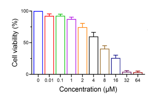
Cell viability of KGN cells following treatment with various concentrations of 4-Hydroperoxy cyclophosphamide (4-HC, 0.01-64 μM) for 24 h (n = 3).
4-Hydroperoxy cyclophosphamide purchased from MedChemExpress. Usage Cited in: Apoptosis. 2025 Jun;30(5-6):1331-1350. [Abstract]
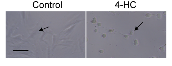
Significant induction of damage to KGN cells by the 6 µM concentration of 4-Hydroperoxy cyclophosphamide (4-HC) for 24 h.
4-Hydroperoxy cyclophosphamide purchased from MedChemExpress. Usage Cited in: Apoptosis. 2025 Jun;30(5-6):1331-1350. [Abstract]
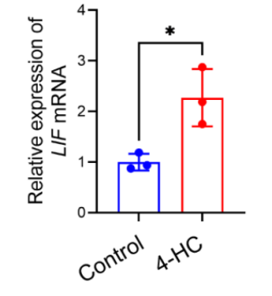
Changes in LIF levels in KGN cells following 4-HC exposure for 24 h being measured by RT-qPCR experiments (n = 3). RT-qPCR analysis indicated a significant upregulation of LIF mRNA level in models compared to controls after 24 h of co-culture with 6 µM 4-Hydroperoxy cyclophosphamide (4-HC).
-
Free Radic Biol Med
2025 Mar 16:230:112-126. PMID: 39947495 -
Anal Chem
Exposome-Scale Investigation of Cl-/Br-Containing Chemicals Using High-Resolution Mass Spectrometry, Multistage Machine Learning, and Cloud Computing. [Abstract]2025 Jun 3;97(21):11099-11109. PMID: 40401576
4-Hydroperoxy cyclophosphamide purchased from MedChemExpress. Usage Cited in: Anal Chem. 2025 Jun 3;97(21):11099-11109. [Abstract]
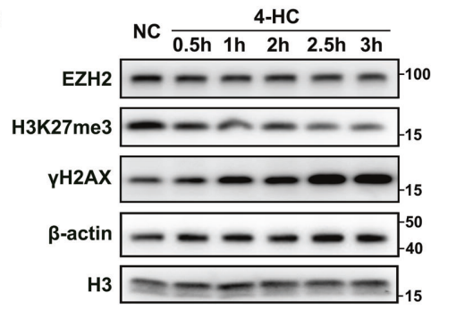
Western blot results for EZH2, H3K27me3, and γH2AX levels in the negative control (NC) and 4-Hydroperoxy cyclophosphamide (4-HC, 2.5 μM; 0.5-3 h) treatment groups. β-actin and H3 were blotted as loading controls. The results showed that with prolonged incubation time with 4-HC, the levels of H3K27me3 and EZH2 in primary GC cells decreased, while the level of γH2AX was significantly increased.
-
J Ethnopharmacol
Zuogui Wan alleviates intergenerational premature ovarian insufficiency through C9-mediated complement-coagulation cascade. [Abstract]2026 Mar 1:358:120979. PMID: 41325864 -
J Ethnopharmacol
Discovery of petroleum ether extract of eclipta targeting p53/Fas pathway for the treatment of chemotherapy-induced alopecia: Network pharmacology and experimental validation. [Abstract]2024 Oct 28:333:118405. PMID: 38844249 -
J Agric Food Chem
Optimized Extraction Process of Fu Brick Tea Polysaccharides Using Ultrasound-Assisted Enzymatic Strategy and Their Enhanced Antioxidant and Immunoprotective Activities. [Abstract]2026 Jun 16. PMID: 42301720 -
Int J Cancer
Establishment of patient-derived organoids for guiding personalized therapies in breast cancer patients. [Abstract]2024 Jul 15;155(2):324-338. PMID: 38533706 -
Biology (Basel)
Urolithin A Protects Ovarian Reserve Via Inhibiting PI3K/Akt Signaling and Preventing Chemotherapy-Induced Follicle Apoptosis. [Abstract]2025 Jul 8;14(7):829. PMID: 40723388 -
Eng Comput
A global sensitivity analysis of a mechanistic model of neoadjuvant chemotherapy for triple negative breast cancer constrained by in vitro and in vivo imaging data. [Abstract]2024;40(3):1469-1499. PMID: 39620056 -
-
bioRxiv
PAX3-FOXO1 drives targetable cell state-dependent metabolic vulnerabilities in rhabdomyosarcoma. [Abstract]2025 Jan 19:2025.01.15.633227. PMID: 39868247 -


Solvent & Solubility
In Vitro:
DMSO : 50 mg/mL (170.60 mM; Need ultrasonic; Hygroscopic DMSO has a significant impact on the solubility of product, please use newly opened DMSO)
Please refer to the solubility information to select the appropriate solvent. Once prepared, please aliquot and store the solution to prevent product inactivation from repeated freeze-thaw cycles.
Storage method and period of stock solution: -80°C, 6 months; -20°C, 1 month (stored under nitrogen). When stored at -80°C, please use it within 6 months. When stored at -20°C, please use it within 1 month.
Please refer to the solubility information to select the appropriate solvent. Once prepared, please aliquot and store the solution to prevent product inactivation from repeated freeze-thaw cycles.
Storage method and period of stock solution: -80°C, 6 months; -20°C, 1 month (stored under nitrogen). When stored at -80°C, please use it within 6 months. When stored at -20°C, please use it within 1 month.
Concentration (start) × Volume (start) = Concentration (final) × Volume (final)
In Vivo:
Select the appropriate dissolution method based on your experimental animal and administration route.
- For the following dissolution methods, please ensure to first prepare a clear stock solution using an In Vitro approach and then sequentially add co-solvents:
- To ensure reliable experimental results, the clarified stock solution can be appropriately stored based on storage conditions. As for the working solution for In Vivo experiments, it is recommended to prepare freshly and use it on the same day.
- The percentages shown for the solvents indicate their volumetric ratio in the final prepared solution. If precipitation or phase separation occurs during preparation, heat and/or sonication can be used to aid dissolution.
Add each solvent one by one: 10% DMSO 40% PEG300 5% Tween-80 45% Saline
Solubility: 2.5 mg/mL (8.53 mM); Clear solution; Need ultrasonic
This protocol yields a clear solution of 2.5 mg/mL.
Taking 1 mL working solution as an example, add 100 μL DMSO stock solution (25.0 mg/mL) to 400 μL PEG300, and mix evenly; then add 50 μL Tween-80 and mix evenly; then add 450 μL Saline to adjust the volume to 1 mL.
Preparation of Saline: Dissolve 0.9 g sodium chloride in ddH₂O and dilute to 100 mL to obtain a clear Saline solution.
Add each solvent one by one: 10% DMSO 90% (20% SBE-β-CD in Saline)
Solubility: ≥ 2.5 mg/mL (8.53 mM); Clear solution
This protocol yields a clear solution of ≥ 2.5 mg/mL (saturation unknown).
Taking 1 mL working solution as an example, add 100 μL DMSO stock solution (25.0 mg/mL) to 900 μL 20% SBE-β-CD in Saline, and mix evenly.
Preparation of 20% SBE-β-CD in Saline (4°C, storage for one week): 2 g SBE-β-CD powder is dissolved in 10 mL Saline, completely dissolve until clear.
In Vivo Dissolution Calculator
Please enter the basic information of animal experiments:
-
-
-
-
Recommended: Prepare an additional quantity of animals to account for potential losses during experiments.
Please enter your animal formula composition:
-
%DMSO +
Recommended: Keep the proportion of DMSO in working solution below 2% if your animal is weak.
-
%+
-
+%Tween-80 + +
-
%Saline +
The co-solvents required include: DMSO, . All of co-solvents are available by MedChemExpress (MCE). , Tween 80. All of co-solvents are available by MedChemExpress (MCE).
Working solution concentration: 0.22 mg/mL
Method for preparing stock solution: mg drug dissolved in μL DMSO. Stock solution concentration: mg/mL. * In solvent : -80°C, 6 months; -20°C, 1 month (stored under nitrogen)
1. Take μL DMSO stock solution;
2. Add μL .
μL , mix evenly;
3. Then add μL Tween 80, mix evenly;
4. Then add μL
Please ensure that the stock solution in the first step is dissolved to a clear state, and add co-solvents in sequence. You can use ultrasonic heating (ultrasonic cleaner, recommended frequency 20-40 kHz), vortexing, etc. to assist dissolution.

Protocols
-
Collagen-Induced Arthritis
Collagen-induced arthritis (CIA) is an autoimmune murine model of rheumatoid arthritis in which immunization with type II collagen (CII) emulsified in an adjuvant induces a T cell- and autoantibody-driven inflammatory arthritis characterized by synovial hyperplasia, immune cell infiltration, and joint destruction. The model typically relies on genetically susceptible mouse strains (e. g. , DBA/1) and reproduces key features of human rheumatoid arthritis, including anti-collagen immune responses and progressive joint inflammation. Disease onset generally occurs within ~3-4 weeks after immunization, depending on antigen/adjuvant combinations and protocol variation. The immunopathology is driven by adaptive immune activation against CII, leading to systemic and local joint inflammation mediated by pro-inflammatory cytokines and effector immune cells, making CIA a standard preclinical platform for evaluating immunomodulatory and anti-arthritic interventions.
-
Apoptosis
Apoptosis, also called programmed cell death, is generally characterized by distinct morphological characteristics.
-
TUNEL staining for apoptotic DNA fragmentation
TUNEL staining detects DNA strand breaks by using terminal deoxynucleotidyl transferase to add labeled nucleotides to exposed 3′-OH DNA termini, generating either microscopic staining in fixed cells or tissue sections, or fluorescence/cytometric signal in cell suspensions. TUNEL positivity reflects DNA fragmentation but should not be interpreted alone as definitive apoptosis, because TUNEL can also label necrotic, autolytic, mechanically damaged, or DNA-repair-associated DNA breaks.
-
Annexin V plus membrane-impermeant dye apoptosis staining
Annexin V-based apoptosis assays rely on the detection of phosphatidylserine (PS) externalization from the inner leaflet of the plasma membrane to the outer leaflet, an early biochemical hallmark of apoptosis. Fluorescently labeled Annexin V binds PS in a calcium-dependent manner, enabling identification of early apoptotic cells by flow cytometry or fluorescence microscopy. When combined with a membrane-impermeant DNA-binding dye (e. g. , propidium iodide), this approach allows discrimination between viable (Annexin V−/dye−), early apoptotic (Annexin V+/dye−), and late apoptotic or necrotic (Annexin V+/dye+) cell populations by assessing membrane integrity and PS exposure.
-
ROS/oxidative-stress fluorescent staining
ROS/oxidative-stress fluorescent staining uses cell-permeant fluorogenic probes that become fluorescent after oxidation inside cells or tissues; commonly used examples include DCFH-DA/DCFDA for broad cellular oxidant detection, DHE for superoxide-related signal detection, MitoSOX for mitochondrial superoxide-related signal detection, and CellROX probes for oxidative-stress-associated fluorescence readouts. The assay detects probe oxidation rather than a single ROS species unless the probe and analysis method have been chemically validated for that species. DCFH-DA enters cells, is deacetylated by intracellular esterases to DCFH, and produces fluorescent DCF after oxidation, so the readout is used as an operational measure of total cellular oxidative stress rather than a species-specific ROS measurement. DHE and MitoSOX can report superoxide-related oxidation, but red fluorescence alone can include non-specific ethidium-like oxidation products; HPLC or optimized spectral approaches are
-
Apoptosis Solutions
Apoptosis is a regulated, generally non-lytic cell-death pathway that removes unwanted, damaged, infected, or abnormal cells through coordinated morphological changes, caspase activation, DNA fragmentation, and membrane remodeling. The intrinsic apoptosis pathway is controlled mainly by mitochondrial outer membrane permeabilization, BCL-2 family proteins, cytochrome c release, apoptosome formation, caspase-9 activation, and downstream executioner caspase-3/7 activation. The extrinsic apoptosis pathway is initiated by death receptors such as Fas, TNFR, and TRAIL receptors, which recruit adaptor proteins and activate caspase-8 before engaging executioner caspases or mitochondrial amplification through BID cleavage. Apoptosis is linked to many phenotypes, including cancer cell killing, tissue homeostasis, immune regulation, neurodegeneration, infection response, and treatment-induced cytotoxicity; unresolved questions include how apoptosis interacts with necroptosis, pyroptosis, ferroptos
-
Research Protocol for Cancer Immunology
Cancer immunology studies how the immune system recognizes, suppresses, edits, or fails to eliminate malignant cells through tumor antigen release, antigen presentation, T-cell priming, immune trafficking, tumor-cell killing, and feedback inhibition in the tumor microenvironment. The cancer-immunity cycle links tumor antigenicity, dendritic-cell priming, CD8+ T-cell infiltration, cytotoxic function, and immune-checkpoint regulation to tumor rejection or immune escape. Immune-checkpoint pathways such as PD-1/PD-L1 and CTLA-4 suppress antitumor T-cell activity and can be therapeutically blocked, but many tumors remain resistant because of poor antigen presentation, weak T-cell infiltration, suppressive myeloid cells, regulatory T cells, and tumor-intrinsic immune-exclusion programs. Unresolved questions include which immune-cell states predict response, how tumor-intrinsic pathways exclude immune cells, how myeloid suppression limits checkpoint blockade, and which combination strategies


Purity & Documentation
-
Data Sheet (280 KB)
-
SDS (393 KB)
- English - EN (393 KB)
- Français - FR (393 KB)
- Deutsch - DE (393 KB)
- Norwegian - NO (393 KB)
- Español - ES (393 KB)
- Swedish - SV (393 KB)
- Italian - IT (393 KB)
- Korean - KR (393 KB)
- Portuguese - PT (393 KB)
-
Handling Instructions (2659 KB)
References
[1]. Strauss G, et al. 4-hydroperoxy-cyclophosphamide mediates caspase-independent T-cell apoptosis involving oxidative stress-induced nuclear relocation of mitochondrial apoptogenic factors AIF and EndoG. Cell Death Differ. 2008 Feb;15(2):332-43. [Content Brief]
[2]. Niu HQ, et al. Combination of 4-hydroperoxy cyclophosphamide and methotrexate inhibits IL-6/sIL-6R-induced RANKL expression in fibroblast-like synoviocytes via suppression of the JAK2/STAT3 and p38MAPK signaling pathway. Int Immunopharmacol. 2018 Aug;61:45-53. [Content Brief]


Complete Stock Solution Preparation Table
Please refer to the solubility information to select the appropriate solvent. Once prepared, please aliquot and store the solution to prevent product inactivation from repeated freeze-thaw cycles.
Storage method and period of stock solution: -80°C, 6 months; -20°C, 1 month (stored under nitrogen). When stored at -80°C, please use it within 6 months. When stored at -20°C, please use it within 1 month.
| Optional Solvent | Concentration Solvent Mass | 1 mg | 5 mg | 10 mg | 25 mg |
|---|---|---|---|---|---|
| DMSO | 1 mM | 3.4120 mL | 17.0602 mL | 34.1204 mL | 85.3009 mL |
| 5 mM | 0.6824 mL | 3.4120 mL | 6.8241 mL | 17.0602 mL | |
| 10 mM | 0.3412 mL | 1.7060 mL | 3.4120 mL | 8.5301 mL | |
| 15 mM | 0.2275 mL | 1.1373 mL | 2.2747 mL | 5.6867 mL | |
| 20 mM | 0.1706 mL | 0.8530 mL | 1.7060 mL | 4.2650 mL | |
| 25 mM | 0.1365 mL | 0.6824 mL | 1.3648 mL | 3.4120 mL | |
| 30 mM | 0.1137 mL | 0.5687 mL | 1.1373 mL | 2.8434 mL | |
| 40 mM | 0.0853 mL | 0.4265 mL | 0.8530 mL | 2.1325 mL | |
| 50 mM | 0.0682 mL | 0.3412 mL | 0.6824 mL | 1.7060 mL | |
| 60 mM | 0.0569 mL | 0.2843 mL | 0.5687 mL | 1.4217 mL | |
| 80 mM | 0.0427 mL | 0.2133 mL | 0.4265 mL | 1.0663 mL | |
| 100 mM | 0.0341 mL | 0.1706 mL | 0.3412 mL | 0.8530 mL |


4-Hydroperoxy cyclophosphamide Related Classifications
HY-117433 Related Classifications
 Powered by Bioz