



CCG-203971
Based on 2 publication(s) in Google Scholar
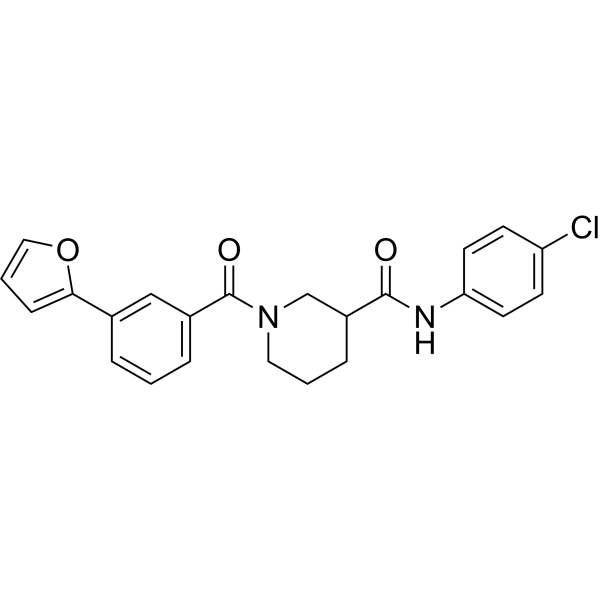
CCG-203971 is a second-generation Rho/MRTF/SRF pathway inhibitor. CCG-203971 potently targets RhoA/C-activated SRE-luciferase (IC50 =6.4 μM). CCG-203971 inhibits PC-3 cell migration with an IC50 of 4.2 μM. Potential anti-metastasis Agent.
For research use only. We do not sell to patients.
- Purity : 99.89%
- CAS No.: 1443437-74-8
- Formula: C23H21ClN2O3
- Molecular Weight:408.88
-
Storage:Powder -20°C, 3 years , 4°C, 2 years ; In solvent -80°C, 2 years , -20°C, 1 year

To place orders, for customer services and technical support, please contact: MedChemExpress USA
Tel: 609-228-6898 E-mail: [email protected] [email protected]


-
 Biological Activity
-
Chemical Information
-
Solvent & Solubility
- Protocols
- Purity & Documentation
- References
-
 Help & FAQs


-
HY-13991
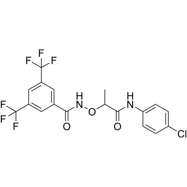
CCG-1423Rho/MRTF/SRF Pathway Inhibitor -
HY-12270
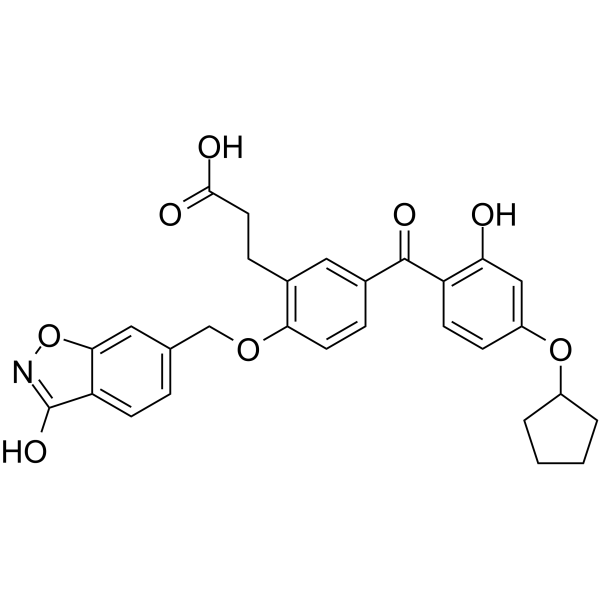
T-5224c-Fos/AP-1 Inhibitor -
HY-121750
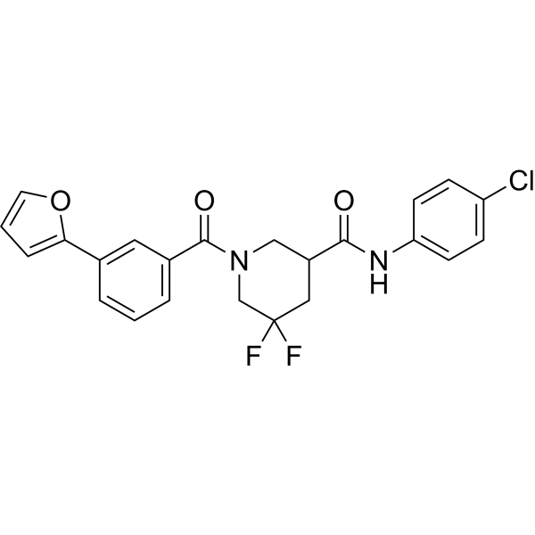
CCG-222740Rho/MRTF/SRF Inhibitor -
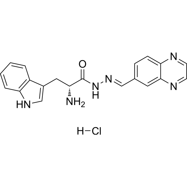
HY-12646
Rhosin hydrochlorideRho GTPases Inhibitor -
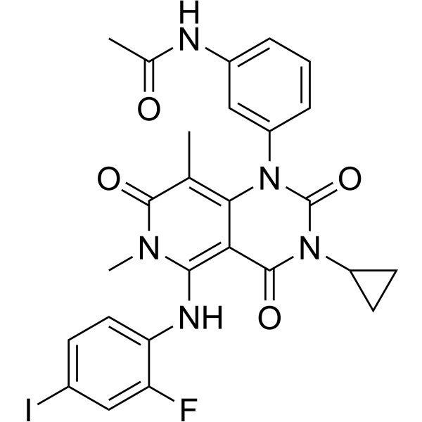
HY-10999
TrametinibMEK Inhibitor -
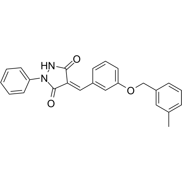
HY-12649
Y16GEF-Rho GTPase Inhibitor -
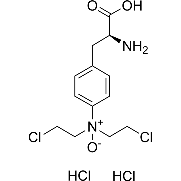
HY-10231
PX-478HIF-1α Inhibitor -
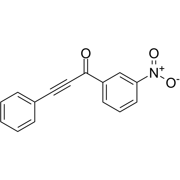
HY-117149
MLS000532223Rho family GTPases Inhibitor -
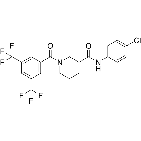
HY-120855
CCG-100602MRTF-A/SRF Inhibitor -
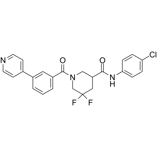
HY-111432
CCG-232601Rho/MRTF/SRF Inhibitor -
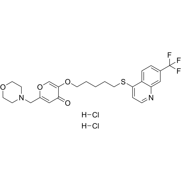
HY-16659
EHT 1864Rac Family Inhibitor -

HY-108361R
CCG-203971 (Standard)Reference Standard
Publications Citing Use of MedChemExpress (MCE) CCG-203971
More Customer Validation & Images

-
IF

-
WB

-
RT-PCR

-
IP

-
IHC


Biological Activity
Description
IC50 & Target
RhoA/MRTF-A[1]
Cellular Effect
|
Cell Line

|
Type | Value | Description | References |
|---|---|---|---|---|
| HCT-116 | IC50 |
76.5 μM
Compound: 3-11k
|
Antiproliferative activity against human HCT-116 cells assessed as inhibition of cell growth measured after 72 hrs by CCK-8 method
Antiproliferative activity against human HCT-116 cells assessed as inhibition of cell growth measured after 72 hrs by CCK-8 method
|
[PMID: 38329974] |
| PC-3 | IC50 |
4.2 μM
Compound: 8a, CCG-203971
|
Antimetastatic activity against human PC3 cells after 24 hrs by scratch wound assay
Antimetastatic activity against human PC3 cells after 24 hrs by scratch wound assay
|
[PMID: 23707258] |
| PC-3 | IC50 |
6.4 μM
Compound: 8a, CCG-203971
|
Inhibition of RhoC-mediated pathway in human PC3 cells assessed as inhibition of SRE-regulated gene transcription by luciferase reporter gene assay
Inhibition of RhoC-mediated pathway in human PC3 cells assessed as inhibition of SRE-regulated gene transcription by luciferase reporter gene assay
|
[PMID: 23707258] |
In Vitro
CCG-203971, a second-generation Ras homolog gene family, member A (RhoA)/myocardin-related transcription factor A (MRTF-A)/serum response factor (SRF) pathway inhibitor, represses both matrix-stiffness and transforming growth factor beta–mediated fibrogenesis as determined by protein and gene expression in a dose-dependent manner. CCG-203971 significantly represses TGF-β- induced MKL1 expression at 25 μM concentration[2]. Human dermal fibroblasts are plated onto 96-well plates and allowed to grow for 3 days in the presence of 30 μM CCG-203971 or DMSO vehicle. Viable cell density is assessed through enzymatic reduction of the water-soluble tetrazolium dye WST-1. Scleroderma dermal fibroblasts proliferate faster than normal cells, and this is inhibited by CCG-203971[3].
MedChemExpress (MCE) has not independently confirmed the accuracy of these methods. They are for reference only.
In Vivo
MedChemExpress (MCE) has not independently confirmed the accuracy of these methods. They are for reference only.
- Scratch/Wound-Healing Migration Assay
- Cell migration
- Zigmond Chamber Chemotaxis Assay
- Under-Agarose Cell Migration Assay
- Neural Crest/Neuronal Cell Migration Explant Assay
- Transwell/Boyden Chamber Migration Assay
- 3D Collagen/Hydrogel Matrix Migration Assay
- Cell-Exclusion Zone Migration Assay
- Dunn/Insall Chamber Chemotaxis Assay
- Orthotopic Cell-Line Xenograft


Chemical Information
-
CAS No. 1443437-74-8
-
Appearance Solid
-
Molecular Weight 408.88
-
Formula C23H21ClN2O3
-
Color White to light yellow
-
SMILES
O=C(C1CN(C(C2=CC=CC(C3=CC=CO3)=C2)=O)CCC1)NC4=CC=C(Cl)C=C4
-
Shipping
Room temperature in continental US; may vary elsewhere.
-
Storage
Powder -20°C 3 years 4°C 2 years In solvent -80°C 2 years -20°C 1 year


Publications (2)
-
Journal Impact Factor
-
Most Recent
-
Bone Res
Matrix stiffness regulates nucleus pulposus cell glycolysis by MRTF-A-dependent mechanotransduction. [Abstract]2025 Feb 14;13(1):23. PMID: 39952914
CCG-203971 purchased from MedChemExpress. Usage Cited in: Bone Res. 2025 Feb 14;13(1):23. [Abstract]
The IF images of MRTF-A (Cy3) and phalloidin (FITC) staining of NPCs treated with CCG-203971 (CCG) (50 μM) for 24 h under rigid substrate condition.
CCG-203971 purchased from MedChemExpress. Usage Cited in: Bone Res. 2025 Feb 14;13(1):23. [Abstract]
The levels of MRTF-A (nucleus), Acan, Col2a1, Col1a1, MMP3 and MMP13 in NPCs treated with 50 μmol/L CCG-203971 (CCG) for 24 h under ridig substrate condition.
CCG-203971 purchased from MedChemExpress. Usage Cited in: Bone Res. 2025 Feb 14;13(1):23. [Abstract]
The mRNA level of Kidins220 in NPCs were treated with 50 μmol/L CCG-203971 (CCG).
CCG-203971 purchased from MedChemExpress. Usage Cited in: Bone Res. 2025 Feb 14;13(1):23. [Abstract]
AMPK was immunoprecipitated from NPCs after CCG-203971 (CCG) (50 μM) treatment, and the presence of AMPK, p-AMPK, and Kidins220 in the immunoprecipitates was evaluated.
CCG-203971 purchased from MedChemExpress. Usage Cited in: Bone Res. 2025 Feb 14;13(1):23. [Abstract]
The IHC images of intervertebral discs at 8 weeks treated with CCG-203971 (CCG) (7.5 mg/kg, i.p.).
-
bioRxiv
Myofibroblast lineage mapping and inhibiting subretinal fibrosis by targeting SMAD3 and MRTF pathways via microRNA-24 functional study. [Abstract]2026 Mar 4:2026.03.03.709397. PMID: 41867749


Solvent & Solubility
In Vitro:
DMSO : 200 mg/mL (489.14 mM; Need ultrasonic; Hygroscopic DMSO has a significant impact on the solubility of product, please use newly opened DMSO)
Please refer to the solubility information to select the appropriate solvent. Once prepared, please aliquot and store the solution to prevent product inactivation from repeated freeze-thaw cycles.
Storage method and period of stock solution: -80°C, 2 years; -20°C, 1 year. When stored at -80°C, please use it within 2 years. When stored at -20°C, please use it within 1 year.
Please refer to the solubility information to select the appropriate solvent. Once prepared, please aliquot and store the solution to prevent product inactivation from repeated freeze-thaw cycles.
Storage method and period of stock solution: -80°C, 2 years; -20°C, 1 year. When stored at -80°C, please use it within 2 years. When stored at -20°C, please use it within 1 year.
Concentration (start) × Volume (start) = Concentration (final) × Volume (final)
In Vivo:
Select the appropriate dissolution method based on your experimental animal and administration route.
- For the following dissolution methods, please ensure to first prepare a clear stock solution using an In Vitro approach and then sequentially add co-solvents:
- To ensure reliable experimental results, the clarified stock solution can be appropriately stored based on storage conditions. As for the working solution for In Vivo experiments, it is recommended to prepare freshly and use it on the same day.
- The percentages shown for the solvents indicate their volumetric ratio in the final prepared solution. If precipitation or phase separation occurs during preparation, heat and/or sonication can be used to aid dissolution.
Add each solvent one by one: 10% DMSO 90% (20% SBE-β-CD in Saline)
Solubility: 2.5 mg/mL (6.11 mM); Suspended solution; Need ultrasonic
This protocol yields a suspended solution of 2.5 mg/mL. Suspended solution can be used for oral and intraperitoneal injection.
Taking 1 mL working solution as an example, add 100 μL DMSO stock solution (25.0 mg/mL) to 900 μL 20% SBE-β-CD in Saline, and mix evenly.
Preparation of 20% SBE-β-CD in Saline (4°C, storage for one week): 2 g SBE-β-CD powder is dissolved in 10 mL Saline, completely dissolve until clear.
Add each solvent one by one: 10% DMSO 40% PEG300 5% Tween-80 45% Saline
Solubility: ≥ 2.08 mg/mL (5.09 mM); Clear solution
This protocol yields a clear solution of ≥ 2.08 mg/mL (saturation unknown).
Taking 1 mL working solution as an example, add 100 μL DMSO stock solution (20.8 mg/mL) to 400 μL PEG300, and mix evenly; then add 50 μL Tween-80 and mix evenly; then add 450 μL Saline to adjust the volume to 1 mL.
Preparation of Saline: Dissolve 0.9 g sodium chloride in ddH₂O and dilute to 100 mL to obtain a clear Saline solution.
In Vivo Dissolution Calculator
Please enter the basic information of animal experiments:
-
-
-
-
Recommended: Prepare an additional quantity of animals to account for potential losses during experiments.
Please enter your animal formula composition:
-
%DMSO +
Recommended: Keep the proportion of DMSO in working solution below 2% if your animal is weak.
-
%+
-
+%Tween-80 + +
-
%Saline +
The co-solvents required include: DMSO, . All of co-solvents are available by MedChemExpress (MCE). , Tween 80. All of co-solvents are available by MedChemExpress (MCE).
Working solution concentration: 0.22 mg/mL
Method for preparing stock solution: mg drug dissolved in μL DMSO. Stock solution concentration: mg/mL.
1. Take μL DMSO stock solution;
2. Add μL .
μL , mix evenly;
3. Then add μL Tween 80, mix evenly;
4. Then add μL
Please ensure that the stock solution in the first step is dissolved to a clear state, and add co-solvents in sequence. You can use ultrasonic heating (ultrasonic cleaner, recommended frequency 20-40 kHz), vortexing, etc. to assist dissolution.


Purity & Documentation
-
Data Sheet (278 KB)
-
SDS (393 KB)
- English - EN (393 KB)
- Français - FR (393 KB)
- Deutsch - DE (393 KB)
- Norwegian - NO (393 KB)
- Español - ES (393 KB)
- Swedish - SV (393 KB)
- Italian - IT (393 KB)
- Korean - KR (393 KB)
- Portuguese - PT (393 KB)
-
Handling Instructions (2659 KB)
References
[1]. Bell JL, et al. Optimization of novel nipecotic bis(amide) inhibitors of the Rho/MKL1/SRF transcriptional pathway as potential anti-metastasis agents. Bioorg Med Chem Lett. 2013 Jul 1;23(13):3826-32. [Content Brief]
[2]. Johnson LA, et al. Novel Rho/MRTF/SRF inhibitors block matrix-stiffness and TGF-β-induced fibrogenesis in human colonic myofibroblasts. Inflamm Bowel Dis. 2014 Jan;20(1):154-65. [Content Brief]
[3]. Haak AJ, et al. Targeting the myofibroblast genetic switch: inhibitors of myocardin-related transcription factor/serum response factor-regulated gene transcription prevent fibrosis in a murine model of skin injury. J Pharmacol Exp Ther. 2014 Jun;349(3):480-6. [Content Brief]


Complete Stock Solution Preparation Table
Please refer to the solubility information to select the appropriate solvent. Once prepared, please aliquot and store the solution to prevent product inactivation from repeated freeze-thaw cycles.
Storage method and period of stock solution: -80°C, 2 years; -20°C, 1 year. When stored at -80°C, please use it within 2 years. When stored at -20°C, please use it within 1 year.
| Optional Solvent | Concentration Solvent Mass | 1 mg | 5 mg | 10 mg | 25 mg |
|---|---|---|---|---|---|
| DMSO | 1 mM | 2.4457 mL | 12.2285 mL | 24.4571 mL | 61.1426 mL |
| 5 mM | 0.4891 mL | 2.4457 mL | 4.8914 mL | 12.2285 mL | |
| 10 mM | 0.2446 mL | 1.2229 mL | 2.4457 mL | 6.1143 mL | |
| 15 mM | 0.1630 mL | 0.8152 mL | 1.6305 mL | 4.0762 mL | |
| 20 mM | 0.1223 mL | 0.6114 mL | 1.2229 mL | 3.0571 mL | |
| 25 mM | 0.0978 mL | 0.4891 mL | 0.9783 mL | 2.4457 mL | |
| 30 mM | 0.0815 mL | 0.4076 mL | 0.8152 mL | 2.0381 mL | |
| 40 mM | 0.0611 mL | 0.3057 mL | 0.6114 mL | 1.5286 mL | |
| 50 mM | 0.0489 mL | 0.2446 mL | 0.4891 mL | 1.2229 mL | |
| 60 mM | 0.0408 mL | 0.2038 mL | 0.4076 mL | 1.0190 mL | |
| 80 mM | 0.0306 mL | 0.1529 mL | 0.3057 mL | 0.7643 mL | |
| 100 mM | 0.0245 mL | 0.1223 mL | 0.2446 mL | 0.6114 mL |


CCG-203971 Related Classifications
HY-108361 Related Classifications
 Powered by Bioz