



Calmodulin-Dependent Protein Kinase II (290-309)
Calmodulin-Dependent Protein Kinase II (290-309) is a potent CaMK antagonist with an IC50 of 52 nM for inhibition of Ca2+/calmodulin-dependent protein kinase II.
For research use only. We do not sell to patients.
- CAS No.: 115044-69-4
- Formula: C103H185N31O24S
- Molecular Weight:2273.83
-
Storage:
Please store the product under the recommended conditions in the Certificate of Analysis.


To place orders, for customer services and technical support, please contact: MedChemExpress USA
Tel: 609-228-6898 E-mail: [email protected] [email protected]


-
 Biological Activity
-
Chemical Information
- Protocols
- Purity & Documentation
- References
-
 Help & FAQs


-
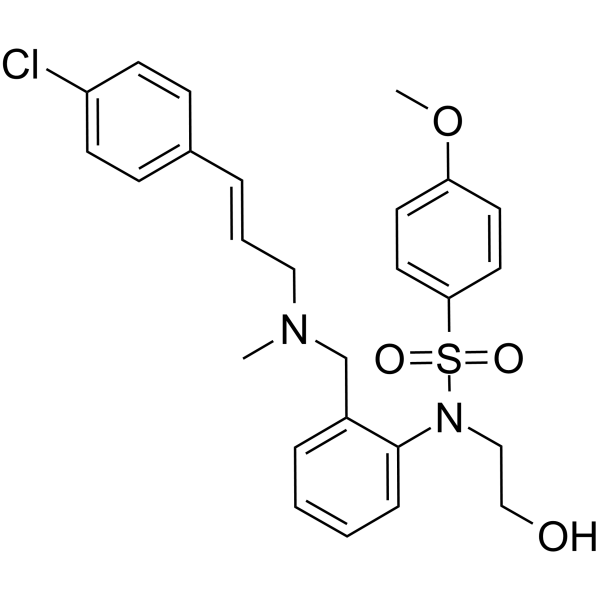
HY-15465
KN-93CaMK II Inhibitor -
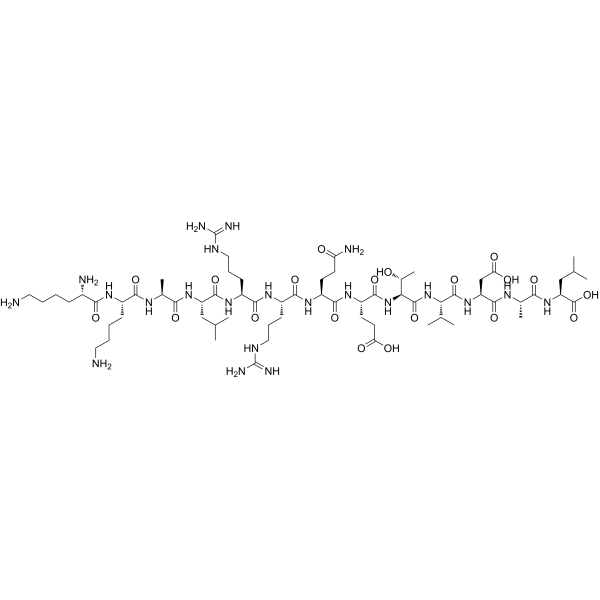
HY-P0225
Autocamtide 2CaMKII Substrate -
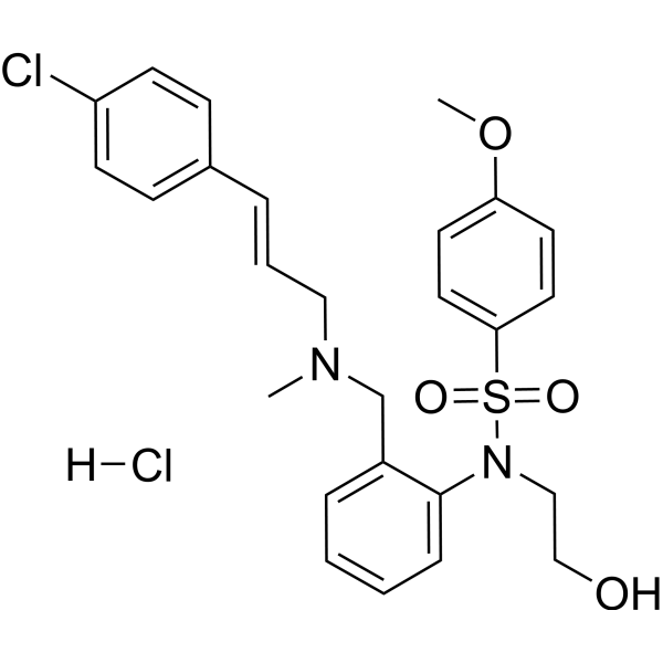
HY-15465A
KN-93 hydrochlorideCaMK II Inhibitor -
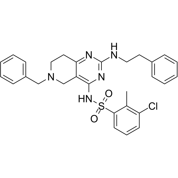
HY-18271
CaMKII-IN-1CaMKII Inhibitor -

HY-P0271
Syntide 2CaMK Substrate -
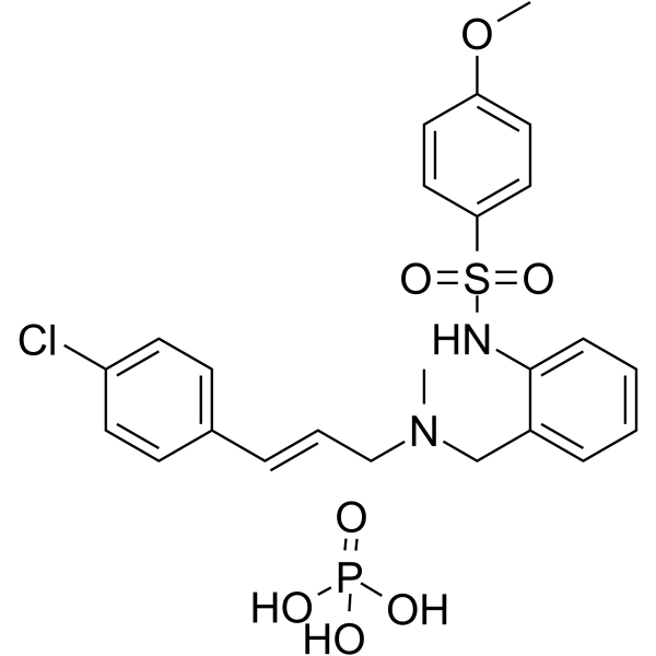
HY-15517A
KN-92 phosphateInactive KN 93 Analog -
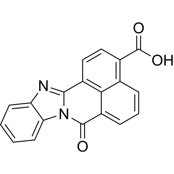
HY-19805
STO-609CaMKK Inhibitor -
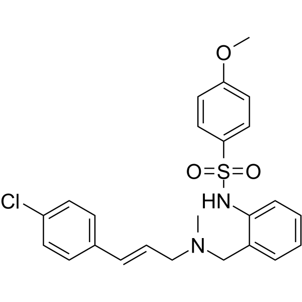
HY-15517
KN-92KN-93 Negative Control -
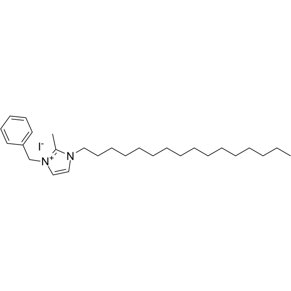
HY-100576
NH125eEF-2K/CaMKIII Inhibitor -
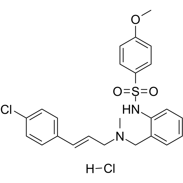
HY-15517B
KN-92 hydrochlorideKN-93 Inactive Derivative -
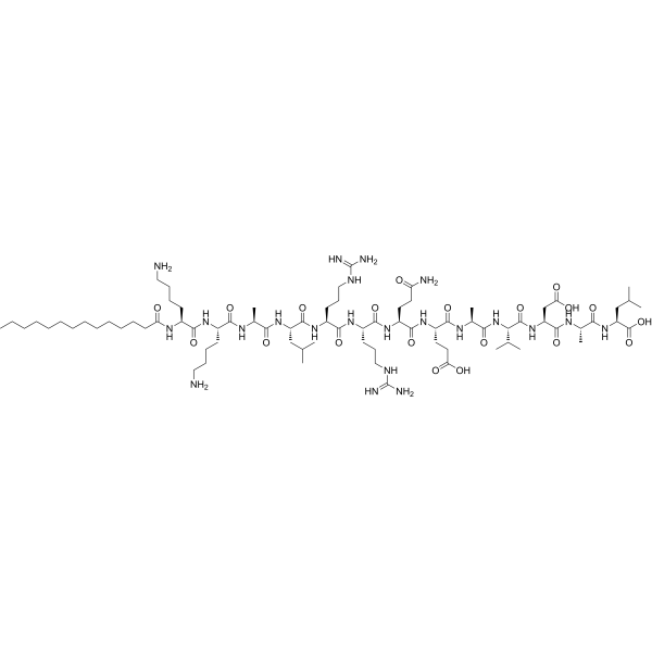
HY-P0215
Autocamtide-2-related inhibitory peptide, myristoylatedCaMKII Inhibitor -

HY-P1029
MLCK inhibitor peptide 18MLCK Inhibitor


Biological Activity
Description
IC50 & Target
|
CaMK II |
In Vitro
Peptide 290 to 309 is found to be a potent calmodulin antagonist with an IC50 of 52 nM for inhibition of Ca2+/calmodulin-dependent protein kinase. Neither truncation from the amino terminus (peptide 296-309) nor extension in the carboxyl-terminal direction (peptide 294-319) markedly affects calmodulin binding, whereas shortening the peptide from the carboxyl terminus (peptide 290-302) or from both ends (peptide 295-304) results in the elimination of this activity[1].
MedChemExpress (MCE) has not independently confirmed the accuracy of these methods. They are for reference only. Further protocols information, click here.


Chemical Information
-
CAS No. 115044-69-4
-
Molecular Weight 2273.83
-
Formula C103H185N31O24S
-
Sequence
Leu-Lys-Lys-Phe-Asn-Ala-Arg-Arg-Lys-Leu-Lys-Gly-Ala-Ile-Leu-Thr-Thr-Met-Leu-Ala
-
Sequence Shortening
LKKFNARRKLKGAILTTMLA
-
Shipping
Room temperature in continental US; may vary elsewhere.
-
Storage
Please store the product under the recommended conditions in the Certificate of Analysis.

Protocols
-
Cell Cytotoxicity Assay
Cytotoxicity assays are usually based on the assessment of cell membrane damage, which can also be indirectly detected by measuring cell viability. Detection methods include MTT assay, CKK-8 assay, LDH assay and ATP assay, etc.
-
Ca2+ Staining Technique
Ca2+ staining is an experimental technique that utilizes specific fluorescent probes (such as Fluo-4 AM, Fura-2, etc.) to qualitatively or quantitatively detect dynamic changes in intracellular Ca2+ concentrations; this is achieved by monitoring the changes in fluorescent signals generated when these probes bind to free intracellular calcium ions. The underlying principle relies primarily on the presence of chelating groups within the probe's molecular structure that possess high affinity for calcium ions.


Purity & Documentation
References

Calculators
Concentration (start) × Volume (start) = Concentration (final) × Volume (final)


Calmodulin-Dependent Protein Kinase II (290-309) Related Classifications
HY-P1479 Related Classifications
 Powered by Bioz