



Corynoxine B
Based on 6 publication(s) in Google Scholar
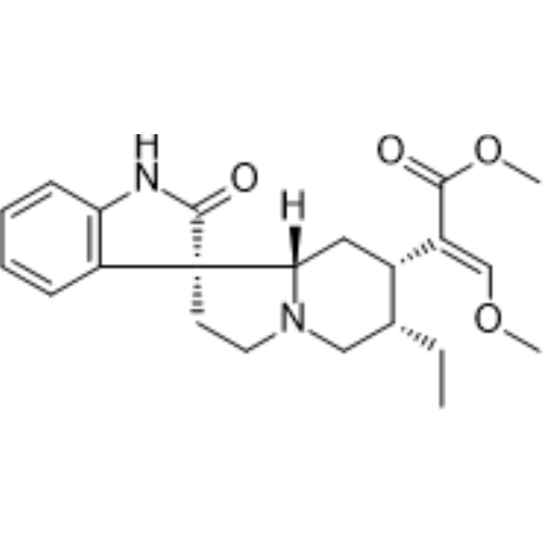
Corynoxine B is an alkaloid-based autophagy inducer and α-synuclein aggregation inhibitor that ameliorates Mn-induced dysregulation of autophagy and enhances α-synuclein (α-syn) clearance in Parkinson's disease mice.
For research use only. We do not sell to patients.
- Purity : 99.76%
- CAS No.: 17391-18-3
- Formula: C22H28N2O4
- Molecular Weight:384.47
-
Storage:Powder -20°C, 3 years , 4°C, 2 years ; In solvent -80°C, 2 years , -20°C, 1 year

To place orders, for customer services and technical support, please contact: MedChemExpress USA
Tel: 609-228-6898 E-mail: [email protected] [email protected]


-
 Biological Activity
-
Chemical Information
-
Solvent & Solubility
- Protocols
- Purity & Documentation
- References
-
 Help & FAQs


-
HY-13821
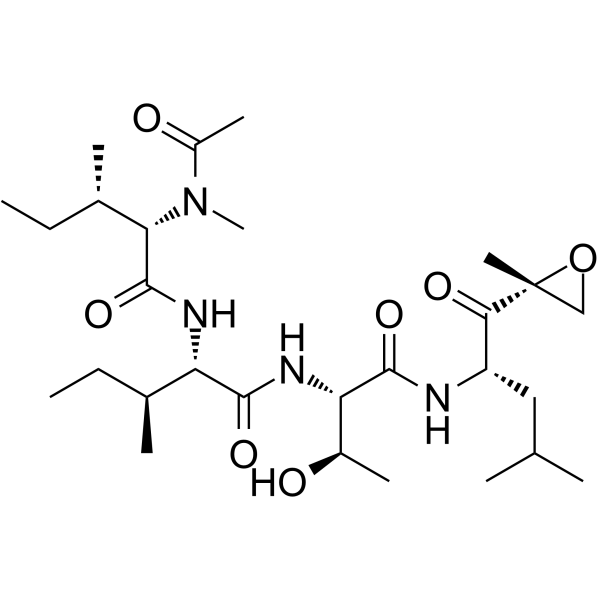
EpoxomicinProteasome Inhibitor -
HY-44134
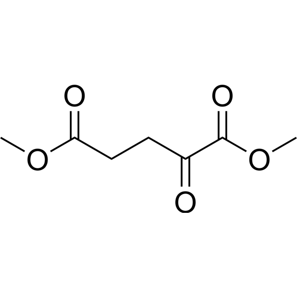
Dimethyl 2-oxoglutarate2-oxoglutarate Derivative -
HY-U00434
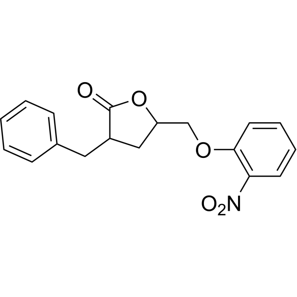
3BDOIFITM3 Modulator -

HY-N0901AR
Corynoxine B (Standard)Autophagy Inducer -
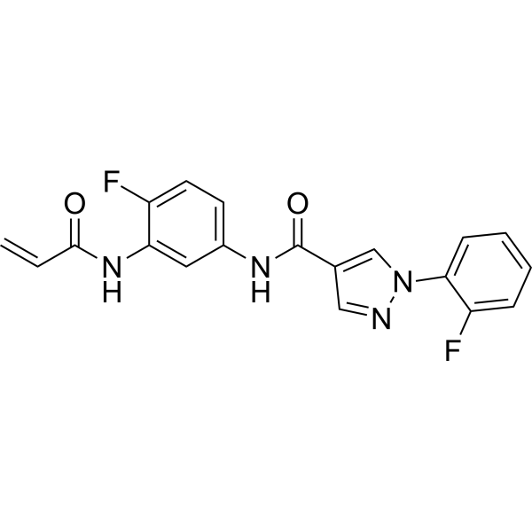
HY-128892
EN6Autophagy Activator -
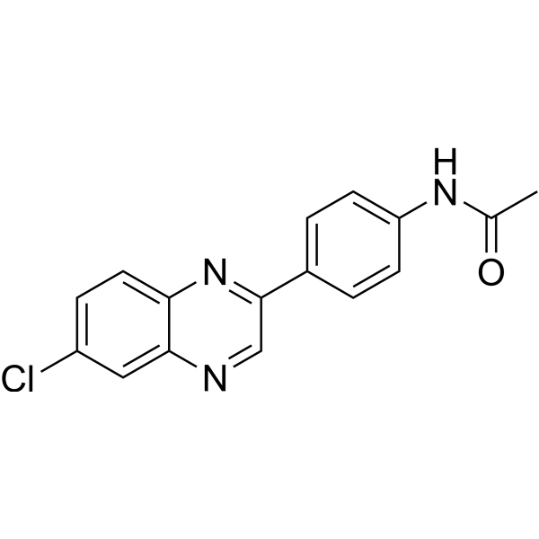
HY-134923
CA77.1CMA Activator -
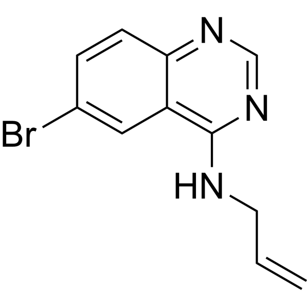
HY-100200
SMER28Autophagy Positive Regulator -
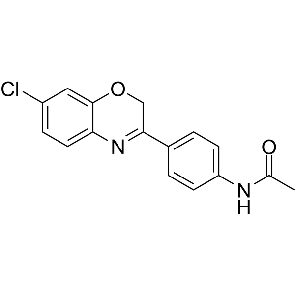
HY-112483
QX77CMA activator -
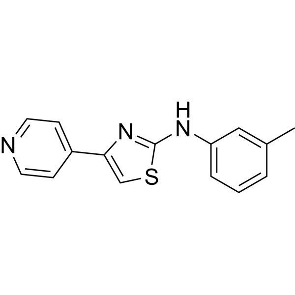
HY-100746
STF-62247Autophagy Inducer -
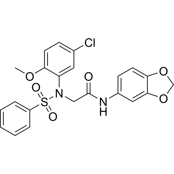
HY-111383
LX2343Beta-secretase Inhibitor -

HY-44134R
Dimethyl 2-oxoglutarate (Standard)Nitrogen Carrier -

HY-100200R
SMER28 (Standard)Autophagy Positive Regulator
Publications Citing Use of MedChemExpress (MCE) Corynoxine B
More- Adv Sci (Weinh). 2024 Nov;11(41):e2308823. [Abstract]
- Acta Pharmacol Sin. 2022 Oct;43(10):2511-2526. [Abstract]
- J Pharm Pharmacol. 2025 Aug 19:rgaf069. [Abstract]
- Food Chem Toxicol. 2019 Feb:124:336-348. [Abstract]
- Drug Test Anal. 2026 Jul 14.
- Plant Biotechnol (Tokyo). 2025 Jun 25;42(2):145-154. [Abstract]
 Customer Validation & Images

-
IHC
-
WB
-
IHC
All α-synuclein Isoforms
More

Biological Activity
Description
IC50 & Target
|
α-synuclein Aggregation |
Cellular Effect
|
Cell Line

|
Type | Value | Description | References |
|---|---|---|---|---|
| HL-60 | IC50 |
>40 μM
Compound: 5
|
Cytotoxicity against human HL60 cells after 48 hrs by MTT assay
Cytotoxicity against human HL60 cells after 48 hrs by MTT assay
|
[PMID: 21070010] |
| SW480 | IC50 |
>40 μM
Compound: 5
|
Cytotoxicity against human SW480 cells after 48 hrs by MTT assay
Cytotoxicity against human SW480 cells after 48 hrs by MTT assay
|
[PMID: 21070010] |
In Vitro
Corynoxine B (25-100 μM, 2 h) can improve Mn induced autophagy dysregulation and neurotoxicity in SH-SY5Y human neuroblastoma cells[1]. Corynoxine B can enhance the expression of BECN1 protein and restore autophagy inhibition caused by overexpression of SNCA/α-synuclein (α-syn) in PC12 cells[2].
MedChemExpress (MCE) has not independently confirmed the accuracy of these methods. They are for reference only. Further protocols information, click here.
-
Cell Line:SH-SY5Y cell
-
Concentration:25-100 μM
-
Incubation Time:2 h
-
Result:Improved cell vitality
-
Cell Line:Inducible PC12 cell lines (iPC12) overexpressing SNCA (WT and A53T)
-
Concentration:
-
Incubation Time:24 h
-
Result:Increased LC3-II and BECN1 protein levels
In Vivo
MedChemExpress (MCE) has not independently confirmed the accuracy of these methods. They are for reference only.
-
Animal Model:A53T α-syn transgenic mouse model[3].
-
Dosage:5-20 mg/kg
-
Administration:Intraperitoneal injection (i.p.)
-
Result:Promoted α-syn clearance


Chemical Information
-
CAS No. 17391-18-3
-
Appearance Solid
-
Molecular Weight 384.47
-
Formula C22H28N2O4
-
Color White to off-white
-
SMILES
O=C(NC1=C2C=CC=C1)[C@]32[C@@](C[C@H](/C(C(OC)=O)=C\OC)[C@H](CC)C4)([H])N4CC3
-
Structure Classification
-
Initial Source
-
Shipping
Room temperature in continental US; may vary elsewhere.
-
Storage
Powder -20°C 3 years 4°C 2 years In solvent -80°C 2 years -20°C 1 year


Publications (6)
-
Journal Impact Factor
-
Most Recent
-
Adv Sci (Weinh)
VPS34 Governs Oocyte Developmental Competence by Regulating Mito/Autophagy: A Novel Insight into the Significance of RAB7 Activity and Its Subcellular Location. [Abstract]2024 Nov;11(41):e2308823. PMID: 39287146 -
Acta Pharmacol Sin
Corynoxine B derivative CB6 prevents Parkinsonian toxicity in mice by inducing PIK3C3 complex-dependent autophagy. [Abstract]2022 Oct;43(10):2511-2526. PMID: 35217810 -
J Pharm Pharmacol
Advanced oxidation protein products accelerates paracetamol-induced liver injury through AMPK-mTOR signaling pathway in chronic kidney disease. [Abstract]2025 Aug 19:rgaf069. PMID: 40828098 -
Food Chem Toxicol
Corynoxine B ameliorates HMGB1-dependent autophagy dysfunction during manganese exposure in SH-SY5Y human neuroblastoma cells. [Abstract]2019 Feb:124:336-348. PMID: 30578841
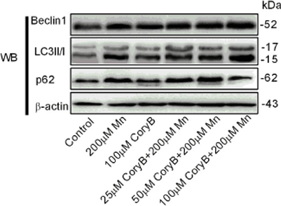
Corynoxine B purchased from MedChemExpress. Usage Cited in: Food Chem Toxicol. 2019 Feb:124:336-348. [Abstract]
The expression levels of Beclin1, LC3II/I, and p62 are determined by western blotting in the control, 200 μM Mn treatment, and Cory B pretreatment groups.
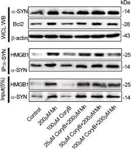
Corynoxine B purchased from MedChemExpress. Usage Cited in: Food Chem Toxicol. 2019 Feb:124:336-348. [Abstract]
Expression levels of α-SYN and Bcl2 in the whole-cell lysates are determined by western blotting in the control, 200 μM Mn treatment, and Cory B pretreatment groups.
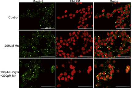
Corynoxine B purchased from MedChemExpress. Usage Cited in: Food Chem Toxicol. 2019 Feb:124:336-348. [Abstract]
The colocalization of HMGB1 (red) and Beclin1 (green), and Bcl2 (red) and Beclin1 (green) in the control, 200 μM Mn treatment, and 100 μM Cory B pretreatment groups.
-
-
Plant Biotechnol (Tokyo)
A sterile plant culture system of Uncaria rhynchophylla as a biosynthetic model of monoterpenoid indole alkaloids. [Abstract]2025 Jun 25;42(2):145-154. PMID: 40636431


Solvent & Solubility
In Vitro:
DMSO : 83.33 mg/mL (216.74 mM; Need ultrasonic; Hygroscopic DMSO has a significant impact on the solubility of product, please use newly opened DMSO)
Please refer to the solubility information to select the appropriate solvent. Once prepared, please aliquot and store the solution to prevent product inactivation from repeated freeze-thaw cycles.
Storage method and period of stock solution: -80°C, 2 years; -20°C, 1 year. When stored at -80°C, please use it within 2 years. When stored at -20°C, please use it within 1 year.
Please refer to the solubility information to select the appropriate solvent. Once prepared, please aliquot and store the solution to prevent product inactivation from repeated freeze-thaw cycles.
Storage method and period of stock solution: -80°C, 2 years; -20°C, 1 year. When stored at -80°C, please use it within 2 years. When stored at -20°C, please use it within 1 year.
Concentration (start) × Volume (start) = Concentration (final) × Volume (final)
In Vivo:
Select the appropriate dissolution method based on your experimental animal and administration route.
- For the following dissolution methods, please ensure to first prepare a clear stock solution using an In Vitro approach and then sequentially add co-solvents:
- To ensure reliable experimental results, the clarified stock solution can be appropriately stored based on storage conditions. As for the working solution for In Vivo experiments, it is recommended to prepare freshly and use it on the same day.
- The percentages shown for the solvents indicate their volumetric ratio in the final prepared solution. If precipitation or phase separation occurs during preparation, heat and/or sonication can be used to aid dissolution.
Add each solvent one by one: 10% DMSO 40% PEG300 5% Tween-80 45% Saline
Solubility: ≥ 2.08 mg/mL (5.41 mM); Clear solution
This protocol yields a clear solution of ≥ 2.08 mg/mL (saturation unknown).
Taking 1 mL working solution as an example, add 100 μL DMSO stock solution (20.8 mg/mL) to 400 μL PEG300, and mix evenly; then add 50 μL Tween-80 and mix evenly; then add 450 μL Saline to adjust the volume to 1 mL.
Preparation of Saline: Dissolve 0.9 g sodium chloride in ddH₂O and dilute to 100 mL to obtain a clear Saline solution.
In Vivo Dissolution Calculator
Please enter the basic information of animal experiments:
-
-
-
-
Recommended: Prepare an additional quantity of animals to account for potential losses during experiments.
Please enter your animal formula composition:
-
%DMSO +
Recommended: Keep the proportion of DMSO in working solution below 2% if your animal is weak.
-
%+
-
+%Tween-80 + +
-
%Saline +
The co-solvents required include: DMSO, . All of co-solvents are available by MedChemExpress (MCE). , Tween 80. All of co-solvents are available by MedChemExpress (MCE).
Working solution concentration: 0.22 mg/mL
Method for preparing stock solution: mg drug dissolved in μL DMSO. Stock solution concentration: mg/mL.
1. Take μL DMSO stock solution;
2. Add μL .
μL , mix evenly;
3. Then add μL Tween 80, mix evenly;
4. Then add μL
Please ensure that the stock solution in the first step is dissolved to a clear state, and add co-solvents in sequence. You can use ultrasonic heating (ultrasonic cleaner, recommended frequency 20-40 kHz), vortexing, etc. to assist dissolution.

Protocols
-
Human pluripotent stem cell midbrain dopaminergic neuron differentiation
Human pluripotent stem cells are directed toward midbrain dopaminergic neurons by first inducing a neural floor-plate-like progenitor state, then patterning cells with ventralizing SHH signaling and midbrain/WNT-FGF cues, and finally maturing progenitors into neurons expressing dopaminergic markers such as TH, NURR1/NR4A2, PITX3, DAT/SLC6A3, VMAT2/SLC18A2, GIRK2/KCNJ6, FOXA2, LMX1A, and EN1. The main readouts are loss of pluripotency, acquisition of FOXA2+/LMX1A+ midbrain floor-plate progenitors, emergence of βIII-tubulin+/MAP2+ neurons, and production of TH+ dopaminergic neurons with molecular, dopamine-release, and electrophysiological features of midbrain dopaminergic identity.
-
Autophagy
Autophagy is a process in which eukaryotic cells use lysosomes to degrade their own cytoplasmic proteins and damaged organelles under the regulation of autophagy related gene (Atg). Microtubule-associated proteins light chain 3 (LC3) is recognized as autophagy marker, which transfers from cytoplasmic LC3 (LC3-I) to membrane type (LC3-II). LC3-II/I ratio could be detected by Western Blot and fluorescence microscopy.
-
Protocol for Pharmacokinetic Study
Pharmacokinetic studies quantify how an organism handles a drug over time through absorption, distribution, metabolism, and excretion, and the core experimental readout is the concentration-time profile of parent drug and, when relevant, metabolites in biological matrices such as plasma, whole blood, urine, bile, or tissue. Pharmacokinetic analysis links dose, route, exposure, clearance, half-life, distribution, bioavailability, and systemic exposure to drug efficacy and toxicity hypotheses rather than measuring a signaling pathway directly. The literature links pharmacokinetics to drug-development phenotypes by showing that drug metabolism and pharmacokinetics influence compound progression, exposure-response interpretation, safety margins, dosing strategy, and failure risk during discovery and development. DMPK science contributes to compound optimization by integrating physicochemical properties, in vitro metabolism, transporter behavior, in vivo exposure, and pharmacodynamic contex
-
Lysosome and acidic-vesicle live-cell staining
Lysosome and acidic-vesicle live-cell staining detects acidic intracellular compartments by using membrane-permeant acidotropic probes that accumulate in low-pH vesicles, including lysosomes, late endosomes, autolysosomes, and acidic phagosomes. LysoTracker staining is commonly used as an intensity-based readout of acidic lysosomal compartment abundance or enlargement, while acridine orange produces green fluorescence in less concentrated compartments and red fluorescence after concentration-dependent accumulation in acidic vesicular organelles. Loss or reduction of acridine-orange red signal can be used as a readout of lysosomal membrane permeabilization or reduced acidic-vesicle integrity. This protocol is designed for live cultured cells and can be adapted for fluorescence microscopy, high-content imaging, plate-reader readout, or flow cytometry when the selected literature supports the readout. Because these dyes report acidotropic accumulation rather than lysosome identity alone,
-
Macroautophagy Solutions
Macroautophagy is a conserved lysosome-dependent degradation pathway in which cytoplasmic material is sequestered into double-membrane autophagosomes and delivered to lysosomes for degradation and recycling. The pathway supports cellular homeostasis during nutrient limitation, organelle stress, protein-aggregate accumulation, infection, differentiation, and tissue remodeling by coupling cargo sequestration, autophagosome maturation, lysosomal fusion, and degradation of cargo-derived macromolecules. The core molecular sequence includes initiation by nutrient- and stress-regulated autophagy machinery, autophagosome nucleation, LC3/ATG8-family conjugation to autophagosomal membranes, cargo selection through receptors such as SQSTM1/p62, autophagosome-lysosome fusion, and lysosomal degradation. LC3 was identified as a mammalian homolog of yeast Atg8 that localizes to autophagosomal membranes after processing, and p62/SQSTM1 was shown to connect ubiquitinated cargo with autophagic degradati


Purity & Documentation
-
Data Sheet (280 KB)
-
SDS (479 KB)
- English - EN (479 KB)
- Français - FR (479 KB)
- Deutsch - DE (479 KB)
- Norwegian - NO (479 KB)
- Español - ES (479 KB)
- Swedish - SV (479 KB)
- Italian - IT (479 KB)
- Korean - KR (479 KB)
- Portuguese - PT (479 KB)
-
Handling Instructions (2659 KB)
References
[1]. Yan D, et al. Corynoxine B ameliorates HMGB1-dependent autophagy dysfunction during manganese exposure in SH-SY5Y human neuroblastoma cells. Food Chem Toxicol. 2019 Feb;124:336-348. [Content Brief]
[2]. Chen L, et al. Corynoxine Protects Dopaminergic Neurons Through Inducing Autophagy and Diminishing Neuroinflammation in Rotenone-Induced Animal Models of Parkinson's Disease. Front Pharmacol. 2021 Apr 13;12:642900. [Content Brief]
[3]. Song JX, et al. HMGB1 is involved in autophagy inhibition caused by SNCA/α-synuclein overexpression: a process modulated by the natural autophagy inducer corynoxine B. Autophagy. 2014 Jan;10(1):144-54. doi: 10.4161/auto.26751. Epub 2013 Jan 1. Erratum in: Autophagy. 2015;11(9):1708. [Content Brief]
[4]. Zhu Q, et al. Corynoxine B targets at HMGB1/2 to enhance autophagy for α-synuclein clearance in fly and rodent models of Parkinson's disease. Acta Pharm Sin B. 2023 Jun;13(6):2701-2714. [Content Brief]


Complete Stock Solution Preparation Table
Please refer to the solubility information to select the appropriate solvent. Once prepared, please aliquot and store the solution to prevent product inactivation from repeated freeze-thaw cycles.
Storage method and period of stock solution: -80°C, 2 years; -20°C, 1 year. When stored at -80°C, please use it within 2 years. When stored at -20°C, please use it within 1 year.
| Optional Solvent | Concentration Solvent Mass | 1 mg | 5 mg | 10 mg | 25 mg |
|---|---|---|---|---|---|
| DMSO | 1 mM | 2.6010 mL | 13.0049 mL | 26.0098 mL | 65.0246 mL |
| 5 mM | 0.5202 mL | 2.6010 mL | 5.2020 mL | 13.0049 mL | |
| 10 mM | 0.2601 mL | 1.3005 mL | 2.6010 mL | 6.5025 mL | |
| 15 mM | 0.1734 mL | 0.8670 mL | 1.7340 mL | 4.3350 mL | |
| 20 mM | 0.1300 mL | 0.6502 mL | 1.3005 mL | 3.2512 mL | |
| 25 mM | 0.1040 mL | 0.5202 mL | 1.0404 mL | 2.6010 mL | |
| 30 mM | 0.0867 mL | 0.4335 mL | 0.8670 mL | 2.1675 mL | |
| 40 mM | 0.0650 mL | 0.3251 mL | 0.6502 mL | 1.6256 mL | |
| 50 mM | 0.0520 mL | 0.2601 mL | 0.5202 mL | 1.3005 mL | |
| 60 mM | 0.0433 mL | 0.2167 mL | 0.4335 mL | 1.0837 mL | |
| 80 mM | 0.0325 mL | 0.1626 mL | 0.3251 mL | 0.8128 mL | |
| 100 mM | 0.0260 mL | 0.1300 mL | 0.2601 mL | 0.6502 mL |


Corynoxine B Related Classifications
HY-N0901A Related Classifications
 Powered by Bioz