



DL-Glutamine

Based on 1 Customer Validation
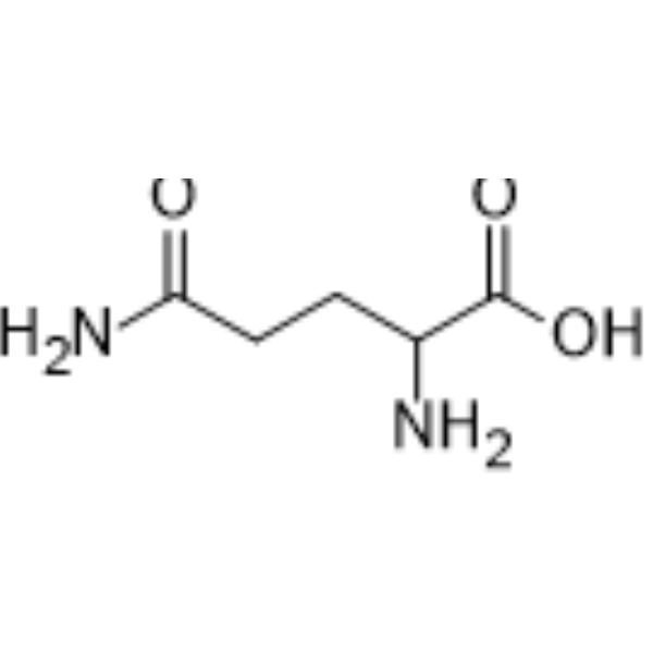
DL-Glutamine ((±)-Glutamine; DL-Gl) is an amino acid metabolite. DL-Glutamine is a plasma metabolic biomarker associated with Treg/Th17 balance in an OVA-induced eosinophilic asthma mouse model. DL-glutamine positively correlates with muscle quality parameters such as muscle water-holding capacity and shear force, and negatively correlates with oxidative stress markers and apoptotic gene expression. DL-Glutamine is associated with periodontal pathogens and oral microbial communities in saliva/serum. DL-Glutamine can be used in research related to asthma, muscle quality, and periodontal disease.
For research use only. We do not sell to patients.
- Purity : 99.91%
- CAS No.: 6899-04-3
- Formula: C5H10N2O3
- Molecular Weight:146.15
-
Storage:Powder -20°C, 3 years , 4°C, 2 years ; In solvent -80°C, 2 years , -20°C, 1 year

To place orders, for customer services and technical support, please contact: MedChemExpress USA
Tel: 609-228-6898 E-mail: [email protected] [email protected]


-
 Biological Activity
-
Chemical Information
-
Solvent & Solubility
- Protocols
- Purity & Documentation
- References
-
 Help & FAQs


-
HY-B1411
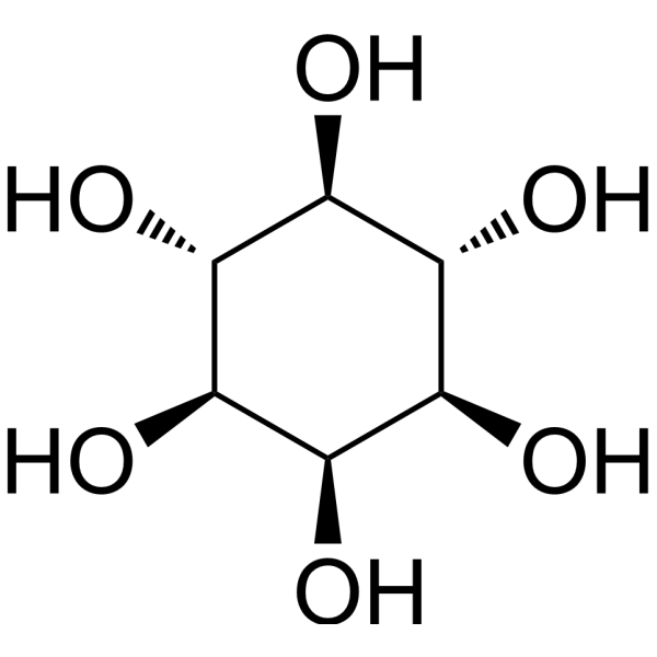
i-InositolEndogenous Metabolite -
HY-N0473
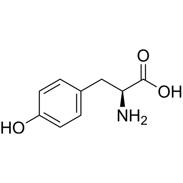
L-TyrosineCitrate Synthase Inhibitor -
HY-W016562
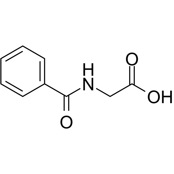
Hippuric acidMetabolite -
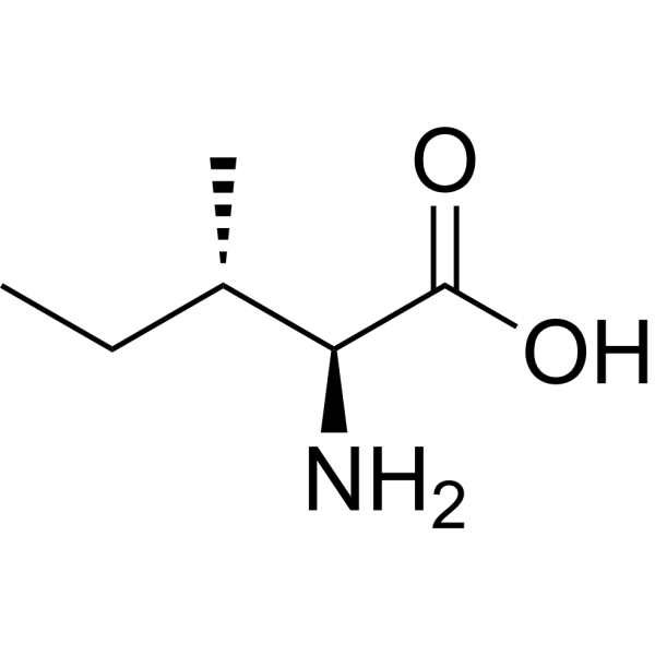
HY-N0771
L-IsoleucineEndogenous Metabolite, L-enantiomer of Isoleucine -

HY-B1346R
DL-Glutamine (Standard)Reference Standard -

HY-N0473R
L-Tyrosine (Standard)Citrate Synthase Inhibitor -
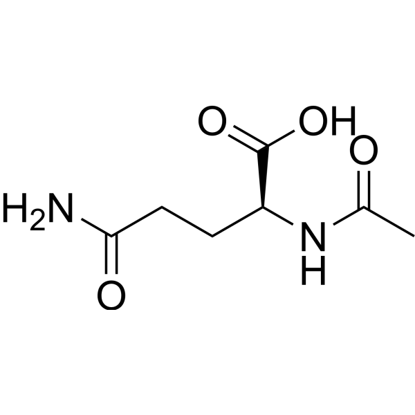
HY-B1065
AceglutamideAutophagy Inducer -
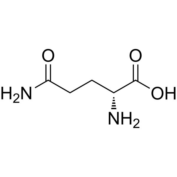
HY-100587
D-GlutamineIsomer of Glutamine -
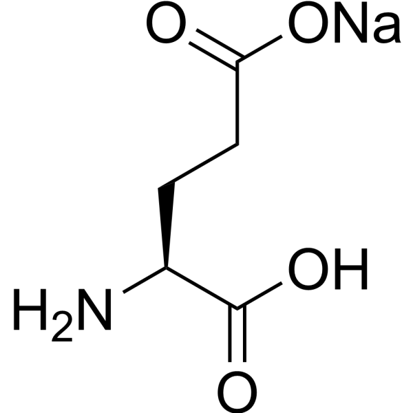
HY-14608A
L-Glutamic acid monosodium saltGlutamate Receptors Agonist -

HY-N0771R
L-Isoleucine (Standard)Endogenous Metabolite, L-enantiomer of Isoleucine -

HY-B1065R
Aceglutamide (Standard)Autophagy Inducer -

HY-100587R
D-Glutamine (Standard)Isomer of Glutamine
All Endogenous Metabolite Isoforms
More

Biological Activity
Description
IC50 & Target
[2]|
Human Endogenous Metabolite |
Microbial Metabolite |
In Vitro
DL-Glutamine is positively correlated with the salivary bacterium O. uli in the C/MIP multi-omics integration network, and forms significant microbe-metabolite association edges with periodontal pathogens in plaque/saliva[2].
DL-Glutamine is negatively correlated with oxidative stress markers (gpx) and apoptosis genes (casp3/8/9), and positively correlated with muscle quality in the muscle of silver carp under transport stress through integrated transcriptomic-metabolomic analysis[3].
MedChemExpress (MCE) has not independently confirmed the accuracy of these methods. They are for reference only. Further protocols information, click here.
In Vivo
MedChemExpress (MCE) has not independently confirmed the accuracy of these methods. They are for reference only.


Chemical Information
-
CAS No. 6899-04-3
-
Appearance Solid
-
Molecular Weight 146.15
-
Formula C5H10N2O3
-
Color White to off-white
-
SMILES
NC(CCC(N)=O)C(O)=O
-
Synonyms
(±)-Glutamine; DL-Gl
-
Structure Classification
-
Initial Source
-
Shipping
Room temperature in continental US; may vary elsewhere.
-
Storage
Powder -20°C 3 years 4°C 2 years In solvent -80°C 2 years -20°C 1 year


Solvent & Solubility
In Vitro:
H2O : 33.33 mg/mL (228.05 mM; Need ultrasonic)
DMSO : < 1 mg/mL (insoluble or slightly soluble)
Please refer to the solubility information to select the appropriate solvent. Once prepared, please aliquot and store the solution to prevent product inactivation from repeated freeze-thaw cycles.
Storage method and period of stock solution: -80°C, 2 years; -20°C, 1 year. When stored at -80°C, please use it within 2 years. When stored at -20°C, please use it within 1 year.
* Note: If you choose water as the stock solution, please dilute it to the working solution, then filter and sterilize it with a 0.22 μm filter before use.
Please refer to the solubility information to select the appropriate solvent. Once prepared, please aliquot and store the solution to prevent product inactivation from repeated freeze-thaw cycles.
Storage method and period of stock solution: -80°C, 2 years; -20°C, 1 year. When stored at -80°C, please use it within 2 years. When stored at -20°C, please use it within 1 year.
* Note: If you choose water as the stock solution, please dilute it to the working solution, then filter and sterilize it with a 0.22 μm filter before use.
Concentration (start) × Volume (start) = Concentration (final) × Volume (final)
In Vivo:
For the following dissolution methods, please prepare the working solution directly:
It is recommended to prepare fresh solutions and use them promptly within a short period of time.
The percentages shown for the solvents indicate their volumetric ratio in the final prepared solution. If precipitation or phase separation occurs during preparation, heat and/or sonication can be used to aid dissolution.
Add each solvent one by one: PBS
Solubility: 50 mg/mL (342.11 mM); Clear solution; Need ultrasonic

Protocols
-
RT-PCR
Reverse transcription technology uses RNA as a template to synthesize DNA. RT-PCR is simple, specific and sensitive, and can be used to detect gene expression levels and expression differences in cells; detect RNA virus content; clone cDNA sequences of specific genes.
-
RNA extraction experimental
By lysing cells, releasing RNA, and removing impurities such as proteins and DNA, high-purity RNA products are finally obtained. The commonly used traditional method is the guanidine isothiocyanate/phenol/chloroform method (Trizol), which is suitable for a variety of animal materials including animal tissues, microorganisms, cultured cells, etc., and most plant materials.
-
Apoptosis
Apoptosis, also called programmed cell death, is generally characterized by distinct morphological characteristics.
-
Naïve CD4+ T-cell subset differentiation/polarization
Naïve CD4+ T-cell subset differentiation/polarization is an in vitro assay in which purified naïve CD4+ T cells are activated through TCR and CD28 costimulation and cultured with defined cytokines and neutralizing antibodies to generate Th0, Th1, Th2, Th17, or induced Treg-like populations. Differentiation is detected by subset-associated cytokines and transcription factors: IFN-γ/T-bet for Th1, IL-4/GATA3 for Th2, IL-17A/RORγt for Th17, and Foxp3 for induced Treg cells. The assay readout is usually generated by intracellular cytokine staining after restimulation, transcription-factor staining by flow cytometry, ELISA of secreted cytokines, or gene-expression analysis. The result reflects cytokine-directed lineage commitment or polarization rather than antigen-specific immune protection by itself.
-
ROS/oxidative-stress fluorescent staining
ROS/oxidative-stress fluorescent staining uses cell-permeant fluorogenic probes that become fluorescent after oxidation inside cells or tissues; commonly used examples include DCFH-DA/DCFDA for broad cellular oxidant detection, DHE for superoxide-related signal detection, MitoSOX for mitochondrial superoxide-related signal detection, and CellROX probes for oxidative-stress-associated fluorescence readouts. The assay detects probe oxidation rather than a single ROS species unless the probe and analysis method have been chemically validated for that species. DCFH-DA enters cells, is deacetylated by intracellular esterases to DCFH, and produces fluorescent DCF after oxidation, so the readout is used as an operational measure of total cellular oxidative stress rather than a species-specific ROS measurement. DHE and MitoSOX can report superoxide-related oxidation, but red fluorescence alone can include non-specific ethidium-like oxidation products; HPLC or optimized spectral approaches are
-
Apoptosis Solutions
Apoptosis is a regulated, generally non-lytic cell-death pathway that removes unwanted, damaged, infected, or abnormal cells through coordinated morphological changes, caspase activation, DNA fragmentation, and membrane remodeling. The intrinsic apoptosis pathway is controlled mainly by mitochondrial outer membrane permeabilization, BCL-2 family proteins, cytochrome c release, apoptosome formation, caspase-9 activation, and downstream executioner caspase-3/7 activation. The extrinsic apoptosis pathway is initiated by death receptors such as Fas, TNFR, and TRAIL receptors, which recruit adaptor proteins and activate caspase-8 before engaging executioner caspases or mitochondrial amplification through BID cleavage. Apoptosis is linked to many phenotypes, including cancer cell killing, tissue homeostasis, immune regulation, neurodegeneration, infection response, and treatment-induced cytotoxicity; unresolved questions include how apoptosis interacts with necroptosis, pyroptosis, ferroptos
-
Real Time qPCR (Q-PCR)
Real-time quantitative PCR (qPCR) quantifies an amplifiable nucleic-acid target by monitoring fluorescence during PCR cycling rather than measuring product only after amplification. The increase in fluorescence tracks accumulation of PCR product, and the quantification cycle (Cq; historically also Ct/CP) is related to the initial amount of target: samples containing more starting target generally reach the defined fluorescence threshold in fewer cycles.
-
How to Choose the Right Model Animal
Choosing the right model animal is a validity-driven decision in which the species, strain, sex, age, genetic background, disease-induction method, outcome measures, and welfare burden must match the scientific question rather than laboratory tradition or convenience. A model should be selected by judging face validity, construct validity, and predictive validity: whether it resembles the human phenotype, whether it reproduces relevant mechanisms, and whether results are likely to predict human biology or treatment response. Animal studies often fail to translate because of species differences, weak disease resemblance, poor experimental design, inadequate reporting, publication bias, and underuse of randomization, blinding, and sample-size justification. Unresolved questions include how to rank competing models objectively, how much human-disease complexity must be reproduced for a given objective, and when non-animal systems such as organoids, ex vivo tissue, or computational models


Purity & Documentation
-
Data Sheet (271 KB)
-
SDS (254 KB)
- English - EN (254 KB)
- Français - FR (254 KB)
- Deutsch - DE (254 KB)
- Norwegian - NO (254 KB)
- Español - ES (254 KB)
- Swedish - SV (254 KB)
- Italian - IT (254 KB)
- Korean - KR (254 KB)
- Portuguese - PT (254 KB)
-
Handling Instructions (2659 KB)
References
[2]. Alamri MM, et al. Multiomics analyses in young grade C molar incisor pattern periodontitis. J Dent. 2026 Nov;174:106871. [Content Brief]
[3]. Meng Y, et al. Comprehensive transcriptomic and metabolomic analysis provides insight into transportation stress effects on fish muscle quality. NPJ science of food. 2026 Jan 07;10(1):44. [Content Brief]


Complete Stock Solution Preparation Table
Please refer to the solubility information to select the appropriate solvent. Once prepared, please aliquot and store the solution to prevent product inactivation from repeated freeze-thaw cycles.
Storage method and period of stock solution: -80°C, 2 years; -20°C, 1 year. When stored at -80°C, please use it within 2 years. When stored at -20°C, please use it within 1 year.
| Optional Solvent | Concentration Solvent Mass | 1 mg | 5 mg | 10 mg | 25 mg |
|---|---|---|---|---|---|
| H2O | 1 mM | 6.8423 mL | 34.2114 mL | 68.4229 mL | 171.0571 mL |
| 5 mM | 1.3685 mL | 6.8423 mL | 13.6846 mL | 34.2114 mL | |
| 10 mM | 0.6842 mL | 3.4211 mL | 6.8423 mL | 17.1057 mL | |
| 15 mM | 0.4562 mL | 2.2808 mL | 4.5615 mL | 11.4038 mL | |
| 20 mM | 0.3421 mL | 1.7106 mL | 3.4211 mL | 8.5529 mL | |
| 25 mM | 0.2737 mL | 1.3685 mL | 2.7369 mL | 6.8423 mL | |
| 30 mM | 0.2281 mL | 1.1404 mL | 2.2808 mL | 5.7019 mL | |
| 40 mM | 0.1711 mL | 0.8553 mL | 1.7106 mL | 4.2764 mL | |
| 50 mM | 0.1368 mL | 0.6842 mL | 1.3685 mL | 3.4211 mL | |
| 60 mM | 0.1140 mL | 0.5702 mL | 1.1404 mL | 2.8510 mL | |
| 80 mM | 0.0855 mL | 0.4276 mL | 0.8553 mL | 2.1382 mL | |
| 100 mM | 0.0684 mL | 0.3421 mL | 0.6842 mL | 1.7106 mL |
* Note: If you choose water as the stock solution, please dilute it to the working solution, then filter and sterilize it with a 0.22 μm filter before use.


DL-Glutamine Related Classifications
HY-B1346 Related Classifications
 Powered by Bioz
Keywords
- DL-Glutamine
- 6899-04-3
- (±)-Glutamine
- DL-Gl
- Endogenous Metabolite
- silver carp muscle
- molar incisor pattern periodontitis
- eosinophilic asthma
- salivary bacterium O.uli
- periodontal pathogens
- Treg/Th17 balance
- plasma metabolite biomarker
- periodontitis pathogenesis
- oral microbial communities
- amino acid metabolite
- Inhibitor
- inhibitor
- inhibit