



LRRK2 inhibitor 1

Based on 1 Customer Validation
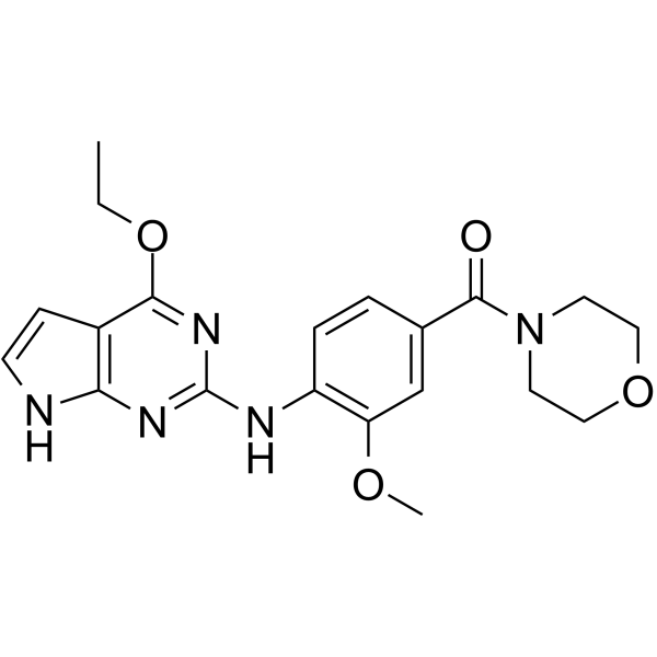
LRRK2 inhibitor 1 is a LRRK2 inhibitor. LRRK2 inhibitor 1 blocks STAT1 phosphorylation and the expression of interferon-stimulated genes, and inhibits intracellular innate immune responses. LRRK2 inhibitor 1 enhances oncolytic virus infection, replication and viral protein expression in tumor cells, promotes tumor cell apoptosis, and cooperates with oncolytic viruses to reduce tumor cell viability. LRRK2 inhibitor 1 enhances the oncolytic activity of multiple oncolytic viruses, and inhibits the growth of glioma xenografts when used in combination with oncolytic viruses. LRRK2 inhibitor 1 can be used in research related to various cancers such as lung cancer, colorectal cancer and liver cancer.
For research use only. We do not sell to patients.
- Purity : 99.80%
- CAS No.: 1802525-61-6
- Formula: C20H23N5O4
- Molecular Weight:397.43
-
Storage:Powder -20°C, 3 years , 4°C, 2 years ; In solvent -80°C, 2 years , -20°C, 1 year

To place orders, for customer services and technical support, please contact: MedChemExpress USA
Tel: 609-228-6898 E-mail: [email protected] [email protected]


-
 Biological Activity
-
Chemical Information
-
Solvent & Solubility
- Protocols
- Purity & Documentation
- References
-
 Help & FAQs


-
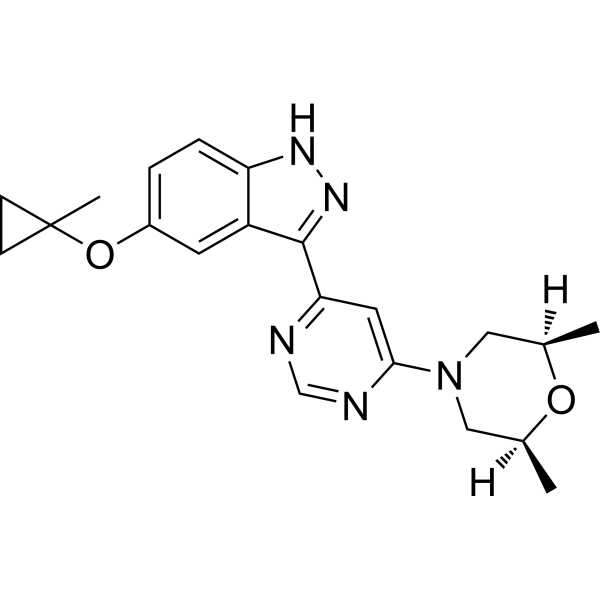
HY-100411
MLi-2LRRK2 Inhibitor -
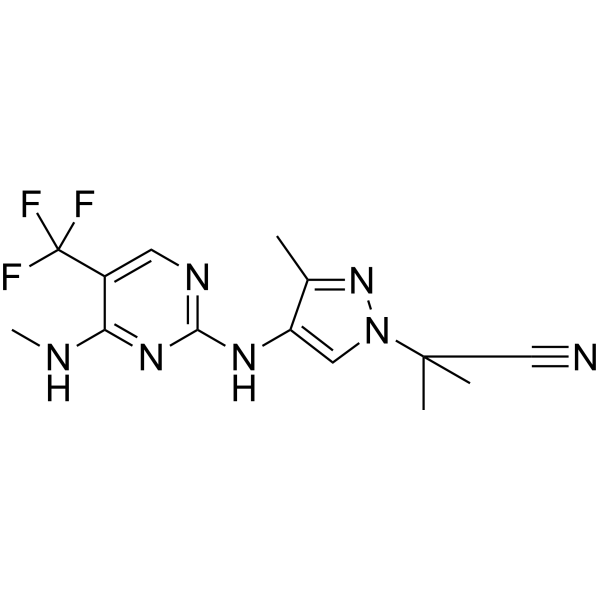
HY-15796
GNE0877LRRK2 Inhibitor -
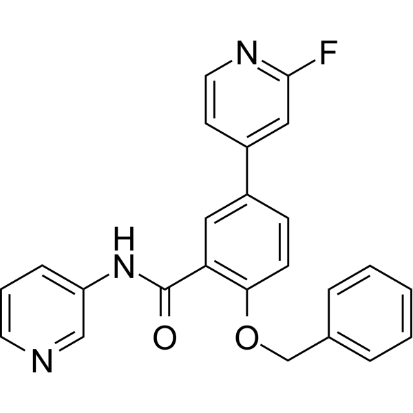
HY-13237
GSK2578215ALRRK2 Inhibitor -

HY-12477
PF-06447475LRRK2 Inhibitor -
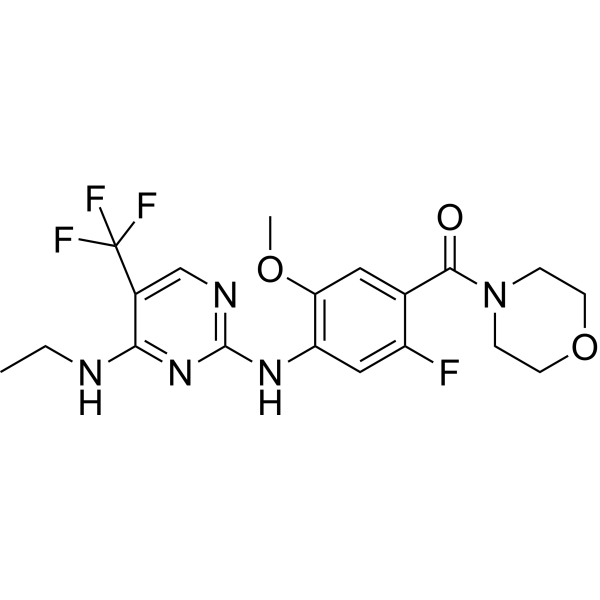
HY-18163
GNE-7915LRRK2 Inhibitor -
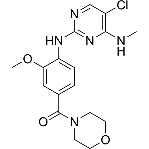
HY-13488
HG-10-102-01LRRK2 Inhibitor -
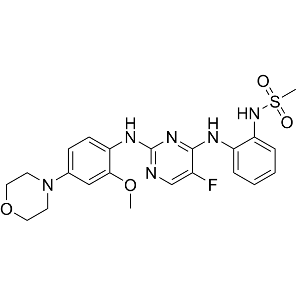
HY-15800A
CZC-25146LRRK2 Inhibitor -
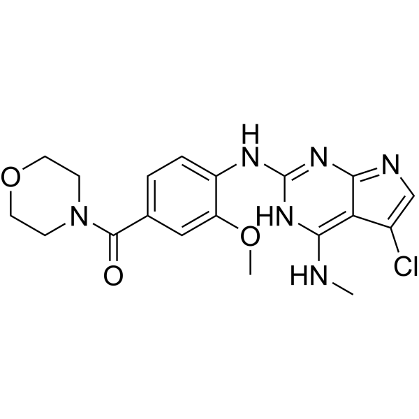
HY-16936
JH-II-127LRRK2 Inhibitor -
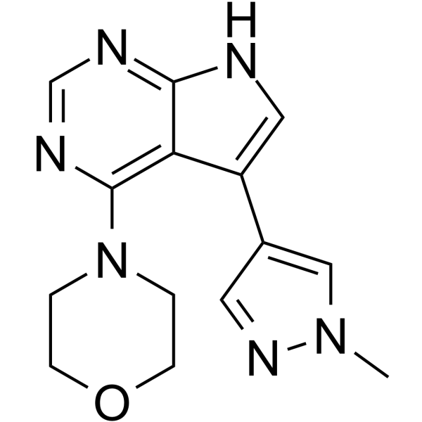
HY-112855
PF-06454589LRRK2 Inhibitor -
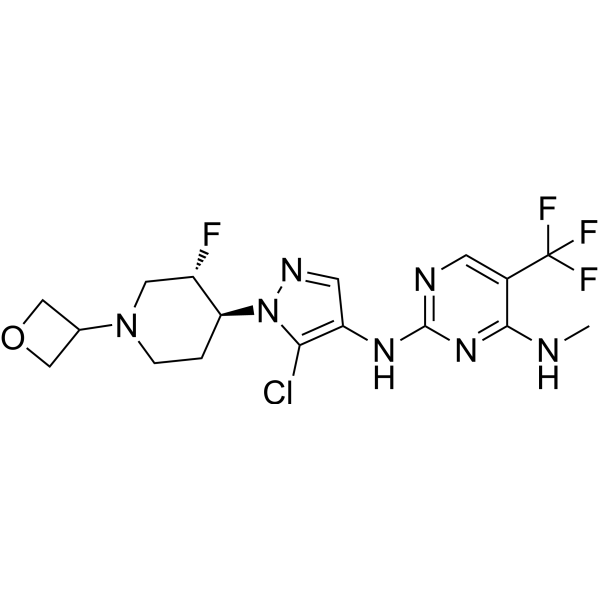
HY-12282
GNE-9605LRRK2 Inhibitor -
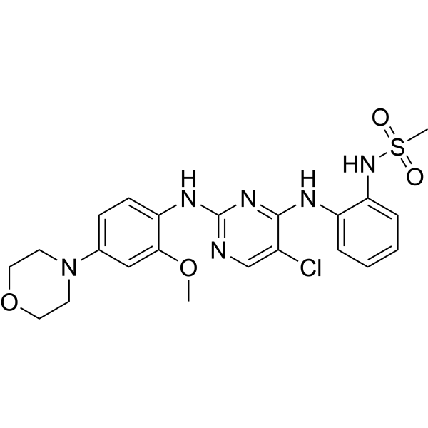
HY-B0792
CZC-54252LRRK2 Inhibitor -
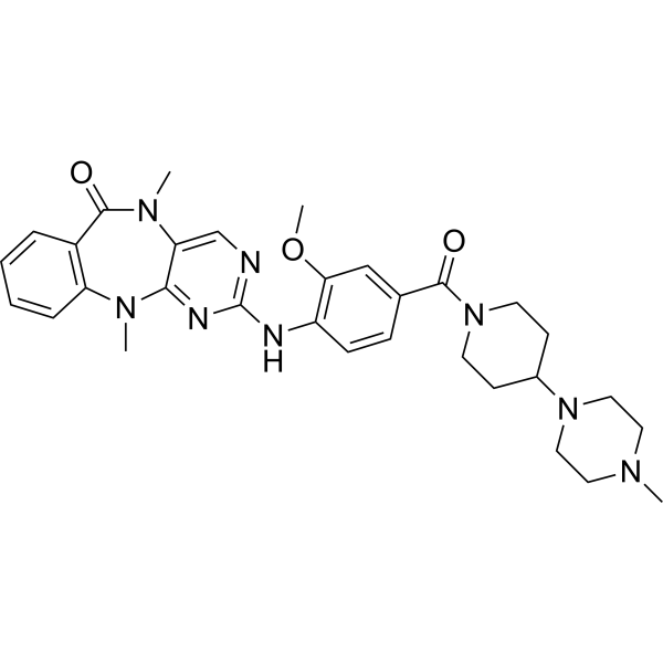
HY-10875
LRRK2-IN-1LRRK2 Inhibitor


Biological Activity
Description
IC50 & Target
[1]|
STAT1 |
In Vitro
LRRK2 inhibitor 1 (1-10 μM; 48 h) significantly enhances the killing effect of oHSV-GFP virus on various human cancer cell lines, including human lung cancer cell line A549 and human colorectal cancer cell line HT-29, and reduces cell viability[1].
LRRK2 inhibitor 1 (5 μM) significantly promotes the replication of oHSV-GFP virus in human glioblastoma cell lines U-251MG, U-87MG, U-118MG and GBM, enhances viral protein expression, and improves viral infectivity[1].
Combined treatment with LRRK2 inhibitor 1 (2.5 μM; 48 h) and oHSV-GFP virus significantly reduces the viability of human glioblastoma cell lines U-251MG and LN-18[1].
LRRK2 inhibitor 1 (2.5 μM; 48 h) significantly enhances the killing effect of HSV-1 recombinant viruses D34.5, OVH, HSV-2 virus and adenovirus HADV5 on the human glioblastoma cell line U-251MG, and reduces cell viability[1].
LRRK2 inhibitor 1 (5 μM; 12 h) enhances the infectivity of oHSV-GFP in the human glioblastoma cell line U-87MG by inhibiting phosphorylation of the STAT pathway and ISG gene expression, thereby suppressing intracellular innate immune responses[1].
Combination treatment with LRRK2 inhibitor 1 (5 μM; 48 h) and oHSV-GFP virus significantly promotes early and late apoptosis in human glioblastoma cell lines U-251MG and U-118MG, thereby enhancing anti-tumor activity[1].
MedChemExpress (MCE) has not independently confirmed the accuracy of these methods. They are for reference only. Further protocols information, click here.
-
Cell Line:A549、HT-29、Hep G2、MDA-MB-231、HeLa、SK-OV-3、A-375、HEp-2、U-20S、Panc 10.5、FaDu、AGS、A-498
-
Concentration:10 μM、5 μM、2 μM、1 μM
-
Incubation Time:4 h (pre-incubation); 48 h (total treatment)
-
Result:Reduced A549 cell viability from 98.1% to 64.59%.
Reduced A-498 cell viability from 85.6% to 72.8%.
Reduced HT-29 cell viability from 91.0% to 38.3%.
Reduced Hep G2 cell viability from 58.9% to 18.1%.
Reduced MDA-MB-231 cell viability from 81.4% to 26.0%.
Reduced HeLa cell viability from 63.8% to 14.5%.
Reduced SK-OV-3 cell viability from 98.6% to 64.3%.
Reduced A-375 cell viability from 100.1% to 19.9%.
Reduced HEp-2 cell viability from 98.0% to 68.0%.
Reduced U-20S cell viability from 97.3% to 78.8%.
Reduced Panc 10.5 cell viability from 91.8% to 61.3%.
Reduced FaDu cell viability from 95.1% to 22.3%.
Reduced AGS cell viability from 100.1% to 24.2%.
-
Cell Line:U-251MG、LN-18
-
Concentration:2.5 μM
-
Incubation Time:4 h (pre-incubation); 48 h (total treatment)
-
Result:Reduced cell viability significantly compared to virus-only group.
Achieved IC50 shift of 10.42-fold in U-251MG cells.
Achieved IC50 shift of 11.73-fold in LN-18 cells.
-
Cell Line:U-251MG、U-118MG
-
Concentration:5 μM
-
Incubation Time:4 h (pre-incubation); 48 h (total treatment)
-
Result:Increased early apoptosis rate of U-251MG cells from 32.1% to 59.5%.
Increased late apoptosis rate of U-251MG cells from 19.9% to 25.6%.
Increased early apoptosis rate of U-118MG cells from 6.0% to 41.6%.
Increased late apoptosis rate of U-118MG cells from 9.0% to 15.6%.
-
Cell Line:U-251MG
-
Concentration:2.5 μM
-
Incubation Time:4 h (pre-incubation); 48 h (total treatment)
-
Result:Reduced U-251MG cell viability from 96.4% to 33.1% when combined with HSV-1 recombinant virus D34.5.
Reduced U-251MG cell viability from 107.0% to 20.1% when combined with HSV-1 recombinant virus OVH.
Reduced U-251MG cell viability from 66.1% to 32.3% when combined with HSV-2 virus.
Reduced U-251MG cell viability from 87.8% to 57.2% when combined with adenovirus HADV5.
In Vivo
LRRK2 inhibitor 1 (2 mg/kg; i.p.; once daily) combined with oHSV-GFP significantly inhibits the growth of xenografts derived from the human glioma PDX model GBM-1, with no significant impact on body weight and favorable safety profile[1].
MedChemExpress (MCE) has not independently confirmed the accuracy of these methods. They are for reference only.
-
Animal Model:Nod Scid (6-week-old female)[1]
-
Dosage:2 mg/kg (LRRK2-IN-1); 1×10⁷ PFU/次 (oHSV-GFP)
-
Administration:i.p. (daily); i.t. (every 2 days, 6 times total)
-
Result:Did not reduce tumor volume compared with control group.
Significantly reduced tumor volume when combined with oHSV-GFP, with no significant effect on mouse body weight and good safety.
-
Animal Model:Nod Scid (6-week-old female)[1]
-
Dosage:2 mg/kg (LRRK2-IN-1); 1×10⁷ PFU/次 (oHSV-GFP)
-
Administration:i.p. (daily); i.t. (every 2 days, 3 times total)
-
Result:Caused only slight tumor volume reduction compared with control group.
Significantly reduced tumor volume when combined with oHSV-GFP, with no significant effect on mouse body weight and good safety.


Chemical Information
-
CAS No. 1802525-61-6
-
Appearance Solid
-
Molecular Weight 397.43
-
Formula C20H23N5O4
-
Color White to off-white
-
SMILES
CCOC1=NC(NC2=C(OC)C=C(C(N3CCOCC3)=O)C=C2)=NC4=C1C=CN4
-
Shipping
Room temperature in continental US; may vary elsewhere.
-
Storage
Powder -20°C 3 years 4°C 2 years In solvent -80°C 2 years -20°C 1 year


Solvent & Solubility
In Vitro:
DMSO : 62.5 mg/mL (157.26 mM; Need ultrasonic; Hygroscopic DMSO has a significant impact on the solubility of product, please use newly opened DMSO)
Please refer to the solubility information to select the appropriate solvent. Once prepared, please aliquot and store the solution to prevent product inactivation from repeated freeze-thaw cycles.
Storage method and period of stock solution: -80°C, 2 years; -20°C, 1 year. When stored at -80°C, please use it within 2 years. When stored at -20°C, please use it within 1 year.
Please refer to the solubility information to select the appropriate solvent. Once prepared, please aliquot and store the solution to prevent product inactivation from repeated freeze-thaw cycles.
Storage method and period of stock solution: -80°C, 2 years; -20°C, 1 year. When stored at -80°C, please use it within 2 years. When stored at -20°C, please use it within 1 year.
Concentration (start) × Volume (start) = Concentration (final) × Volume (final)
In Vivo:
Select the appropriate dissolution method based on your experimental animal and administration route.
- For the following dissolution methods, please ensure to first prepare a clear stock solution using an In Vitro approach and then sequentially add co-solvents:
- To ensure reliable experimental results, the clarified stock solution can be appropriately stored based on storage conditions. As for the working solution for In Vivo experiments, it is recommended to prepare freshly and use it on the same day.
- The percentages shown for the solvents indicate their volumetric ratio in the final prepared solution. If precipitation or phase separation occurs during preparation, heat and/or sonication can be used to aid dissolution.
Add each solvent one by one: 10% DMSO 40% PEG300 5% Tween-80 45% Saline
Solubility: ≥ 2.08 mg/mL (5.23 mM); Clear solution
This protocol yields a clear solution of ≥ 2.08 mg/mL (saturation unknown).
Taking 1 mL working solution as an example, add 100 μL DMSO stock solution (20.8 mg/mL) to 400 μL PEG300, and mix evenly; then add 50 μL Tween-80 and mix evenly; then add 450 μL Saline to adjust the volume to 1 mL.
Preparation of Saline: Dissolve 0.9 g sodium chloride in ddH₂O and dilute to 100 mL to obtain a clear Saline solution.
Add each solvent one by one: 10% DMSO 90% (20% SBE-β-CD in Saline)
Solubility: 2.08 mg/mL (5.23 mM); Suspended solution; Need ultrasonic
This protocol yields a suspended solution of 2.08 mg/mL. Suspended solution can be used for oral and intraperitoneal injection.
Taking 1 mL working solution as an example, add 100 μL DMSO stock solution (20.8 mg/mL) to 900 μL 20% SBE-β-CD in Saline, and mix evenly.
Preparation of 20% SBE-β-CD in Saline (4°C, storage for one week): 2 g SBE-β-CD powder is dissolved in 10 mL Saline, completely dissolve until clear.
In Vivo Dissolution Calculator
Please enter the basic information of animal experiments:
-
-
-
-
Recommended: Prepare an additional quantity of animals to account for potential losses during experiments.
Please enter your animal formula composition:
-
%DMSO +
Recommended: Keep the proportion of DMSO in working solution below 2% if your animal is weak.
-
%+
-
+%Tween-80 + +
-
%Saline +
The co-solvents required include: DMSO, . All of co-solvents are available by MedChemExpress (MCE). , Tween 80. All of co-solvents are available by MedChemExpress (MCE).
Working solution concentration: 0.22 mg/mL
Method for preparing stock solution: mg drug dissolved in μL DMSO. Stock solution concentration: mg/mL.
1. Take μL DMSO stock solution;
2. Add μL .
μL , mix evenly;
3. Then add μL Tween 80, mix evenly;
4. Then add μL
Please ensure that the stock solution in the first step is dissolved to a clear state, and add co-solvents in sequence. You can use ultrasonic heating (ultrasonic cleaner, recommended frequency 20-40 kHz), vortexing, etc. to assist dissolution.

Protocols
-
Western Blot
Western blotting (WB) is a commonly used experimental method in molecular biology, biochemistry, and immunogenetics for identifying and quantifying target proteins. It combines gel electrophoresis with immunoassay, enabling researchers to analyze protein expression, post-translational modifications, and molecular weight.
-
RNA extraction experimental
By lysing cells, releasing RNA, and removing impurities such as proteins and DNA, high-purity RNA products are finally obtained. The commonly used traditional method is the guanidine isothiocyanate/phenol/chloroform method (Trizol), which is suitable for a variety of animal materials including animal tissues, microorganisms, cultured cells, etc., and most plant materials.
-
Research Protocol for Infectious Diseases
Infectious-disease experiments test how pathogens interact with host barriers, innate immune receptors, inflammatory signaling, pathogen replication, and tissue injury; pattern-recognition receptors such as TLRs, RIG-I-like receptors, NOD-like receptors, and inflammasomes detect microbial molecules and activate NF-κB, interferon, and cytokine responses. The central hypothesis is that infection severity reflects the balance between pathogen burden and host response: protective inflammation restricts pathogen growth, whereas excessive or mislocalized inflammation contributes to tissue damage and disease phenotype. Unresolved questions include which host pathways are protective versus pathogenic, why some infection models fail to translate to human disease, and which combined readouts best predict clinically relevant infection outcomes.
-
Mammalian live/dead viability and cytotoxicity staining
Live/dead viability and cytotoxicity staining assays are based on the simultaneous detection of intracellular esterase activity in metabolically active (viable) cells and membrane integrity loss in non-viable cells. In commonly used dual-staining approaches, membrane-permeant fluorogenic substrates are converted by intracellular esterases into fluorescent products in live cells, while impermeant DNA-binding dyes selectively enter cells with compromised plasma membranes and label nucleic acids in dead or dying cells, enabling discrimination between viable and non-viable populations by fluorescence microscopy or flow cytometry.
-
Annexin V plus membrane-impermeant dye apoptosis staining
Annexin V-based apoptosis assays rely on the detection of phosphatidylserine (PS) externalization from the inner leaflet of the plasma membrane to the outer leaflet, an early biochemical hallmark of apoptosis. Fluorescently labeled Annexin V binds PS in a calcium-dependent manner, enabling identification of early apoptotic cells by flow cytometry or fluorescence microscopy. When combined with a membrane-impermeant DNA-binding dye (e. g. , propidium iodide), this approach allows discrimination between viable (Annexin V−/dye−), early apoptotic (Annexin V+/dye−), and late apoptotic or necrotic (Annexin V+/dye+) cell populations by assessing membrane integrity and PS exposure.
-
CCK-8/WST-8 Cell Proliferation Assay
The CCK-8/WST-8 assay is based on the reduction of the water-soluble tetrazolium salt WST-8 to a water-soluble formazan product by cellular dehydrogenases in metabolically active cells, where the generated formazan amount is proportional to the number of living cells and is quantified by measuring absorbance in the visible range, providing a colorimetric readout for cell viability and proliferation assessment. This class of tetrazolium-based assays improves upon earlier MTT-based systems by producing a water-soluble formazan, eliminating the need for organic solubilization steps and enabling direct spectrophotometric measurement in culture medium.
-
Liver Cancer Modeling
Liver cancer can be classified into primary liver cancer and secondary liver cancer. Secondary liver cancer is the metastatic liver cancer. Primary liver cancer includes hepatocellular carcinoma (HCC), intrahepatic cholangiocarcinoma (ICC) and fibrolamellar HCC, of which HCC is the most common form, accounting for approximately 90% of primary liver cancers[1]. HCC mouse models include chemical agent-induced models, transplanted tumor models, and genetic engineered models.
-
Protocol For Protein Expression And Purification
Recombinant protein expression in Escherichia coli followed by purification of a His-tagged soluble protein by immobilized metal affinity chromatography (IMAC), with optional MBP fusion and TEV tag removal when the construct includes these elements. The biological readout is production of the encoded target protein, detected as an inducible band at the expected molecular mass by SDS-PAGE and quantified by total protein assay or chromatographic absorbance; the purification readout is enrichment of the target protein in elution fractions after selective binding of polyhistidine residues to immobilized Ni2+/metal-chelate resin and elution by imidazole-containing buffer. Expression is driven by an inducible bacterial expression system, commonly T7/lac-based, in which IPTG or lactose/auto-induction activates transcription and translation of the cloned gene; lower induction temperature, lower inducer concentration, induction timing, and solubility-enhancing fusion tags can influence the frac
-
Cell Viability Determination by MTT Colorimetric Assay
The following protocol uses the MTT colorimetric assay as a classic literature-established method for assessing cell viability/metabolic activity in cultured mammalian cells. MTT[3-(4,5-dimethylthiazol-2-yl)-2,5-diphenyltetrazolium bromide] is reduced by metabolically active cells to a colored formazan product; the amount of formazan is quantified spectrophotometrically and provides an indirect measure of metabolically active viable cells. Importantly, MTT reduction reflects cellular oxidoreductase/metabolic activity rather than an absolute direct count of living cells, so changes in cellular metabolism can alter the signal independently of cell number.
-
Kinase activity and phosphorylation assays
Kinase activity assays measure the ability of kinases to transfer phosphate groups from ATP to specific substrates, while phosphorylation assays detect the presence and levels of phosphorylated proteins. Common methods include radiolabeled ATP incorporation (e. g. ,), ADP release detection via bioluminescence (e. g. ,[3]), enzyme-linked immunosorbent assays (ELISA) for phospho-specific epitopes (e. g. ,[6]), and microtiter-based formats for high-throughput screening (e. g. ,[8]). The ADP-Glo assay quantifies kinase activity by measuring ADP produced during phosphorylation using a luciferase-based system. Radiometric assays involve autoradiography or scintillation counting after incorporation of 32P-labeled ATP into substrate proteins. ELISA-based approaches rely on phospho-specific antibodies to detect activated kinases in cell lysates or purified samples.
-
Apoptosis
Apoptosis, also called programmed cell death, is generally characterized by distinct morphological characteristics.
-
TUNEL staining for apoptotic DNA fragmentation
TUNEL staining detects DNA strand breaks by using terminal deoxynucleotidyl transferase to add labeled nucleotides to exposed 3′-OH DNA termini, generating either microscopic staining in fixed cells or tissue sections, or fluorescence/cytometric signal in cell suspensions. TUNEL positivity reflects DNA fragmentation but should not be interpreted alone as definitive apoptosis, because TUNEL can also label necrotic, autolytic, mechanically damaged, or DNA-repair-associated DNA breaks.
-
Cell Cytotoxicity Assay
Cytotoxicity assays are usually based on the assessment of cell membrane damage, which can also be indirectly detected by measuring cell viability. Detection methods include MTT assay, CKK-8 assay, LDH assay and ATP assay, etc.
-
Apoptosis Solutions
Apoptosis is a regulated, generally non-lytic cell-death pathway that removes unwanted, damaged, infected, or abnormal cells through coordinated morphological changes, caspase activation, DNA fragmentation, and membrane remodeling. The intrinsic apoptosis pathway is controlled mainly by mitochondrial outer membrane permeabilization, BCL-2 family proteins, cytochrome c release, apoptosome formation, caspase-9 activation, and downstream executioner caspase-3/7 activation. The extrinsic apoptosis pathway is initiated by death receptors such as Fas, TNFR, and TRAIL receptors, which recruit adaptor proteins and activate caspase-8 before engaging executioner caspases or mitochondrial amplification through BID cleavage. Apoptosis is linked to many phenotypes, including cancer cell killing, tissue homeostasis, immune regulation, neurodegeneration, infection response, and treatment-induced cytotoxicity; unresolved questions include how apoptosis interacts with necroptosis, pyroptosis, ferroptos
-
Protocol for Kinase activity and phosphorylation assays
Kinase activity assays measure transfer of phosphate from ATP to a protein or peptide substrate, generating phosphorylated substrate, ADP, or incorporated radiolabeled phosphate as the readout; phosphorylation assays measure site-specific phosphorylation in cells or tissues as a proxy for kinase-pathway activation, inhibition, or substrate regulation. Phosphorylation can be detected by phospho-specific Western blot, immunoprecipitation kinase assay, phospho-immunofluorescence, phospho-flow cytometry, luminescent ADP detection, radiolabeled ATP incorporation, or reporter-based pathway assays, and these readouts can be applied to cancer cells, primary neurons, mouse tumors, organoids, inflammatory macrophages, ferroptosis studies, and mitophagy studies when the kinase target is biologically relevant.
-
MTT Cell Proliferation Assay
The MTT assay is a colorimetric endpoint assay for estimating viable cell number, cell growth, cytotoxicity, or cell activation in cultured mammalian cells. Living cells reduce the yellow tetrazolium salt MTT into purple/blue formazan, while dead cells do not generate the same signal; the resulting color can be quantified with a multiwell spectrophotometer. MTT reduction is commonly interpreted as a readout of metabolic activity that often correlates with viable cell number, but it should not be treated as a direct cell-counting method unless the assay is optimized for the cell type and experimental condition. Studies show that MTT reduction can involve mitochondrial and non-mitochondrial reducing systems, and formazan may accumulate in intracellular lipid droplets rather than simply marking mitochondria.


Purity & Documentation
-
Data Sheet (282 KB)
-
SDS (254 KB)
- English - EN (254 KB)
- Français - FR (254 KB)
- Deutsch - DE (254 KB)
- Norwegian - NO (254 KB)
- Español - ES (254 KB)
- Swedish - SV (254 KB)
- Italian - IT (254 KB)
- Korean - KR (254 KB)
- Portuguese - PT (254 KB)
-
Handling Instructions (2659 KB)
References


Complete Stock Solution Preparation Table
Please refer to the solubility information to select the appropriate solvent. Once prepared, please aliquot and store the solution to prevent product inactivation from repeated freeze-thaw cycles.
Storage method and period of stock solution: -80°C, 2 years; -20°C, 1 year. When stored at -80°C, please use it within 2 years. When stored at -20°C, please use it within 1 year.
| Optional Solvent | Concentration Solvent Mass | 1 mg | 5 mg | 10 mg | 25 mg |
|---|---|---|---|---|---|
| DMSO | 1 mM | 2.5162 mL | 12.5808 mL | 25.1617 mL | 62.9042 mL |
| 5 mM | 0.5032 mL | 2.5162 mL | 5.0323 mL | 12.5808 mL | |
| 10 mM | 0.2516 mL | 1.2581 mL | 2.5162 mL | 6.2904 mL | |
| 15 mM | 0.1677 mL | 0.8387 mL | 1.6774 mL | 4.1936 mL | |
| 20 mM | 0.1258 mL | 0.6290 mL | 1.2581 mL | 3.1452 mL | |
| 25 mM | 0.1006 mL | 0.5032 mL | 1.0065 mL | 2.5162 mL | |
| 30 mM | 0.0839 mL | 0.4194 mL | 0.8387 mL | 2.0968 mL | |
| 40 mM | 0.0629 mL | 0.3145 mL | 0.6290 mL | 1.5726 mL | |
| 50 mM | 0.0503 mL | 0.2516 mL | 0.5032 mL | 1.2581 mL | |
| 60 mM | 0.0419 mL | 0.2097 mL | 0.4194 mL | 1.0484 mL | |
| 80 mM | 0.0315 mL | 0.1573 mL | 0.3145 mL | 0.7863 mL | |
| 100 mM | 0.0252 mL | 0.1258 mL | 0.2516 mL | 0.6290 mL |


LRRK2 inhibitor 1 Related Classifications
HY-111493 Related Classifications
 Powered by Bioz