



Lasofoxifene tartrate
Based on 6 publication(s) in Google Scholar
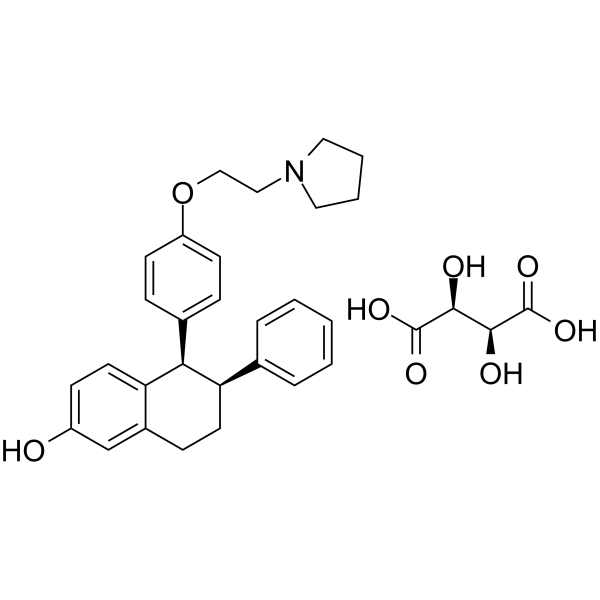
Lasofoxifene (CP-336156) tartrate is an orally active and selective estrogen receptor modulator (SERM). Lasofoxifene tartrate exhibits an anti-osteoporotic function and also inhibits primary tumor growth and metastases. Lasofoxifene tartrate can be used for the research of breast cancer and postmenopausal osteoporosis.
For research use only. We do not sell to patients.
- Purity : 99.93%
- CAS No.: 190791-29-8
- Formula: C32H37NO8
- Molecular Weight:563.64
-
Storage:
4°C, sealed storage, away from moisture
* In solvent : -80°C, 6 months; -20°C, 1 month (sealed storage, away from moisture)

To place orders, for customer services and technical support, please contact: MedChemExpress USA
Tel: 609-228-6898 E-mail: [email protected] [email protected]


-
 Biological Activity
-
Chemical Information
-
Solvent & Solubility
- Protocols
- Purity & Documentation
- References
-
 Help & FAQs


-
HY-B0005
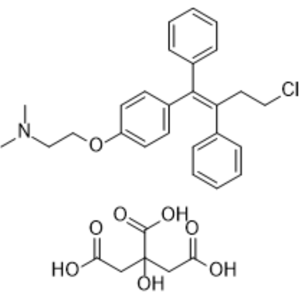
Toremifene citrateApoptosis Inducer -
HY-16950
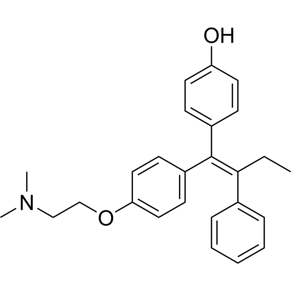
4-HydroxytamoxifenEstrogen Receptor Modulator -

HY-B0723
OspemifeneEstrogen Receptor Modulator -
HY-13738A
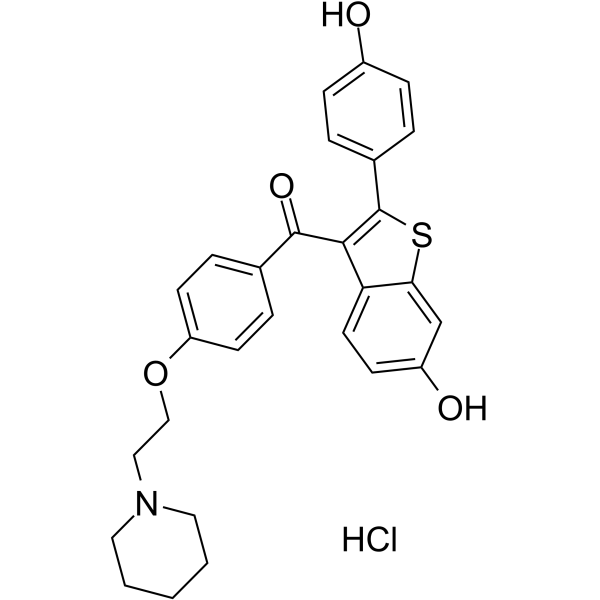
Raloxifene hydrochlorideEstrogen Receptor Modulator -
HY-19822
ElacestrantEstrogen Receptor Degrader -
HY-A0036
Bazedoxifene acetateSERM -
HY-A0031
BazedoxifeneSERM -
HY-13757
Tamoxifen CitrateEstrogen Receptor Modulator -
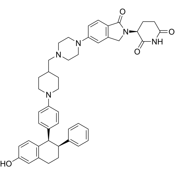
HY-138642
VepdegestrantPROTAC ER Degrader -
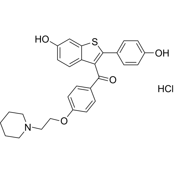
HY-13738AR
Raloxifene hydrochloride (Standard) -
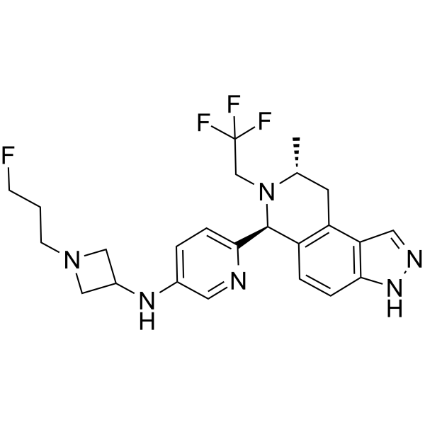
HY-136255
CamizestrantEstrogen Receptor Antagonist -
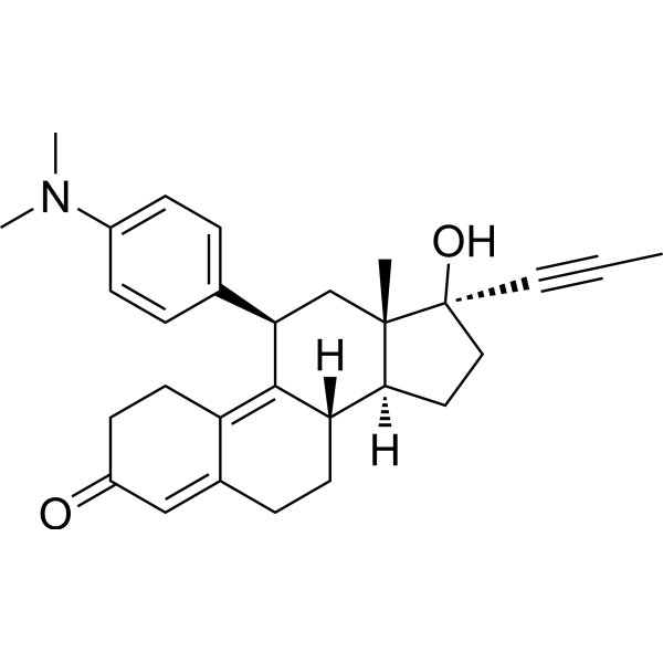
HY-13683
MifepristoneProgesterone Receptor Antagonist
Publications Citing Use of MedChemExpress (MCE) Lasofoxifene tartrate
More

Biological Activity
Description
IC50 & Target
Target: Estrogen Receptor[1]
In Vitro
Lasofoxifene tartrate (1 nM-1 μM; 48 h) shows antagonist activity on ER+ breast cancer cells without being affected by the expression level of activating ERα mutants relative to wild-type (WT) ERα[2].
MedChemExpress (MCE) has not independently confirmed the accuracy of these methods. They are for reference only. Further protocols information, click here.
In Vivo
Lasofoxifene tartrate (4 mg/mice; s.c.; 5 day/week; for 43 d) protects against generalised bone loss in CIA by increasing trabecular bone mineral density (BMD), cortical thickness in mice[1].
Lasofoxifene tartrate (5, and 10 mg/kg; s.c.; 5 day/week; for 70 d) exerts function of inhibiting primary tumor growth and reducing metastases to the lung and the liver in mice[3].
MedChemExpress (MCE) has not independently confirmed the accuracy of these methods. They are for reference only.
-
Animal Model:Post-menopausal RA model on OVX (ovariectomised) DBA/1 mice (female DBA/1 mice, 8-10 weeks old, CIA-treated)[1]
-
Dosage:4 mg/mouse/day
-
Administration:Subcutaneous injection; 5 days a week from the first signs of arthritis (day 18); 43 days
-
Result:Reduced in arthritis severity, including synovial inflammation and destruction of joints reduction.
The mean arthritis frequency was 47% while the vehicle group was 81% at 42 days post immunization.
-
Animal Model:NSG mices with xenograft tumors model (MIND, mammary intraductal): WT, Y537S and D538G ERα render tumors[3]
-
Dosage:1, 5, or 10 mg/kg
-
Administration:Subcutaneous injection; 5 days per week; for 70 days
-
Result:Elicited a superior inhibitory effect at a dose of 10 mg/kg, resulted potential tumor shrinkage in Y537S and D538G tumors.
And also reduced tumor weight to 60% for Y537S and 50% for D538G at 5 and 10 mg/kg, respectively.


Chemical Information
-
CAS No. 190791-29-8
-
Appearance Solid
-
Molecular Weight 563.64
-
Formula C32H37NO8
-
Color White to off-white
-
SMILES
OC1=CC=C2[C@@H](C3=CC=C(OCCN4CCCC4)C=C3)[C@@H](C5=CC=CC=C5)CCC2=C1.O=C(O)[C@@H](O)[C@H](O)C(O)=O
-
Synonyms
CP-336156 tartrate
-
Shipping
Room temperature in continental US; may vary elsewhere.
-
Storage
4°C, sealed storage, away from moisture
* In solvent : -80°C, 6 months; -20°C, 1 month (sealed storage, away from moisture)


Publications (6)
-
Journal Impact Factor
-
Most Recent
-
NPJ Breast Cancer
Discovery of a novel small molecule degrader of wild type and mutant estrogen receptors using DNA encoded libraries. [Abstract]2025 Nov 12;11(1):125. PMID: 41224743 -
NPJ Breast Cancer
Unconventional isoquinoline-based SERMs elicit fulvestrant-like transcriptional programs in ER+ breast cancer cells. [Abstract]2022 Dec 14;8(1):130. PMID: 36517522 -
Mol Cancer Ther
The Dysregulated Pharmacology of Clinically Relevant ESR1 Mutants is Normalized by Ligand-activated WT Receptor. [Abstract]2020 Jul;19(7):1395-1405. PMID: 32381587 -
Gynecol Oncol
Constitutively active ESR1 mutations in gynecologic malignancies and clinical response to estrogen-receptor directed therapies. [Abstract]2019 Jul;154(1):199-206. PMID: 30987772 -
-
bioRxiv
Targeting Unique Ligand Binding Domain Structural Features Downregulates DKK1 in Y537S ESR1 Mutant Breast Cancer Cells. [Abstract]2024 Jun 2:2024.05.28.596307. PMID: 38854123


Solvent & Solubility
In Vitro:
DMSO : 83.33 mg/mL (147.84 mM; Need ultrasonic; Hygroscopic DMSO has a significant impact on the solubility of product, please use newly opened DMSO)
Please refer to the solubility information to select the appropriate solvent. Once prepared, please aliquot and store the solution to prevent product inactivation from repeated freeze-thaw cycles.
Storage method and period of stock solution: -80°C, 6 months; -20°C, 1 month (sealed storage, away from moisture). When stored at -80°C, please use it within 6 months. When stored at -20°C, please use it within 1 month.
Please refer to the solubility information to select the appropriate solvent. Once prepared, please aliquot and store the solution to prevent product inactivation from repeated freeze-thaw cycles.
Storage method and period of stock solution: -80°C, 6 months; -20°C, 1 month (sealed storage, away from moisture). When stored at -80°C, please use it within 6 months. When stored at -20°C, please use it within 1 month.
Concentration (start) × Volume (start) = Concentration (final) × Volume (final)
In Vivo:
Select the appropriate dissolution method based on your experimental animal and administration route.
- For the following dissolution methods, please ensure to first prepare a clear stock solution using an In Vitro approach and then sequentially add co-solvents:
- To ensure reliable experimental results, the clarified stock solution can be appropriately stored based on storage conditions. As for the working solution for In Vivo experiments, it is recommended to prepare freshly and use it on the same day.
- The percentages shown for the solvents indicate their volumetric ratio in the final prepared solution. If precipitation or phase separation occurs during preparation, heat and/or sonication can be used to aid dissolution.
Add each solvent one by one: 10% DMSO 40% PEG300 5% Tween-80 45% Saline
Solubility: ≥ 2.08 mg/mL (3.69 mM); Clear solution
This protocol yields a clear solution of ≥ 2.08 mg/mL (saturation unknown).
Taking 1 mL working solution as an example, add 100 μL DMSO stock solution (20.8 mg/mL) to 400 μL PEG300, and mix evenly; then add 50 μL Tween-80 and mix evenly; then add 450 μL Saline to adjust the volume to 1 mL.
Preparation of Saline: Dissolve 0.9 g sodium chloride in ddH₂O and dilute to 100 mL to obtain a clear Saline solution.
Add each solvent one by one: 10% DMSO 90% (20% SBE-β-CD in Saline)
Solubility: ≥ 2.08 mg/mL (3.69 mM); Clear solution
This protocol yields a clear solution of ≥ 2.08 mg/mL (saturation unknown).
Taking 1 mL working solution as an example, add 100 μL DMSO stock solution (20.8 mg/mL) to 900 μL 20% SBE-β-CD in Saline, and mix evenly.
Preparation of 20% SBE-β-CD in Saline (4°C, storage for one week): 2 g SBE-β-CD powder is dissolved in 10 mL Saline, completely dissolve until clear.
In Vivo Dissolution Calculator
Please enter the basic information of animal experiments:
-
-
-
-
Recommended: Prepare an additional quantity of animals to account for potential losses during experiments.
Please enter your animal formula composition:
-
%DMSO +
Recommended: Keep the proportion of DMSO in working solution below 2% if your animal is weak.
-
%+
-
+%Tween-80 + +
-
%Saline +
The co-solvents required include: DMSO, . All of co-solvents are available by MedChemExpress (MCE). , Tween 80. All of co-solvents are available by MedChemExpress (MCE).
Working solution concentration: 0.22 mg/mL
Method for preparing stock solution: mg drug dissolved in μL DMSO. Stock solution concentration: mg/mL. * In solvent : -80°C, 6 months; -20°C, 1 month (sealed storage, away from moisture)
1. Take μL DMSO stock solution;
2. Add μL .
μL , mix evenly;
3. Then add μL Tween 80, mix evenly;
4. Then add μL
Please ensure that the stock solution in the first step is dissolved to a clear state, and add co-solvents in sequence. You can use ultrasonic heating (ultrasonic cleaner, recommended frequency 20-40 kHz), vortexing, etc. to assist dissolution.

Protocols
-
Research Protocol for Endocrine Diseases
Endocrine diseases often arise from disrupted hormone production, hormone signaling, or target-tissue responsiveness; for diabetes-focused endocrine disease models, insulin signaling regulates glucose uptake, hepatic glucose output, lipid metabolism, and β-cell compensation. Type 2 diabetes develops through interacting defects in insulin resistance, β-cell dysfunction, adipose inflammation, hepatic glucose overproduction, altered incretin signaling, and ectopic lipid metabolism. A major unresolved question is whether endocrine dysfunction is driven primarily by target-tissue insulin resistance, intrinsic β-cell failure, immune/inflammatory stress, or combined multi-organ failure that differs by disease stage.
-
Patient-Derived Xenograft (PDX)
Patient-derived xenograft (PDX) models are generated by engrafting primary human tumor tissue directly into immunodeficient mice, allowing in vivo propagation of patient tumor biology without initial in vitro adaptation. These models are used to preserve key histopathological and molecular characteristics of the original tumor and enable assessment of tumor growth dynamics and therapeutic response in a living organism. The biological readout is tumor engraftment and subsequent growth in the murine host, which reflects the ability of human tumor cells to survive, vascularize, and expand in an immunocompromised microenvironment.
-
Patient-Derived Orthotopic Xenograft (PDOX)
Patient-derived orthotopic xenograft (PDOX) modeling implants fresh patient tumor tissue or patient-derived tumor cells into the anatomically corresponding organ or tissue site of immunodeficient mice, usually by surgical orthotopic implantation, to preserve patient tumor histology, local microenvironmental context, invasion, metastatic behavior, and treatment-response features better than subcutaneous implantation. PDOX readouts include tumor engraftment, orthotopic tumor growth, local invasion, metastasis, recurrence after resection, histologic similarity to the donor tumor, biomarker retention, molecular concordance, survival, and response or resistance to therapy. PDOX models are used for preclinical drug testing and individualized therapy evaluation, but engraftment success varies by tumor type and specimen quality.
-
Subcutaneous Cell-Line-Derived Xenograft
Subcutaneous cell-line-derived xenograft (CDX) models are established by implanting cultured human cancer cell lines into immunodeficient mice, where the injected cells form localized tumors that can be monitored in vivo as a measure of tumorigenic potential, growth kinetics, and treatment response. These models are widely used in oncology research because they allow reproducible tumor formation and enable comparative assessment of tumor growth between different cell lines or genetic manipulations in a controlled in vivo microenvironment. Subcutaneous implantation of cancer cells in immunodeficient mice is a standard approach for evaluating tumor growth behavior and therapeutic response across multiple cancer types, including prostate, esophageal, pancreatic, and colon cancer models.
-
Breast Cancer Modeling
Breast cancer is a heterogeneous cancer, and it has been distinguished into four subtypes: luminal A, luminal B, HER2-positive and basal-like. Molecular mutations, epigenetic alterations, hormone exposure and immune microenvironment are related to the progression of breast cancer.
-
Orthotopic Cell-Line Xenograft
Orthotopic cell-line xenograft models involve implantation of human cancer cell lines into the anatomically corresponding organ of immunodeficient mice to reproduce tumor growth within a native microenvironment, enabling more clinically relevant tumor behavior compared with subcutaneous models. These models are widely used because orthotopic placement better recapitulates tumor progression, including invasion and metastatic spread, which are often underrepresented in heterotopic implantation systems. Compared with conventional xenografts, orthotopic implantation is described as more technically complex but provides improved simulation of tumor-microenvironment interactions and metastatic behavior, making it particularly valuable for translational oncology research. Surgical orthotopic implantation approaches have been emphasized as enabling faithful reproduction of clinical cancer features, including metastasis and disease progression patterns that align with the tumor’s organ of origi


Purity & Documentation
-
Data Sheet (280 KB)
-
SDS (725 KB)
- English - EN (725 KB)
- Français - FR (725 KB)
- Deutsch - DE (725 KB)
- Norwegian - NO (725 KB)
- Español - ES (725 KB)
- Swedish - SV (725 KB)
- Italian - IT (725 KB)
- Korean - KR (725 KB)
- Portuguese - PT (725 KB)
-
Handling Instructions (2659 KB)
References
[1]. Andersson A, et al. Selective oestrogen receptor modulators lasofoxifene and bazedoxifene inhibit joint inflammation and osteoporosis in ovariectomised mice with collagen-induced arthritis. Rheumatology (Oxford). 2016 Mar;55(3):553-63. [Content Brief]
[2]. Andreano KJ, et al. The Dysregulated Pharmacology of Clinically Relevant ESR1 Mutants is Normalized by Ligand-activated WT Receptor. Mol Cancer Ther. 2020 Jul. 19(7):1395-1405. [Content Brief]
[3]. Lainé M, et al. Lasofoxifene as a potential treatment for therapy-resistant ER-positive metastatic breast cancer. Breast Cancer Res. 2021 May 12. 23(1):54. [Content Brief]


Complete Stock Solution Preparation Table
Please refer to the solubility information to select the appropriate solvent. Once prepared, please aliquot and store the solution to prevent product inactivation from repeated freeze-thaw cycles.
Storage method and period of stock solution: -80°C, 6 months; -20°C, 1 month (sealed storage, away from moisture). When stored at -80°C, please use it within 6 months. When stored at -20°C, please use it within 1 month.
| Optional Solvent | Concentration Solvent Mass | 1 mg | 5 mg | 10 mg | 25 mg |
|---|---|---|---|---|---|
| DMSO | 1 mM | 1.7742 mL | 8.8709 mL | 17.7418 mL | 44.3546 mL |
| 5 mM | 0.3548 mL | 1.7742 mL | 3.5484 mL | 8.8709 mL | |
| 10 mM | 0.1774 mL | 0.8871 mL | 1.7742 mL | 4.4355 mL | |
| 15 mM | 0.1183 mL | 0.5914 mL | 1.1828 mL | 2.9570 mL | |
| 20 mM | 0.0887 mL | 0.4435 mL | 0.8871 mL | 2.2177 mL | |
| 25 mM | 0.0710 mL | 0.3548 mL | 0.7097 mL | 1.7742 mL | |
| 30 mM | 0.0591 mL | 0.2957 mL | 0.5914 mL | 1.4785 mL | |
| 40 mM | 0.0444 mL | 0.2218 mL | 0.4435 mL | 1.1089 mL | |
| 50 mM | 0.0355 mL | 0.1774 mL | 0.3548 mL | 0.8871 mL | |
| 60 mM | 0.0296 mL | 0.1478 mL | 0.2957 mL | 0.7392 mL | |
| 80 mM | 0.0222 mL | 0.1109 mL | 0.2218 mL | 0.5544 mL | |
| 100 mM | 0.0177 mL | 0.0887 mL | 0.1774 mL | 0.4435 mL |


Lasofoxifene tartrate Related Classifications
HY-A0038 Related Classifications
 Powered by Bioz