



Lurbinectedin
Based on 9 publication(s) in Google Scholar
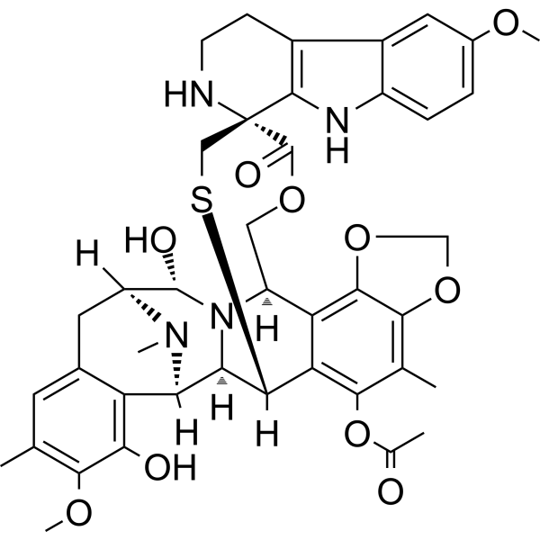
Lurbinectedin (PM01183) is a DNA minor groove covalent binder with potent anti-tumour activity; inhibits RMG1 and RMG2 cell growth with IC50 values of 1.25 and 1.16 nM, respectively.
For research use only. We do not sell to patients.
- Purity : 99.70%
- CAS No.: 497871-47-3
- Formula: C41H44N4O10S
- Molecular Weight:784.87
-
Storage:Powder -20°C, 3 years ; In solvent -80°C, 6 months , -20°C, 1 month

To place orders, for customer services and technical support, please contact: MedChemExpress USA
Tel: 609-228-6898 E-mail: [email protected] [email protected]


-
 Biological Activity
-
Chemical Information
-
Solvent & Solubility
- Protocols
- Purity & Documentation
- References
-
 Help & FAQs


-
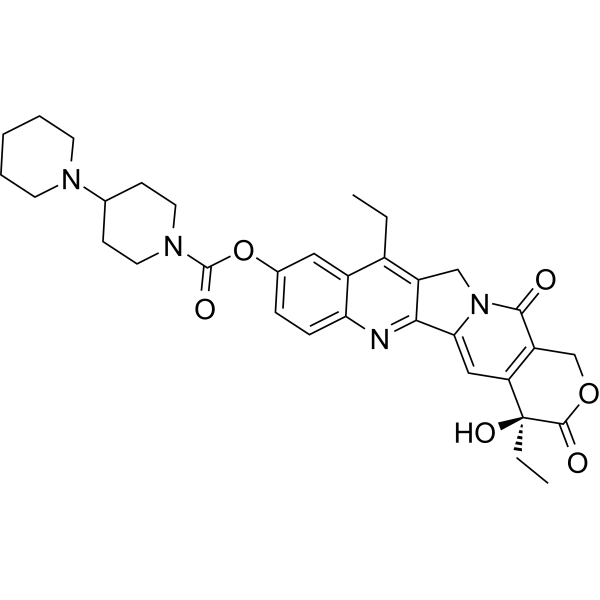
HY-16562
IrinotecanTopoisomerase I Inhibitor -
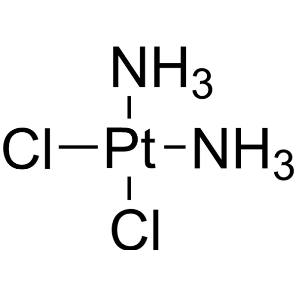
HY-17394
CisplatinDNA Alkylator -
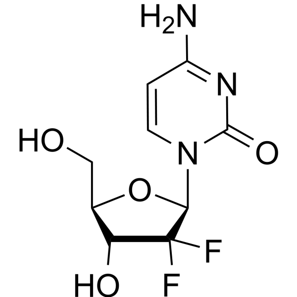
HY-17026
GemcitabineDNA Synthesis Inhibitor -
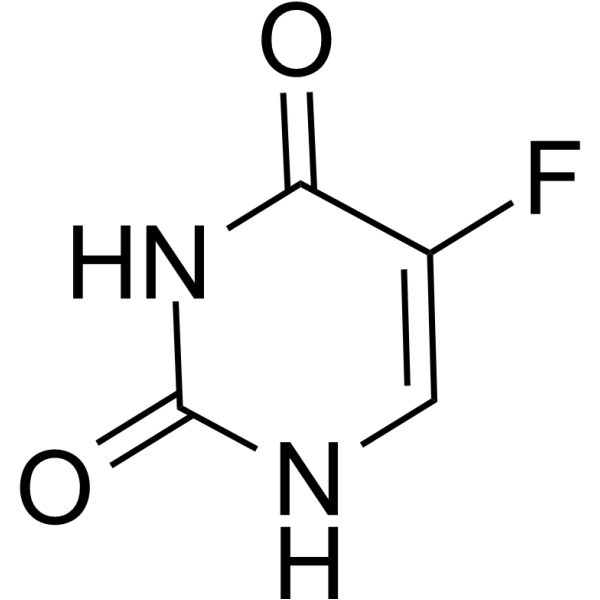
HY-90006
5-FluorouracilThymidylate Synthase Inhibitor -
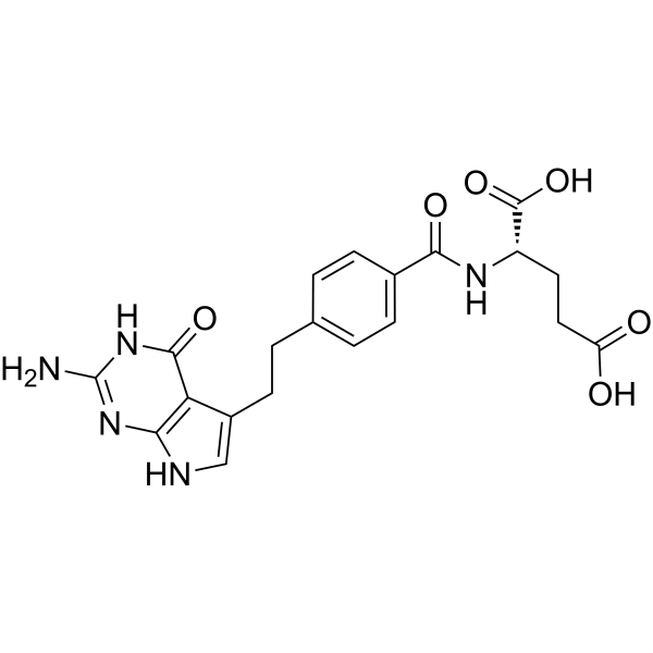
HY-10820
Pemetrexed -
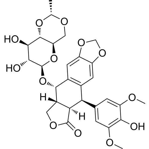
HY-13629
EtoposideTopoisomerase II Inhibitor -
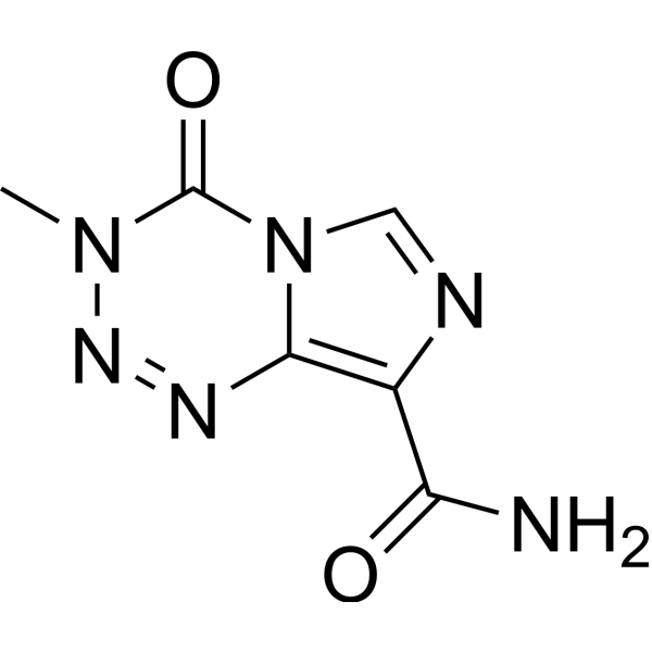
HY-17364
TemozolomideDNA Alkylator -
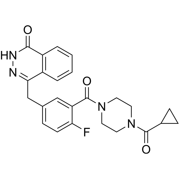
HY-10162
OlaparibPARP Inhibitor -
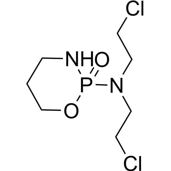
HY-17420
CyclophosphamideAlkylating Agent -
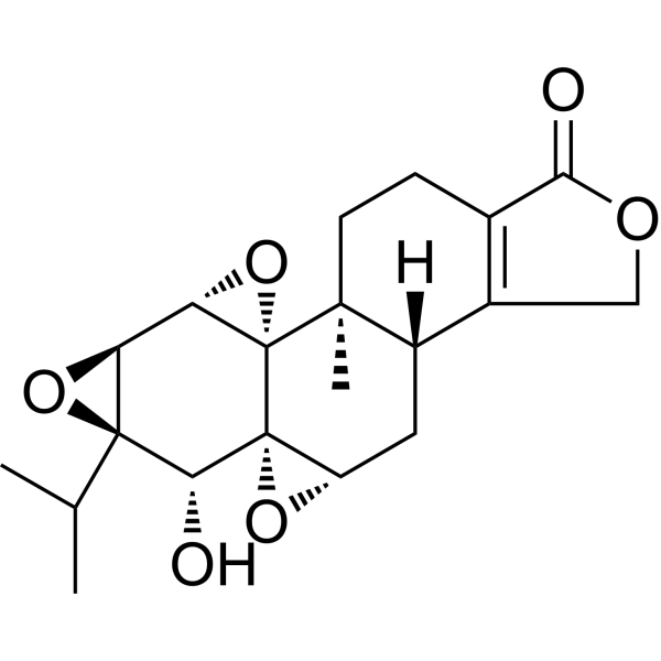
HY-32735
TriptolideImmunosuppressive Agent -
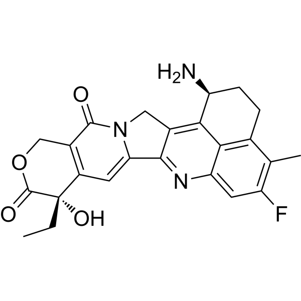
HY-13631
ExatecanTopoisomerase I Inhibitor -
HY-17393
CarboplatinDNA Synthesis Inhibitor
Publications Citing Use of MedChemExpress (MCE) Lurbinectedin
More- Int J Mol Sci. 2025 Sep 18;26(18):9085. [Abstract]
- iScience. 2024 May 27;27(6):110132. [Abstract]
- Thorac Cancer. 2022 Apr;13(8):1184-1192. [Abstract]
- bioRxiv. 2026 Jun 29:2026.06.26.734921.
- Preprints. 2025 Jul 21.
- Patent. US20250041313A1.
- Patent. US20240207261A1.
- Patent. US20240207261A1.
- bioRxiv. 2023 Sep 17.
 Customer Validation & Images

-
Cell Proliferation/Viability Assay
-
Apoptosis Analysis
-
Cell Imaging/Staining
-
WB
-
Bio/Physico-chemical Assay
All DNA/RNA Synthesis Isoforms
More

Biological Activity
Description
IC50 & Target
IC50: 1.25 nM (RMG1), 1.16 nM (RMG2)[1]
In Vitro
PM01183 is a new synthetic tetrahydroisoquinoline alkaloid that is currently in phase I clinical development for the treatment of solid tumours. PM01183–DNA adducts in living cells give rise to double-strand breaks, triggering S-phase accumulation and apoptosis. The potent cytotoxic activity of PM01183 is ascertained in a 23-cell line panel with a mean GI50 value of 2.7 nM[2]. Lurbinectedin exhibits significant antitumor activity toward chemosensitive and chemoresistant human ovarian clear cell carcinoma (CCC) cells in vitro[1].
MedChemExpress (MCE) has not independently confirmed the accuracy of these methods. They are for reference only. Further protocols information, click here.
In Vivo
MedChemExpress (MCE) has not independently confirmed the accuracy of these methods. They are for reference only.
Clinical Trial
| NCT Number | Sponsor | Condition | Start Date |
Phase

|
|---|---|---|---|---|
| NCT01329991 | Plexxikon| | 2011-05 | PHASE1 |


Chemical Information
-
CAS No. 497871-47-3
-
Appearance Solid
-
Molecular Weight 784.87
-
Formula C41H44N4O10S
-
Color White to light yellow
-
SMILES
O[C@@H]([C@@](CC1=CC(C)=C2OC)([H])N(C)[C@]3([H])C1=C2O)N([C@](COC4=O)([H])C5=C(OCO6)C6=C7C)[C@@]3([H])[C@@](SC[C@@]4(NCC8)C9=C8C%10=CC(OC)=CC=C%10N9)([H])C5=C7OC(C)=O
-
Synonyms
PM01183; LY-01017
-
Shipping
Room temperature in continental US; may vary elsewhere.
-
Storage
Powder -20°C 3 years In solvent -80°C 6 months -20°C 1 month


Publications (9)
-
Journal Impact Factor
-
Most Recent
-
Int J Mol Sci
The DNA Minor Groove Binders Trabectedin and Lurbinectedin Are Potent Antitumor Agents in Human Intrahepatic Cholangiocarcinoma. [Abstract]2025 Sep 18;26(18):9085. PMID: 41009648
Lurbinectedin purchased from MedChemExpress. Usage Cited in: Int J Mol Sci. 2025 Sep 18;26(18):9085. [Abstract]
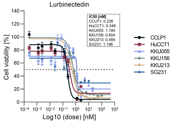
Effect Lurbinectedin on the viability of intrahepatic cholangiocarcinoma cell lines. Cell viability of six human intrahepatic cholangiocarinoma (iCCA) cell lines (CCLP1, HUCCT1, KKU055, KKU156, KKU213, and SG231) exposed to 0-100 nM Lurbinectedin for 48 h was assessed by MTT assay. A summary of the IC50 of the conpound in the six cell lines is depicted next to the graph. Dashed horizontal lines indicate 50% cell viability.
Lurbinectedin purchased from MedChemExpress. Usage Cited in: Int J Mol Sci. 2025 Sep 18;26(18):9085. [Abstract]
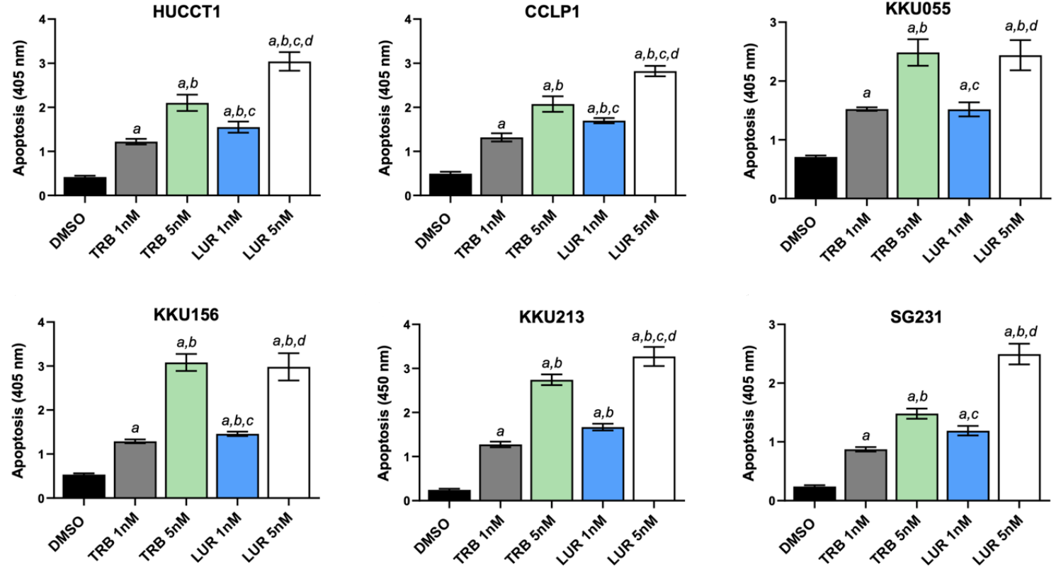
Apoptosis was assessed in HUCCT1, CCLP1, KU055, KKU156, KKU213 and SG231 cell lines treated for 48 hours with trabectedin (TRB) and Lurbinectedin (LUR) at 1 nM and 5 nM concentrations. Cells treated with solvent (DMSO) served as controls.
Lurbinectedin purchased from MedChemExpress. Usage Cited in: Int J Mol Sci. 2025 Sep 18;26(18):9085. [Abstract]
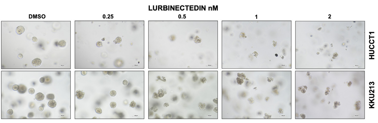
Lurbinectedin (LUR) restrain the growth of HUCCT1 and KKU213 intrahepatic cholangiocarcinoma cells grown as 3D cultures (spheroids). HUCCT1- and KKU213-derived spheroids were treated with DMSO (vehicle) and increasing concentrations of trabectedin (TRB) and lurbinectedin (LUR; 0.25–2 nM; n = 3 wells per condition). Note the decrease in size and number of spheroids following the treatment with Lurbinectedin (LUR), as assessed by the optical microscope. Original magnification: 10×; scale bar: 100 µm.
Lurbinectedin purchased from MedChemExpress. Usage Cited in: Int J Mol Sci. 2025 Sep 18;26(18):9085. [Abstract]
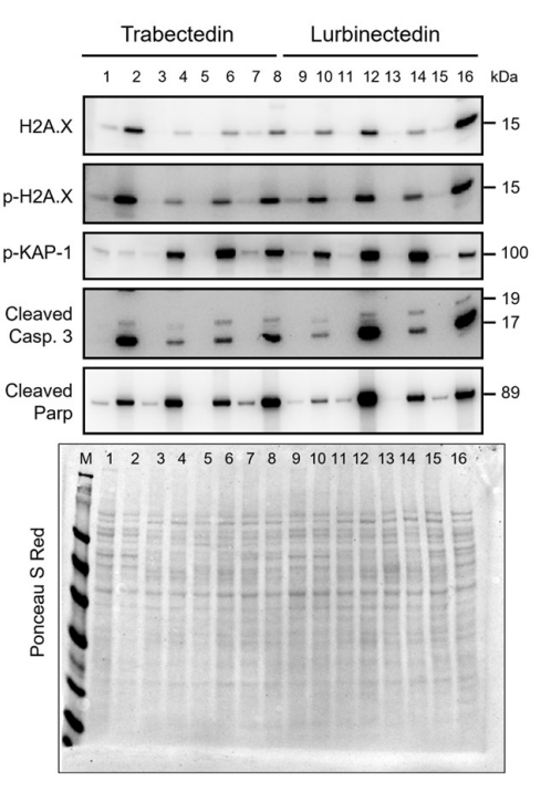
Western blot analysis was applied to assess the levels of several effectors of DNA damage response and apoptosis pathways in HUCCT1, SG231, CCLP1, and KKU055 intrahepatic cholangiocarcinoma (iCCA) cell lines exposed to trabectedin (TRB) and Lurbinectedin (LUR) at 1 nM concentration. Cells were treated for 48 h, and Western blot analysis was conducted at this time point. Representative blots are shown.
Lurbinectedin purchased from MedChemExpress. Usage Cited in: Int J Mol Sci. 2025 Sep 18;26(18):9085. [Abstract]
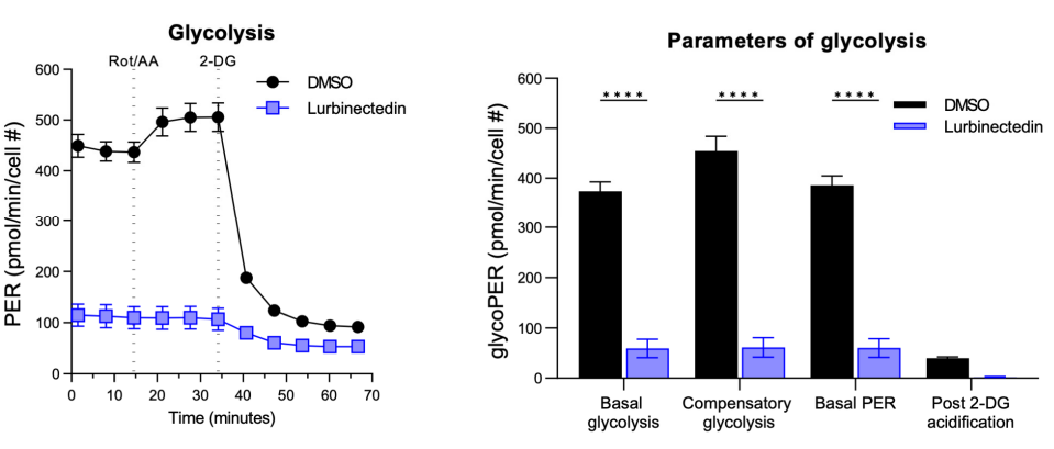
Effect of trabectedin and Lurbinectedin (LUR) on the mitochondrial respiration of human intrahepatic cholangiocarcinoma cells. For the Seahorse XF Mito Stress Test, KKU055 cells were treated with 1 nM Lurbinectedin (LUR), or a matched DMSO concentration for 24 h. The Seahorse Mito Stress Test profile of normalized oxygen consumption rate (OCR) in LUR-treated KKU055 cells is shown. The results showed LUR administration significantly decreased most parameters (basal and compensatory glycolysis, and basal proton efflux rate). All OCR levels were background corrected and normalized to nuclei fluorescent staining. Dotted lines indicate the time point of compound injection. Cell #, cell number; FCCP, carbonyl cyanide-p-trifluoromethoxyphenylhydrazone; Rot/AA, rotenone/antimycin A.
Lurbinectedin purchased from MedChemExpress. Usage Cited in: Int J Mol Sci. 2025 Sep 18;26(18):9085. [Abstract]
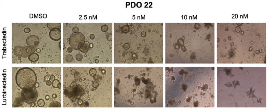
Microscopic images demonstrate that treatment with trabectedin (TRB) and Lurbinectedin (LUR) (2.5-20 nM; 72 h) resulted in a reduction in the compact, well-organized structure of the organoids, evidenced by the loss of the typical cystic shape associated with cholangiocarcinoma organoids.
-
iScience
Teriflunomide/leflunomide synergize with chemotherapeutics by decreasing mitochondrial fragmentation via DRP1 in SCLC. [Abstract]2024 May 27;27(6):110132. PMID: 38993482 -
Thorac Cancer
High mRNA expression of POU2F3 in small cell lung cancer cell lines predicts the effect of lurbinectedin. [Abstract]2022 Apr;13(8):1184-1192. PMID: 35278040 -
-
-
-
-
-


Solvent & Solubility
In Vitro:
DMSO : 20 mg/mL (25.48 mM; Need ultrasonic; Hygroscopic DMSO has a significant impact on the solubility of product, please use newly opened DMSO)
Please refer to the solubility information to select the appropriate solvent. Once prepared, please aliquot and store the solution to prevent product inactivation from repeated freeze-thaw cycles.
Storage method and period of stock solution: -80°C, 6 months; -20°C, 1 month. When stored at -80°C, please use it within 6 months. When stored at -20°C, please use it within 1 month.
Please refer to the solubility information to select the appropriate solvent. Once prepared, please aliquot and store the solution to prevent product inactivation from repeated freeze-thaw cycles.
Storage method and period of stock solution: -80°C, 6 months; -20°C, 1 month. When stored at -80°C, please use it within 6 months. When stored at -20°C, please use it within 1 month.
Concentration (start) × Volume (start) = Concentration (final) × Volume (final)

Protocols
-
Cell Cytotoxicity Assay
Cytotoxicity assays are usually based on the assessment of cell membrane damage, which can also be indirectly detected by measuring cell viability. Detection methods include MTT assay, CKK-8 assay, LDH assay and ATP assay, etc.
-
Protocol for Cell Counting and Cell Density Analysis
Cell counting and cell-density analysis estimate the number of cells in a known volume or field area. Manual hemocytometer counting uses a chamber of defined geometry to convert counted cells into cells/mL, while automated counters and image-analysis workflows detect cell objects from optical, brightfield, fluorescence, impedance, or digital-image features. Trypan blue viability counting is based on dye exclusion: viable cells with intact membranes exclude dye, while non-viable cells with compromised membranes stain blue. The readout is total cell density, viable-cell density, dead-cell density, and percent viability. Cell density can also be estimated from microscopy images by counting objects per image area, from flow cytometry using calibrated volume or reference particles, or from in situ microscopy in bioreactors after calibration against reference methods such as hemocytometer or flow cytometry.
-
Somatic Cell Culture
A method of simulating the in vivo environment in vitro to maintain the cell growth, differentation and main functions.
-
CCK-8/WST-8 Cell Proliferation Assay
The CCK-8/WST-8 assay is based on the reduction of the water-soluble tetrazolium salt WST-8 to a water-soluble formazan product by cellular dehydrogenases in metabolically active cells, where the generated formazan amount is proportional to the number of living cells and is quantified by measuring absorbance in the visible range, providing a colorimetric readout for cell viability and proliferation assessment. This class of tetrazolium-based assays improves upon earlier MTT-based systems by producing a water-soluble formazan, eliminating the need for organic solubilization steps and enabling direct spectrophotometric measurement in culture medium.
-
Cell Counting-Based Growth Curve Assay
Cell counting-based growth curve assays quantify cell proliferation by directly measuring changes in viable cell number over time using manual or automated counting methods such as hemocytometer-based counting or instrument-assisted cell enumeration, enabling construction of growth curves that reflect population expansion dynamics in response to culture conditions. A widely used approach is trypan blue exclusion with hemocytometer counting, where membrane-compromised (non-viable) cells take up the dye, allowing discrimination between viable and non-viable cells while simultaneously enabling total cell number quantification. Repeated sampling across time points allows estimation of proliferation rate, growth phases, and comparative growth kinetics between experimental conditions.
-
MTT Cell Proliferation Assay
The MTT assay is a colorimetric endpoint assay for estimating viable cell number, cell growth, cytotoxicity, or cell activation in cultured mammalian cells. Living cells reduce the yellow tetrazolium salt MTT into purple/blue formazan, while dead cells do not generate the same signal; the resulting color can be quantified with a multiwell spectrophotometer. MTT reduction is commonly interpreted as a readout of metabolic activity that often correlates with viable cell number, but it should not be treated as a direct cell-counting method unless the assay is optimized for the cell type and experimental condition. Studies show that MTT reduction can involve mitochondrial and non-mitochondrial reducing systems, and formazan may accumulate in intracellular lipid droplets rather than simply marking mitochondria.
-
Cell Viability Determination by MTT Colorimetric Assay
The following protocol uses the MTT colorimetric assay as a classic literature-established method for assessing cell viability/metabolic activity in cultured mammalian cells. MTT[3-(4,5-dimethylthiazol-2-yl)-2,5-diphenyltetrazolium bromide] is reduced by metabolically active cells to a colored formazan product; the amount of formazan is quantified spectrophotometrically and provides an indirect measure of metabolically active viable cells. Importantly, MTT reduction reflects cellular oxidoreductase/metabolic activity rather than an absolute direct count of living cells, so changes in cellular metabolism can alter the signal independently of cell number.


Purity & Documentation
-
Data Sheet (288 KB)
-
SDS (393 KB)
- English - EN (393 KB)
- Français - FR (393 KB)
- Deutsch - DE (393 KB)
- Norwegian - NO (393 KB)
- Español - ES (393 KB)
- Swedish - SV (393 KB)
- Italian - IT (393 KB)
- Korean - KR (393 KB)
- Portuguese - PT (393 KB)
-
Handling Instructions (2659 KB)
References
[1]. Takahashi R, et al. Preclinical Investigations of PM01183 (Lurbinectedin) as a Single Agent or in Combination with Other Anticancer Agents for Clear Cell Carcinoma of the Ovary. PLoS One. 2016 Mar 17;11(3):e0151050. [Content Brief]
[2]. Leal JF, et al. PM01183, a new DNA minor groove covalent binder with potent in vitro and in vivo anti-tumour activity. Br J Pharmacol. 2010 Nov;161(5):1099-110. [Content Brief]
[3]. Vidal A, et al. Lurbinectedin (PM01183), a new DNA minor groove binder, inhibits growth of orthotopic primary graft of NSC 119875-resistant epithelial ovarian cancer. Clin Cancer Res. 2012 Oct 1;18(19):5399-411. [Content Brief]


Complete Stock Solution Preparation Table
Please refer to the solubility information to select the appropriate solvent. Once prepared, please aliquot and store the solution to prevent product inactivation from repeated freeze-thaw cycles.
Storage method and period of stock solution: -80°C, 6 months; -20°C, 1 month. When stored at -80°C, please use it within 6 months. When stored at -20°C, please use it within 1 month.
| Optional Solvent | Concentration Solvent Mass | 1 mg | 5 mg | 10 mg | 25 mg |
|---|---|---|---|---|---|
| DMSO | 1 mM | 1.2741 mL | 6.3705 mL | 12.7410 mL | 31.8524 mL |
| 5 mM | 0.2548 mL | 1.2741 mL | 2.5482 mL | 6.3705 mL | |
| 10 mM | 0.1274 mL | 0.6370 mL | 1.2741 mL | 3.1852 mL | |
| 15 mM | 0.0849 mL | 0.4247 mL | 0.8494 mL | 2.1235 mL | |
| 20 mM | 0.0637 mL | 0.3185 mL | 0.6370 mL | 1.5926 mL | |
| 25 mM | 0.0510 mL | 0.2548 mL | 0.5096 mL | 1.2741 mL |


Lurbinectedin Related Classifications
HY-16293 Related Classifications
 Powered by Bioz