



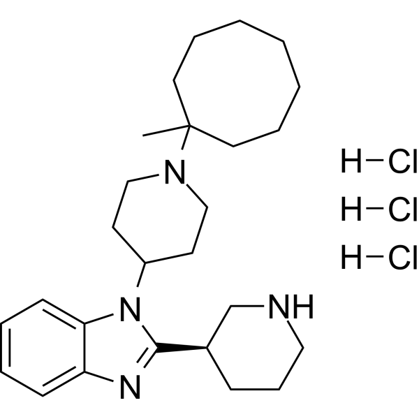
MCOPPB trihydrochloride
Based on 1 publication(s) in Google Scholar
MCOPPB trihydrochloride is a NOP/ORL1 G protein-coupled receptor agonist and autophagy inhibitor that can cross the blood-brain barrier. MCOPPB trihydrochloride clears senescent cells, regulates locomotion, lipid storage and immune responses, and inhibits fibrosis and angiogenesis. MCOPPB trihydrochloride blocks autophagic flux, induces changes in locomotion and lipid storage, and activates the stress-responsive immune transcription network, thereby improving post-infarction cardiac function and exerting anxiolytic effects. MCOPPB trihydrochloride can be applied to research fields such as aging-related diseases and ischemic heart failure.
For research use only. We do not sell to patients.
- Purity : 99.98%
- CAS No.: 1108147-88-1
- Formula: C26H43Cl3N4
- Molecular Weight:518.01
-
Storage:
4°C, sealed storage, away from moisture
* In solvent : -80°C, 6 months; -20°C, 1 month (sealed storage, away from moisture)

To place orders, for customer services and technical support, please contact: MedChemExpress USA
Tel: 609-228-6898 E-mail: [email protected] [email protected]


-
 Biological Activity
-
Chemical Information
-
Solvent & Solubility
- Protocols
- Purity & Documentation
- References
-
 Help & FAQs


-
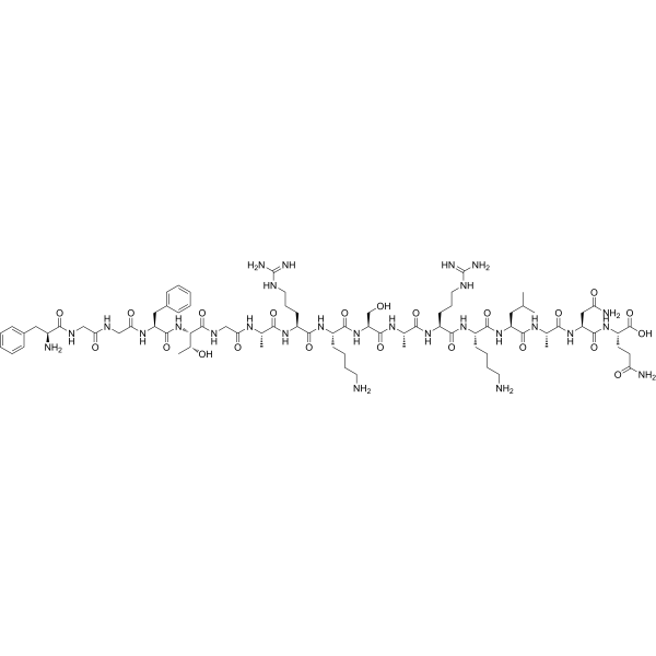
HY-P0183
NociceptinNociceptin Receptor Ligand -
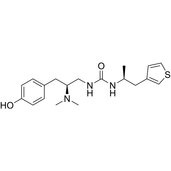
HY-101386
PZM21μ-opioid Receptor Agonist -
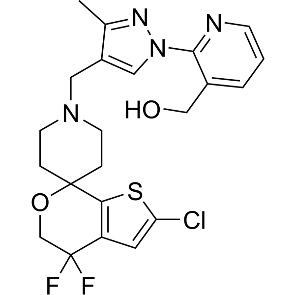
HY-114452
LY2940094NOP Antagonist -
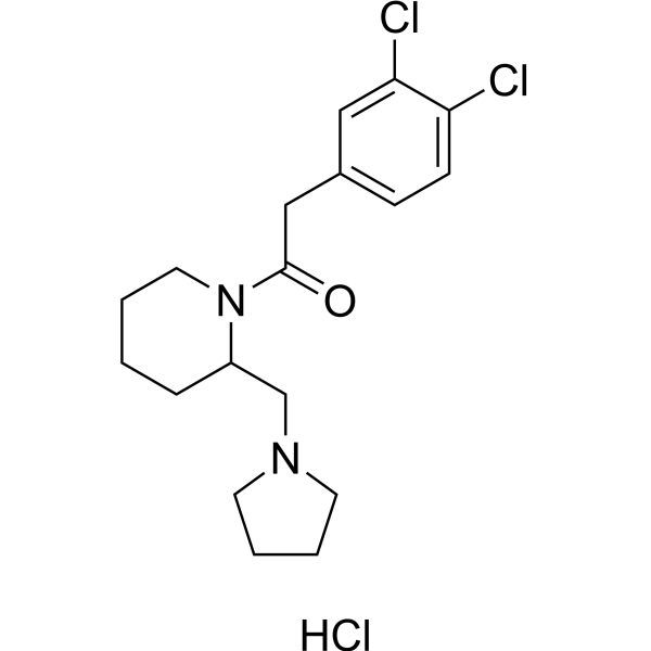
HY-101079
BRL 52537 hydrochlorideKOR Agonist -
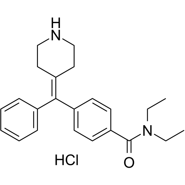
HY-101039A
AR-M 1000390 hydrochlorideδ Opioid Receptor Agonist -
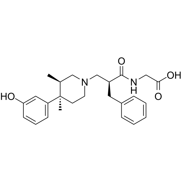
HY-13243
Alvimopanμ-opioid Receptor Antagonist -
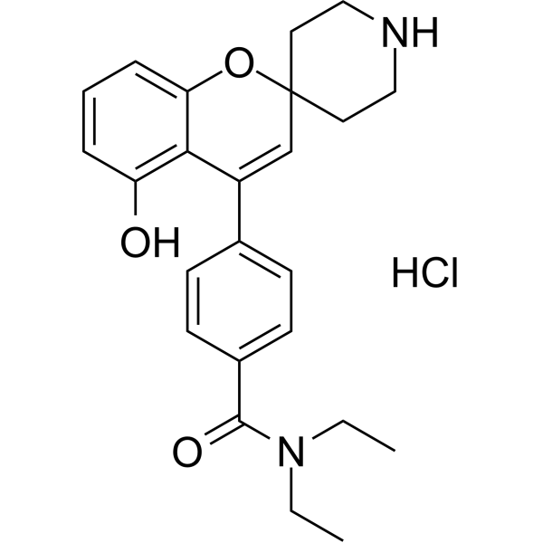
HY-13044
ADL-5859 hydrochlorideOpioid Receptor Agonist -
HY-100122
Bevenopranμ-opioid receptor antagonist t -
HY-B0380
TrimebutineOpioid Receptor Agonist -
HY-115066
GSK1521498 free baseμ-opioid Receptor Antagonist -
HY-10487
JDTic dihydrochloride -
HY-15708
LY2795050κ(kappa)-opioid Receptor (KOR) Antagonist
Publications Citing Use of MedChemExpress (MCE) MCOPPB trihydrochloride
MoreAll Opioid Receptor Isoforms
More

Biological Activity
Description
IC50 & Target
[1]|
NOP Receptor/ORL1 |
In Vitro
MCOPPB trihydrochloride (10 μM; 5 days) preferentially reduces viability of Aphidicolin (HY-N6733)-induced senescent human MRC5 fibroblasts while having less effect on non-senescent proliferating human MRC5 fibroblasts[1].
MCOPPB trihydrochloride (0.125-5 μM; 24 h) reduces viability of doxorubicin-induced senescent human HepG2 and Huh-7 hepatocellular carcinoma cells in a dose-dependent manner, with significant effects observed at concentrations ≥0.75 μM[1].
MCOPPB trihydrochloride (0.5 µM; 3 days total) downregulates NPPA and NPPB gene expression in ET-1-stimulated neonatal rat cardiomyocytes, an effect that is mediated via the ORL1 receptor[3].
MCOPPB trihydrochloride (0.5 µM; 3 days total) reduces ET-1-induced nuclear NFATc4 accumulation in H9C2 cells, an effect that is mediated via the ORL1 receptor[3].
MedChemExpress (MCE) has not independently confirmed the accuracy of these methods. They are for reference only. Further protocols information, click here.
-
Cell Line:aphidicolin-induced senescent human MRC5 fibroblasts, non-senescent proliferating human MRC5 fibroblasts
-
Concentration:10 μM
-
Incubation Time:5 days
-
Result:Showed preferential toxicity towards aphidicolin-induced senescent MRC5 fibroblasts compared to non-senescent proliferating MRC5 fibroblasts.
-
Cell Line:doxorubicin-induced senescent human HepG2 hepatocellular carcinoma cells, doxorubicin-induced senescent human Huh-7 hepatocellular carcinoma cells
-
Concentration:0.125-5 μM
-
Incubation Time:24 h
-
Result:Reduced viability of doxorubicin-induced senescent HepG2 and Huh-7 cells in a dose-dependent manner.
Identified 0.5 μM as the maximal cytostatic dose without cytotoxic effects.
Significantly reduced viability in Huh-7 cells at concentrations ≥0.75 μM compared to controls.
Significantly reduced viability in HepG2 cells at concentrations ≥0.75 μM compared to controls.
-
Cell Line:H9C2 cells
-
Concentration:0.5 µM (MCOPPB trihydrochloride); 100 nM (ET-1); 10 µM (ORL1 antagonist)
-
Incubation Time:24 h (ET-1 added after MCOPPB trihydrochloride); 30 min (ORL1 antagonist added after ET-1); 3 days (total)
-
Result:Decreased the ET-1-induced increase in nuclear NFATc4 protein expression in H9C2 cells.
Reversed the reduction of nuclear NFATc4 protein expression when co-treated with an ORL1 antagonist, significantly increasing nuclear NFATc4 expression.
In Vivo
MCOPPB trihydrochloride (2.5 mg/kg/day; i.p.; 5 days per week; 3 months) significantly improves cardiac function, reduces fibrosis, increases cardiomyocyte diameter, and induces angiogenesis in rats with post-myocardial infarction heart failure, with an ejection fraction of 67% at euthanasia[3].
MedChemExpress (MCE) has not independently confirmed the accuracy of these methods. They are for reference only.
-
Animal Model:C57BL/6 Mice with Age-related senescence model (male, 20 weeks old)[1]
-
Dosage:5 mg/kg (first week); 2.5 mg/kg (subsequent 2 weeks)
-
Administration:i.p.; 5 consecutive days followed by 2 days rest; 21 days
-
Result:Reduced total ambulatory distance and border zone distance in the open-field test, and reduced mobility time in the forced swim test.
Reduced senescent cell burden by ~70% in visceral white adipose tissue and ~70% in hepatic parenchyma.
Induced a ~20% increase in adipocyte size in white adipose tissue, and mild hepatic steatosis with a steatosis score of 0.6.
Decreased the number of F4/80-positive hepatic macrophages by ~40% and double-positive F4/80+ β-gal+ senescent macrophages by ~60%.
Altered liver gene expression profiles, with enrichment in pathways related to immune response to pathogens, TREM1 signaling, mTOR signaling, hepatic stellate activation, and senescence, with TLR4 identified as a key nodal hub.


Chemical Information
-
CAS No. 1108147-88-1
-
Appearance Solid
-
Molecular Weight 518.01
-
Formula C26H43Cl3N4
-
Color White to off-white
-
SMILES
CC1(N2CCC(N3C4=CC=CC=C4N=C3[C@H]5CNCCC5)CC2)CCCCCCC1.[H]Cl.[H]Cl.[H]Cl
-
Shipping
Room temperature in continental US; may vary elsewhere.
-
Storage
4°C, sealed storage, away from moisture
* In solvent : -80°C, 6 months; -20°C, 1 month (sealed storage, away from moisture)


Publications (1)
-
Journal Impact Factor
-
Most Recent
-
RSC Med Chem
Probing non-peptide agonists binding at the human nociceptin/orphanin FQ receptor: a molecular modelling study. [Abstract]2024 Dec 10. PMID: 39790123


Solvent & Solubility
In Vitro:
DMSO : 100 mg/mL (193.05 mM; Need ultrasonic; Hygroscopic DMSO has a significant impact on the solubility of product, please use newly opened DMSO)
Please refer to the solubility information to select the appropriate solvent. Once prepared, please aliquot and store the solution to prevent product inactivation from repeated freeze-thaw cycles.
Storage method and period of stock solution: -80°C, 6 months; -20°C, 1 month (sealed storage, away from moisture). When stored at -80°C, please use it within 6 months. When stored at -20°C, please use it within 1 month.
Please refer to the solubility information to select the appropriate solvent. Once prepared, please aliquot and store the solution to prevent product inactivation from repeated freeze-thaw cycles.
Storage method and period of stock solution: -80°C, 6 months; -20°C, 1 month (sealed storage, away from moisture). When stored at -80°C, please use it within 6 months. When stored at -20°C, please use it within 1 month.
Concentration (start) × Volume (start) = Concentration (final) × Volume (final)
In Vivo:
Select the appropriate dissolution method based on your experimental animal and administration route.
- For the following dissolution methods, please ensure to first prepare a clear stock solution using an In Vitro approach and then sequentially add co-solvents:
- To ensure reliable experimental results, the clarified stock solution can be appropriately stored based on storage conditions. As for the working solution for In Vivo experiments, it is recommended to prepare freshly and use it on the same day.
- The percentages shown for the solvents indicate their volumetric ratio in the final prepared solution. If precipitation or phase separation occurs during preparation, heat and/or sonication can be used to aid dissolution.
Add each solvent one by one: 10% DMSO 40% PEG300 5% Tween-80 45% Saline
Solubility: ≥ 2.5 mg/mL (4.83 mM); Clear solution
This protocol yields a clear solution of ≥ 2.5 mg/mL (saturation unknown).
Taking 1 mL working solution as an example, add 100 μL DMSO stock solution (25.0 mg/mL) to 400 μL PEG300, and mix evenly; then add 50 μL Tween-80 and mix evenly; then add 450 μL Saline to adjust the volume to 1 mL.
Preparation of Saline: Dissolve 0.9 g sodium chloride in ddH₂O and dilute to 100 mL to obtain a clear Saline solution.
Add each solvent one by one: 10% DMSO 90% (20% SBE-β-CD in Saline)
Solubility: ≥ 2.5 mg/mL (4.83 mM); Clear solution
This protocol yields a clear solution of ≥ 2.5 mg/mL (saturation unknown).
Taking 1 mL working solution as an example, add 100 μL DMSO stock solution (25.0 mg/mL) to 900 μL 20% SBE-β-CD in Saline, and mix evenly.
Preparation of 20% SBE-β-CD in Saline (4°C, storage for one week): 2 g SBE-β-CD powder is dissolved in 10 mL Saline, completely dissolve until clear.
In Vivo Dissolution Calculator
Please enter the basic information of animal experiments:
-
-
-
-
Recommended: Prepare an additional quantity of animals to account for potential losses during experiments.
Please enter your animal formula composition:
-
%DMSO +
Recommended: Keep the proportion of DMSO in working solution below 2% if your animal is weak.
-
%+
-
+%Tween-80 + +
-
%Saline +
The co-solvents required include: DMSO, . All of co-solvents are available by MedChemExpress (MCE). , Tween 80. All of co-solvents are available by MedChemExpress (MCE).
Working solution concentration: 0.22 mg/mL
Method for preparing stock solution: mg drug dissolved in μL DMSO. Stock solution concentration: mg/mL. * In solvent : -80°C, 6 months; -20°C, 1 month (sealed storage, away from moisture)
1. Take μL DMSO stock solution;
2. Add μL .
μL , mix evenly;
3. Then add μL Tween 80, mix evenly;
4. Then add μL
Please ensure that the stock solution in the first step is dissolved to a clear state, and add co-solvents in sequence. You can use ultrasonic heating (ultrasonic cleaner, recommended frequency 20-40 kHz), vortexing, etc. to assist dissolution.

Protocols
-
Fibrosis/Collagen Morphometry
Fibrosis and collagen morphometry is based on the quantitative visualization of fibrillar collagen deposition in tissue sections using histochemical stains such as Sirius Red (Picrosirius Red) or Masson's trichrome, followed by image-based or polarization-enhanced analysis to estimate collagen proportional area as a surrogate of extracellular matrix accumulation during fibrotic remodeling. Sirius Red combined with polarized light microscopy enhances detection of collagen fibers due to birefringence properties, enabling more specific visualization of collagen type I and III fibrils compared to conventional bright-field histology, while whole-section or region-restricted digital morphometry reduces field-selection bias in fibrosis assessment. Alternative quantitative approaches include second harmonic generation (SHG) and two-photon excited fluorescence microscopy, which enable label-free detection of fibrillar collagen and have been validated against histological staining and biochemica
-
Collagen: Sirius Red Staining
Sirius Red or picrosirius red staining is a histochemical method for visualizing collagen-rich extracellular matrix in tissue sections, and collagen fibers are detected as red-stained structures under bright-field microscopy with enhanced birefringence under polarized light. Picrosirius red is useful for assessing total collagen organization, distribution, and fibrosis burden, but polarized color should not be interpreted as a definitive collagen type I versus type III readout because color is affected by fiber orientation, thickness, and packing.
-
Human pluripotent stem cell endothelial-cell differentiation
Human pluripotent stem cell endothelial differentiation is based on stepwise developmental patterning: early activation of WNT/GSK3β inhibition promotes mesodermal or vascular progenitor entry, followed by endothelial specification using VEGF-related signaling, BMP4, FGF2, Notch modulation, or cAMP depending on the published protocol. Endothelial differentiation is read out by acquisition of CD31, CD34, VE-cadherin/CD144, KDR/VEGFR2, vWF, Tie2, NOS3, acetylated LDL uptake, tube/network formation, barrier function, and in vivo vessel-forming capacity where tested.
-
Vascular/Branching Fractal Analysis
Vascular/branching fractal analysis quantifies the geometric complexity of vessel trees or vascular networks from segmented 2D images, commonly by converting vessels into binary and/or skeletonized maps and estimating fractal dimension using box-counting or related approaches. Fractal dimension is interpreted as an image-derived readout of vascular branching complexity, space filling, or density, and has been applied to retinal photographs, fluorescein angiography, OCT angiography, capillary perfusion maps, and in vitro Matrigel angiogenesis networks. The assay readout is generated from vessel-positive pixels after image preprocessing, vessel segmentation, binarization, and optional skeletonization; reported outputs include fractal dimension, vessel density, branchpoint density, endpoint density, vessel length density, tortuosity, and generation-based branching metrics when VESGEN-style analysis is used. The biological interpretation is limited to quantitative vascular patterning and s
-
Connective Tissue: Masson's Trichrome/Collagen Trichrome Staining
Masson’s Trichrome (collagen/trichrome staining) is a histological technique that differentially stains tissue compartments using sequential acidic dyes to distinguish collagen from muscle and cytoplasmic components based on dye affinity and tissue permeability differences, enabling visualization of fibrosis and connective tissue architecture in histological sections. The classical formulation typically uses Weigert's iron hematoxylin for nuclear staining, Biebrich scarlet-acid fuchsin for cytoplasm and muscle, and aniline blue (or light green variants) for collagen, producing a characteristic blue/green collagen signal contrasted against red cytoplasm and dark nuclei. The staining principle relies on selective displacement of smaller dye molecules by larger anionic dyes in collagen-rich regions under controlled acidified conditions, which enhances collagen-specific dye retention. This property makes the method widely used for fibrosis assessment in organs such as heart, liver, lung, a
-
Autophagy
Autophagy is a process in which eukaryotic cells use lysosomes to degrade their own cytoplasmic proteins and damaged organelles under the regulation of autophagy related gene (Atg). Microtubule-associated proteins light chain 3 (LC3) is recognized as autophagy marker, which transfers from cytoplasmic LC3 (LC3-I) to membrane type (LC3-II). LC3-II/I ratio could be detected by Western Blot and fluorescence microscopy.
-
Endothelial Tube Formation Assay
Endothelial tube formation assay evaluates the ability of endothelial cells to attach, migrate, align, and organize into capillary-like networks when cultured on gelled basement membrane extract or Matrigel; the readout is the morphology and quantity of tube-like networks, which reflects an in vitro endothelial morphogenesis step related to angiogenesis. Basement membrane extract/Matrigel provides laminin-rich extracellular matrix cues that support endothelial differentiation into capillary-like structures, but it can contain biologically active growth factors, so growth-factor-reduced matrix is preferred when testing defined angiogenic stimulators or inhibitors.
-
Research Protocol for Cardiovascular Diseases
Cardiovascular disease can be modeled as maladaptive cardiac remodeling, where ischemic injury or pressure overload activates inflammatory signaling, fibroblast activation, extracellular-matrix deposition, cardiomyocyte hypertrophy, vascular remodeling, and progressive ventricular dysfunction. The TGF-β/SMAD axis is a central profibrotic pathway after myocardial injury and pressure overload, while innate immune and cytokine pathways regulate leukocyte recruitment, scar formation, and adverse remodeling. Key unresolved questions include which inflammatory signals are reparative versus harmful, when fibrosis is protective versus maladaptive, and whether pathway inhibition improves function without weakening necessary infarct healing or compensatory remodeling.
-
Lysosome and acidic-vesicle live-cell staining
Lysosome and acidic-vesicle live-cell staining detects acidic intracellular compartments by using membrane-permeant acidotropic probes that accumulate in low-pH vesicles, including lysosomes, late endosomes, autolysosomes, and acidic phagosomes. LysoTracker staining is commonly used as an intensity-based readout of acidic lysosomal compartment abundance or enlargement, while acridine orange produces green fluorescence in less concentrated compartments and red fluorescence after concentration-dependent accumulation in acidic vesicular organelles. Loss or reduction of acridine-orange red signal can be used as a readout of lysosomal membrane permeabilization or reduced acidic-vesicle integrity. This protocol is designed for live cultured cells and can be adapted for fluorescence microscopy, high-content imaging, plate-reader readout, or flow cytometry when the selected literature supports the readout. Because these dyes report acidotropic accumulation rather than lysosome identity alone,
-
Macroautophagy Solutions
Macroautophagy is a conserved lysosome-dependent degradation pathway in which cytoplasmic material is sequestered into double-membrane autophagosomes and delivered to lysosomes for degradation and recycling. The pathway supports cellular homeostasis during nutrient limitation, organelle stress, protein-aggregate accumulation, infection, differentiation, and tissue remodeling by coupling cargo sequestration, autophagosome maturation, lysosomal fusion, and degradation of cargo-derived macromolecules. The core molecular sequence includes initiation by nutrient- and stress-regulated autophagy machinery, autophagosome nucleation, LC3/ATG8-family conjugation to autophagosomal membranes, cargo selection through receptors such as SQSTM1/p62, autophagosome-lysosome fusion, and lysosomal degradation. LC3 was identified as a mammalian homolog of yeast Atg8 that localizes to autophagosomal membranes after processing, and p62/SQSTM1 was shown to connect ubiquitinated cargo with autophagic degradati
-
Senescence-associated β-galactosidase staining
Senescence-associated β-galactosidase staining detects β-galactosidase activity that is histochemically visible at pH 6. 0 in senescent cells, where X-gal cleavage produces an insoluble blue precipitate observable by bright-field microscopy. This activity reflects increased lysosomal β-galactosidase/lysosomal mass rather than a senescence-essential enzyme, because GLB1 depletion or genetic lysosomal β-galactosidase deficiency can abolish SA-β-gal staining while cells still undergo senescence. SA-β-gal was originally reported in senescent but not presenescent fibroblasts and keratinocytes, absent from quiescent fibroblasts and terminally differentiated keratinocytes, and increased with donor age in human skin samples. Because SA-β-gal can also appear in some non-senescent or tissue-specific contexts, interpretation should be paired with experimental controls and, when possible, independent senescence markers.
-
Endothelial Cell Migration/Angiogenic Sprouting Assay
Endothelial cell migration and angiogenic sprouting assays are in vitro (and partially ex vivo-adapted) functional models that quantify the ability of endothelial cells to undergo coordinated migration, extracellular matrix invasion, and multicellular organization into capillary-like sprouts in response to pro-angiogenic stimuli such as VEGF, bFGF, or conditioned microenvironments. These assays are used to model early angiogenic events including tip-cell formation, directional migration, and lumen-like sprout extension, which collectively reflect angiogenic activation and vascular morphogenesis processes observed in vivo.


Purity & Documentation
-
Data Sheet (286 KB)
-
SDS (252 KB)
- English - EN (252 KB)
- Français - FR (252 KB)
- Deutsch - DE (252 KB)
- Norwegian - NO (252 KB)
- Español - ES (252 KB)
- Swedish - SV (252 KB)
- Italian - IT (252 KB)
- Korean - KR (252 KB)
- Portuguese - PT (252 KB)
-
Handling Instructions (2659 KB)
References
[1]. Raffaele M, et al. Nociceptin/orphanin FQ opioid receptor (NOP) selective ligand MCOPPB links anxiolytic and senolytic effects. Geroscience. 2022;44(1):463-483. [Content Brief]
[2]. Lacey M, et al. Autophagy revealed as a targetable vulnerability in senescent cells by cell painting phenotypic profiling: a mechanistic study of MCOPPB and related compounds. Geroscience. Published online April 30, 2026. [Content Brief]
[3]. Pathan SS, et al. Activation of a GPCR, ORL1 Receptor: A Novel Therapy to Prevent Heart Failure Progression. J Cardiovasc Dev Dis. 2024;11(11):355. Published 2024 Nov 5. [Content Brief]


Complete Stock Solution Preparation Table
Please refer to the solubility information to select the appropriate solvent. Once prepared, please aliquot and store the solution to prevent product inactivation from repeated freeze-thaw cycles.
Storage method and period of stock solution: -80°C, 6 months; -20°C, 1 month (sealed storage, away from moisture). When stored at -80°C, please use it within 6 months. When stored at -20°C, please use it within 1 month.
| Optional Solvent | Concentration Solvent Mass | 1 mg | 5 mg | 10 mg | 25 mg |
|---|---|---|---|---|---|
| DMSO | 1 mM | 1.9305 mL | 9.6523 mL | 19.3046 mL | 48.2616 mL |
| 5 mM | 0.3861 mL | 1.9305 mL | 3.8609 mL | 9.6523 mL | |
| 10 mM | 0.1930 mL | 0.9652 mL | 1.9305 mL | 4.8262 mL | |
| 15 mM | 0.1287 mL | 0.6435 mL | 1.2870 mL | 3.2174 mL | |
| 20 mM | 0.0965 mL | 0.4826 mL | 0.9652 mL | 2.4131 mL | |
| 25 mM | 0.0772 mL | 0.3861 mL | 0.7722 mL | 1.9305 mL | |
| 30 mM | 0.0643 mL | 0.3217 mL | 0.6435 mL | 1.6087 mL | |
| 40 mM | 0.0483 mL | 0.2413 mL | 0.4826 mL | 1.2065 mL | |
| 50 mM | 0.0386 mL | 0.1930 mL | 0.3861 mL | 0.9652 mL | |
| 60 mM | 0.0322 mL | 0.1609 mL | 0.3217 mL | 0.8044 mL | |
| 80 mM | 0.0241 mL | 0.1207 mL | 0.2413 mL | 0.6033 mL | |
| 100 mM | 0.0193 mL | 0.0965 mL | 0.1930 mL | 0.4826 mL |


MCOPPB trihydrochloride Related Classifications
HY-13101 Related Classifications
 Powered by Bioz
Keywords
- MCOPPB trihydrochloride
- 1108147-88-1
- Opioid Receptor
- Autophagy
- Serine 346
- human Huh-7 hepatocellular carcinoma cells
- neonatal rat cardiomyocytes
- neonatal mouse cardiomyocytes
- blood-brain barrier
- NOP receptor
- human MRC5 fibroblasts
- human HepG2 hepatocellular carcinoma cells
- H9C2 cells
- NOP/ORL1 G protein-coupled receptor
- Inhibitor
- inhibitor
- inhibit