



MG-101
Based on 13 publication(s) in Google Scholar
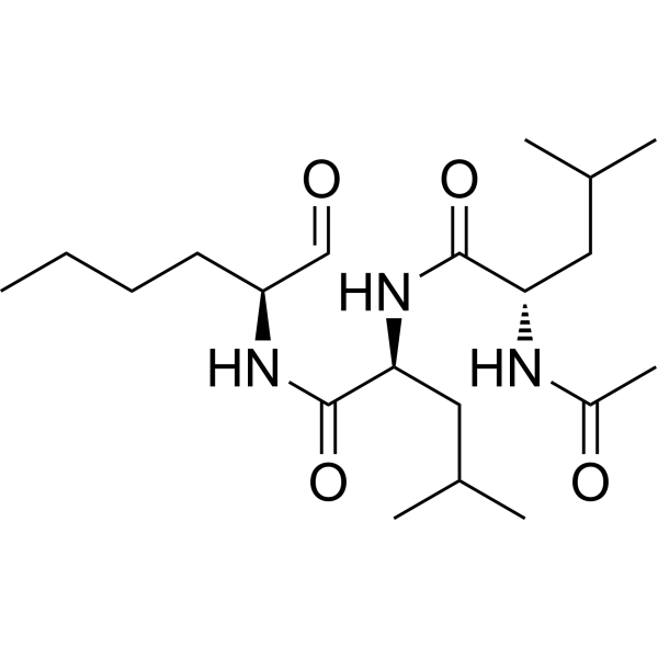
MG-101 (ALLN) is an inhibitor of cysteine proteases which inhibits calpain I, calpain II, cathepsin B and cathepsin L with Kis of 190, 220, 150 and 500 pM, respectively. MG-101 induces apoptosis and inhibits tumor growth, it can be used for the research of colon cancer.
For research use only. We do not sell to patients.
- Purity : 98.0%
- CAS No.: 110044-82-1
- Formula: C20H37N3O4
- Molecular Weight:383.53
-
Storage:Powder -20°C, 3 years , 4°C, 2 years ; In solvent -80°C, 6 months , -20°C, 1 month

To place orders, for customer services and technical support, please contact: MedChemExpress USA
Tel: 609-228-6898 E-mail: [email protected] [email protected]


-
 Biological Activity
-
Chemical Information
-
Solvent & Solubility
- Protocols
- Purity & Documentation
- References
-
 Help & FAQs


-
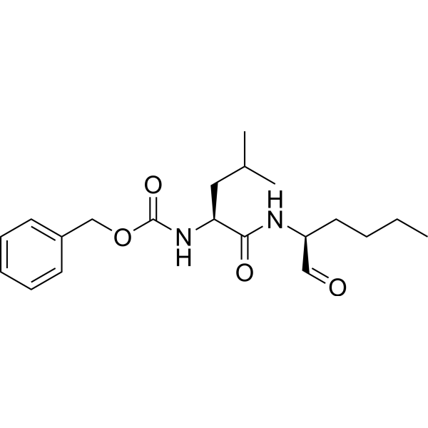
HY-100223
CalpeptinCalpain Inhibitor -

HY-13259
MG-132Proteasome Inhibitor -
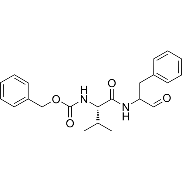
HY-18236
MDL-28170Calpain Inhibitor -
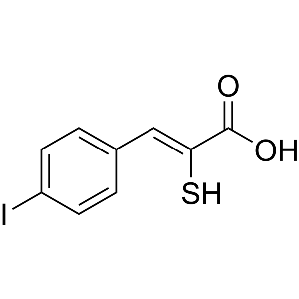
HY-100529
PD150606Calpain Inhibitor -
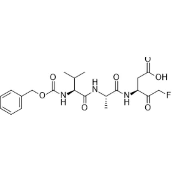
HY-16658B
Z-VAD-FMKPan-Caspase Inhibitor -
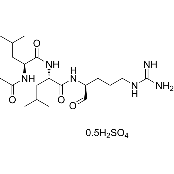
HY-18234A
Leupeptin hemisulfateProtease Inhibitor -

HY-10227
BortezomibProteasome Inhibitor -

HY-100558
Bafilomycin A1V-ATPase Inhibitor -

HY-100579
Ferrostatin-1Ferroptosis Inhibitor -
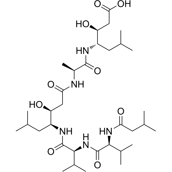
HY-P0018
PepstatinAspartic Protease Inhibitor -
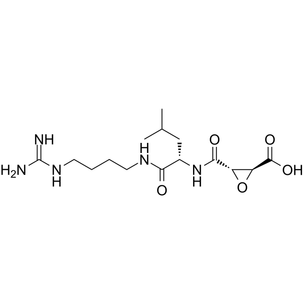
HY-15282
E-64Cysteine Peptidase Inhibitor -
HY-B0496
PMSFCathepsin Inhibitor
Publications Citing Use of MedChemExpress (MCE) MG-101
More- Signal Transduct Target Ther. 2025 Dec 17;10(1):413. [Abstract]
- Science. 2021 Mar 5;371(6533):eabb2224. [Abstract]
- Nat Commun. 2025 Jan 8;16(1):496. [Abstract]
- J Genet Genomics. 2024 Oct;51(10):1020-1029. [Abstract]
- Cell Rep. 2021 Jun 29;35(13):109299. [Abstract]
- Int J Mol Sci. 2022 May 16;23(10):5529. [Abstract]
- Glia. 2022 Dec;70(12):2392-2408. [Abstract]
- Sci Rep. 2025 Jul 21;15(1):26481. [Abstract]
- Sci Rep. 2022 Jul 16;12(1):12197. [Abstract]
- J Cell Mol Med. 2020 Aug;24(16):9287-9299. [Abstract]
- Exp Cell Res. 2023 Dec 15;433(2):113859. [Abstract]
- Biochem Biophys Res Commun. 2018 Sep 3;503(1):297-303. [Abstract]
- bioRxiv. 2020 Jun.
 Customer Validation & Images

-
RT-PCR

-
Cell Proliferation/Viability Assay

-
Bio/Physico-chemical Assay

-
WB

-
Bio/Physico-chemical Assay


Biological Activity
Description
Cellular Effect
|
Cell Line

|
Type | Value | Description | References |
|---|---|---|---|---|
| B16 | IC50 |
14.5 μM
Compound: ALLN
|
Inhibitory concentration of the compound was determined against melanoma B16 cells
Inhibitory concentration of the compound was determined against melanoma B16 cells
|
[PMID: 15634028] |
| HeLa | CC50 |
25.1 μM
Compound: ALLN
|
Cytotoxicity against human HeLa cells assessed as growth inhibition after 48 hrs by MTS assay
Cytotoxicity against human HeLa cells assessed as growth inhibition after 48 hrs by MTS assay
|
[PMID: 23357632] |
| L1210 | IC50 |
3 μM
Compound: ALLN
|
Inhibitory concentration of the compound was determined against L1210 cells
Inhibitory concentration of the compound was determined against L1210 cells
|
[PMID: 15634028] |
In Vitro
MG-101 (0-26 μM; 24 h) affects the growth of colon cancer HCT116 cells[1]. MG-101 (0-26 μM; 24 h) induces apoptosis of various cancer cells[1]. MG-101 (0-26 μM; 24 h) induces Bax-dependent apoptosis in HCT116 cells[1].
MedChemExpress (MCE) has not independently confirmed the accuracy of these methods. They are for reference only. Further protocols information, click here.
-
Cell Line:HCT116 cell line
-
Concentration:0-26 μM
-
Incubation Time:24 hours
-
Result:Anchorage-independently and significantly decreased the viability of HCT116 cells.
-
Cell Line:HCT116, RKO, SW480 and HepG2 cell lines
-
Concentration:0-26 μM
-
Incubation Time:24 hours
-
Result:Dose-dependently decreased the concentration of pro-caspase 3 and increased the concentration of cleaved-PARP in HCT116 cells. Induced apoptosis of HCT116, RKO, SW480 and HepG2 cells.
-
Cell Line:HCT116, HCT116/p53-/- and HCT116/Bax-/- cell lines
-
Concentration:0-26 μM
-
Incubation Time:24 hours
-
Result:Induced apoptosis via a Bax-dependent manner, but not via a p53-dependent manner.
In Vivo
MedChemExpress (MCE) has not independently confirmed the accuracy of these methods. They are for reference only.
-
Animal Model:Female athymic nude mice with HCT116 cells injection[1]
-
Dosage:10 mg/kg
-
Administration:Intraperitoneal injection; 10 mg/kg once daily; for 15 days
-
Result:Significantly decreased tumor weight and volume of treated mice at day 26 compared to control mice, and showed no effect on body weights.


Chemical Information
-
CAS No. 110044-82-1
-
Appearance Solid
-
Molecular Weight 383.53
-
Formula C20H37N3O4
-
Color White to light yellow
-
SMILES
O=C(N[C@H](C=O)CCCC)[C@@H](NC([C@@H](NC(C)=O)CC(C)C)=O)CC(C)C
-
Synonyms
Calpain inhibitor I; Ac-LLnL-CHO; ALLN
-
Shipping
Room temperature in continental US; may vary elsewhere.
-
Storage
Powder -20°C 3 years 4°C 2 years In solvent -80°C 6 months -20°C 1 month


Publications (13)
-
Journal Impact Factor
-
Most Recent
-
Signal Transduct Target Ther
Disruption of heme homeostasis by nuclear receptor Nur77 induces pyroptosis through granzyme B-dependent GSDMC cleavage. [Abstract]2025 Dec 17;10(1):413. PMID: 41407678 -
Science
A structure of human Scap bound to Insig-2 suggests how their interaction is regulated by sterols. [Abstract]2021 Mar 5;371(6533):eabb2224. PMID: 33446483 -
Nat Commun
Phenazine biosynthesis-like domain-containing protein (PBLD) and Cedrelone promote antiviral immune response by activating NF-ĸB. [Abstract]2025 Jan 8;16(1):496. PMID: 39779683
MG-101 purchased from MedChemExpress. Usage Cited in: Nat Commun. 2025 Jan 8;16(1):496. [Abstract]
Real-time PCR analysis the mRNA expression of IFNs and ISGs in PBLD-overexpressing HeLa cells treated with DMSO, MG101 (20 μM) or QNZ (10 μM) or transfected with siNF-κB (10 nM) or siNC, and then infected with BPIV3 (MOI = 1), VSV (MOI = 1) or HSV-1 (MOI = 1), respectively for 24 h.
MG-101 purchased from MedChemExpress. Usage Cited in: Nat Commun. 2025 Jan 8;16(1):496. [Abstract]
To investigate the roles of NF-κB in PBLD mediated IFN-I signaling pathway, we used two pharmacological inhibitors of NF-κB, MG-101 and QNZ. The optimal concentrations of MG-101 (20 μM) and QNZ (10 μM) were determined through cell viability assays.
-
J Genet Genomics
2024 Oct;51(10):1020-1029. PMID: 38663479 -
Cell Rep
2021 Jun 29;35(13):109299. PMID: 34192549 -
Int J Mol Sci
Decreased Expression of Plakophilin-2 and αT-Catenin in Arrhythmogenic Right Ventricular Cardiomyopathy: Potential Markers for Diagnosis. [Abstract]2022 May 16;23(10):5529. PMID: 35628349
MG-101 purchased from MedChemExpress. Usage Cited in: Int J Mol Sci. 2022 May 16;23(10):5529. [Abstract]
Protein levels in HL-1/shLuc, shCTNNA3, and shPKP2 cardiomyocytes after MG101 (5 μM, 24 h) treatment.
-
Glia
Astrocyte-specific loss of lactoferrin influences neuronal structure and function by interfering with cholesterol synthesis. [Abstract]2022 Dec;70(12):2392-2408. PMID: 35946355 -
Sci Rep
Substrate profiling of marine-derived thermotolerant cysteine protease reveals unique cleavage preferences for industrial applications. [Abstract]2025 Jul 21;15(1):26481. PMID: 40691222
MG-101 purchased from MedChemExpress. Usage Cited in: Sci Rep. 2025 Jul 21;15(1):26481. [Abstract]
Dose response curves of two inhibitors, leupeptin and MG101 (0.01 nM-10 μM), against globupain. The substrate Boc-QAR-amc was used. Activity was normalized to a vehicle treated sample.
-
Sci Rep
Hepatitis C virus NS3/4A inhibitors and other drug-like compounds as covalent binders of SARS-CoV-2 main protease. [Abstract]2022 Jul 16;12(1):12197. PMID: 35842458 -
J Cell Mol Med
Calpain-2 plays a pivotal role in the inhibitory effects of propofol against TNF-α-induced autophagy in mouse hippocampal neurons. [Abstract]2020 Aug;24(16):9287-9299. PMID: 32627970
MG-101 purchased from MedChemExpress. Usage Cited in: J Cell Mol Med. 2020 Aug;24(16):9287-9299. [Abstract]
We found that TNF-α-induced calpain activation was attenuated by pre-treatment of neurons with 50 μmol/L calpain inhibitor IV (a calpain-2 selective inhibitor), 200 nmol/L NA101 (a calpain-2 selective inhibitor), 20 μmol/L calpain Inhibitor I (MG-101, a non-selective calpain inhibitor) and 50 μmol/L calpeptin (a non-selective calpain inhibitor).
-
Exp Cell Res
Cardiac ischemic preconditioning promotes cMyBP-C phosphorylation by inhibiting the calpain-mediated proteolysis. [Abstract]2023 Dec 15;433(2):113859. PMID: 38000772 -
Biochem Biophys Res Commun
Effects of intracellular iron overload on cell death and identification of potent cell death inhibitors. [Abstract]2018 Sep 3;503(1):297-303. PMID: 29890135 -


Solvent & Solubility
In Vitro:
DMSO : ≥ 100 mg/mL (260.74 mM; Hygroscopic DMSO has a significant impact on the solubility of product, please use newly opened DMSO)
* "≥" means soluble, but saturation unknown.
Please refer to the solubility information to select the appropriate solvent. Once prepared, please aliquot and store the solution to prevent product inactivation from repeated freeze-thaw cycles.
Storage method and period of stock solution: -80°C, 6 months; -20°C, 1 month. When stored at -80°C, please use it within 6 months. When stored at -20°C, please use it within 1 month.
Please refer to the solubility information to select the appropriate solvent. Once prepared, please aliquot and store the solution to prevent product inactivation from repeated freeze-thaw cycles.
Storage method and period of stock solution: -80°C, 6 months; -20°C, 1 month. When stored at -80°C, please use it within 6 months. When stored at -20°C, please use it within 1 month.
Concentration (start) × Volume (start) = Concentration (final) × Volume (final)
In Vivo:
Select the appropriate dissolution method based on your experimental animal and administration route.
- For the following dissolution methods, please ensure to first prepare a clear stock solution using an In Vitro approach and then sequentially add co-solvents:
- To ensure reliable experimental results, the clarified stock solution can be appropriately stored based on storage conditions. As for the working solution for In Vivo experiments, it is recommended to prepare freshly and use it on the same day.
- The percentages shown for the solvents indicate their volumetric ratio in the final prepared solution. If precipitation or phase separation occurs during preparation, heat and/or sonication can be used to aid dissolution.
Add each solvent one by one: 10% DMSO 40% PEG300 5% Tween-80 45% Saline
Solubility: ≥ 1.67 mg/mL (4.35 mM); Clear solution
This protocol yields a clear solution of ≥ 1.67 mg/mL (saturation unknown).
Taking 1 mL working solution as an example, add 100 μL DMSO stock solution (16.7 mg/mL) to 400 μL PEG300, and mix evenly; then add 50 μL Tween-80 and mix evenly; then add 450 μL Saline to adjust the volume to 1 mL.
Preparation of Saline: Dissolve 0.9 g sodium chloride in ddH₂O and dilute to 100 mL to obtain a clear Saline solution.
Add each solvent one by one: 10% DMSO 90% (20% SBE-β-CD in Saline)
Solubility: 1.67 mg/mL (4.35 mM); Suspended solution; Need ultrasonic
This protocol yields a suspended solution of 1.67 mg/mL. Suspended solution can be used for oral and intraperitoneal injection.
Taking 1 mL working solution as an example, add 100 μL DMSO stock solution (16.7 mg/mL) to 900 μL 20% SBE-β-CD in Saline, and mix evenly.
Preparation of 20% SBE-β-CD in Saline (4°C, storage for one week): 2 g SBE-β-CD powder is dissolved in 10 mL Saline, completely dissolve until clear.
In Vivo Dissolution Calculator
Please enter the basic information of animal experiments:
-
-
-
-
Recommended: Prepare an additional quantity of animals to account for potential losses during experiments.
Please enter your animal formula composition:
-
%DMSO +
Recommended: Keep the proportion of DMSO in working solution below 2% if your animal is weak.
-
%+
-
+%Tween-80 + +
-
%Saline +
The co-solvents required include: DMSO, . All of co-solvents are available by MedChemExpress (MCE). , Tween 80. All of co-solvents are available by MedChemExpress (MCE).
Working solution concentration: 0.22 mg/mL
Method for preparing stock solution: mg drug dissolved in μL DMSO. Stock solution concentration: mg/mL.
1. Take μL DMSO stock solution;
2. Add μL .
μL , mix evenly;
3. Then add μL Tween 80, mix evenly;
4. Then add μL
Please ensure that the stock solution in the first step is dissolved to a clear state, and add co-solvents in sequence. You can use ultrasonic heating (ultrasonic cleaner, recommended frequency 20-40 kHz), vortexing, etc. to assist dissolution.

Protocols
-
Apoptosis
Apoptosis, also called programmed cell death, is generally characterized by distinct morphological characteristics.
-
TUNEL staining for apoptotic DNA fragmentation
TUNEL staining detects DNA strand breaks by using terminal deoxynucleotidyl transferase to add labeled nucleotides to exposed 3′-OH DNA termini, generating either microscopic staining in fixed cells or tissue sections, or fluorescence/cytometric signal in cell suspensions. TUNEL positivity reflects DNA fragmentation but should not be interpreted alone as definitive apoptosis, because TUNEL can also label necrotic, autolytic, mechanically damaged, or DNA-repair-associated DNA breaks.
-
Annexin V plus membrane-impermeant dye apoptosis staining
Annexin V-based apoptosis assays rely on the detection of phosphatidylserine (PS) externalization from the inner leaflet of the plasma membrane to the outer leaflet, an early biochemical hallmark of apoptosis. Fluorescently labeled Annexin V binds PS in a calcium-dependent manner, enabling identification of early apoptotic cells by flow cytometry or fluorescence microscopy. When combined with a membrane-impermeant DNA-binding dye (e. g. , propidium iodide), this approach allows discrimination between viable (Annexin V−/dye−), early apoptotic (Annexin V+/dye−), and late apoptotic or necrotic (Annexin V+/dye+) cell populations by assessing membrane integrity and PS exposure.
-
Patient-Derived Xenograft (PDX)
Patient-derived xenograft (PDX) models are generated by engrafting primary human tumor tissue directly into immunodeficient mice, allowing in vivo propagation of patient tumor biology without initial in vitro adaptation. These models are used to preserve key histopathological and molecular characteristics of the original tumor and enable assessment of tumor growth dynamics and therapeutic response in a living organism. The biological readout is tumor engraftment and subsequent growth in the murine host, which reflects the ability of human tumor cells to survive, vascularize, and expand in an immunocompromised microenvironment.
-
Apoptosis Solutions
Apoptosis is a regulated, generally non-lytic cell-death pathway that removes unwanted, damaged, infected, or abnormal cells through coordinated morphological changes, caspase activation, DNA fragmentation, and membrane remodeling. The intrinsic apoptosis pathway is controlled mainly by mitochondrial outer membrane permeabilization, BCL-2 family proteins, cytochrome c release, apoptosome formation, caspase-9 activation, and downstream executioner caspase-3/7 activation. The extrinsic apoptosis pathway is initiated by death receptors such as Fas, TNFR, and TRAIL receptors, which recruit adaptor proteins and activate caspase-8 before engaging executioner caspases or mitochondrial amplification through BID cleavage. Apoptosis is linked to many phenotypes, including cancer cell killing, tissue homeostasis, immune regulation, neurodegeneration, infection response, and treatment-induced cytotoxicity; unresolved questions include how apoptosis interacts with necroptosis, pyroptosis, ferroptos
-
Patient-Derived Orthotopic Xenograft (PDOX)
Patient-derived orthotopic xenograft (PDOX) modeling implants fresh patient tumor tissue or patient-derived tumor cells into the anatomically corresponding organ or tissue site of immunodeficient mice, usually by surgical orthotopic implantation, to preserve patient tumor histology, local microenvironmental context, invasion, metastatic behavior, and treatment-response features better than subcutaneous implantation. PDOX readouts include tumor engraftment, orthotopic tumor growth, local invasion, metastasis, recurrence after resection, histologic similarity to the donor tumor, biomarker retention, molecular concordance, survival, and response or resistance to therapy. PDOX models are used for preclinical drug testing and individualized therapy evaluation, but engraftment success varies by tumor type and specimen quality.
-
Subcutaneous Cell-Line-Derived Xenograft
Subcutaneous cell-line-derived xenograft (CDX) models are established by implanting cultured human cancer cell lines into immunodeficient mice, where the injected cells form localized tumors that can be monitored in vivo as a measure of tumorigenic potential, growth kinetics, and treatment response. These models are widely used in oncology research because they allow reproducible tumor formation and enable comparative assessment of tumor growth between different cell lines or genetic manipulations in a controlled in vivo microenvironment. Subcutaneous implantation of cancer cells in immunodeficient mice is a standard approach for evaluating tumor growth behavior and therapeutic response across multiple cancer types, including prostate, esophageal, pancreatic, and colon cancer models.
-
Orthotopic Cell-Line Xenograft
Orthotopic cell-line xenograft models involve implantation of human cancer cell lines into the anatomically corresponding organ of immunodeficient mice to reproduce tumor growth within a native microenvironment, enabling more clinically relevant tumor behavior compared with subcutaneous models. These models are widely used because orthotopic placement better recapitulates tumor progression, including invasion and metastatic spread, which are often underrepresented in heterotopic implantation systems. Compared with conventional xenografts, orthotopic implantation is described as more technically complex but provides improved simulation of tumor-microenvironment interactions and metastatic behavior, making it particularly valuable for translational oncology research. Surgical orthotopic implantation approaches have been emphasized as enabling faithful reproduction of clinical cancer features, including metastasis and disease progression patterns that align with the tumor’s organ of origi


Purity & Documentation
-
Data Sheet (278 KB)
-
SDS (393 KB)
- English - EN (393 KB)
- Français - FR (393 KB)
- Deutsch - DE (393 KB)
- Norwegian - NO (393 KB)
- Español - ES (393 KB)
- Swedish - SV (393 KB)
- Italian - IT (393 KB)
- Korean - KR (393 KB)
- Portuguese - PT (393 KB)
-
Handling Instructions (2659 KB)
References


Complete Stock Solution Preparation Table
Please refer to the solubility information to select the appropriate solvent. Once prepared, please aliquot and store the solution to prevent product inactivation from repeated freeze-thaw cycles.
Storage method and period of stock solution: -80°C, 6 months; -20°C, 1 month. When stored at -80°C, please use it within 6 months. When stored at -20°C, please use it within 1 month.
| Optional Solvent | Concentration Solvent Mass | 1 mg | 5 mg | 10 mg | 25 mg |
|---|---|---|---|---|---|
| DMSO | 1 mM | 2.6074 mL | 13.0368 mL | 26.0736 mL | 65.1839 mL |
| 5 mM | 0.5215 mL | 2.6074 mL | 5.2147 mL | 13.0368 mL | |
| 10 mM | 0.2607 mL | 1.3037 mL | 2.6074 mL | 6.5184 mL | |
| 15 mM | 0.1738 mL | 0.8691 mL | 1.7382 mL | 4.3456 mL | |
| 20 mM | 0.1304 mL | 0.6518 mL | 1.3037 mL | 3.2592 mL | |
| 25 mM | 0.1043 mL | 0.5215 mL | 1.0429 mL | 2.6074 mL | |
| 30 mM | 0.0869 mL | 0.4346 mL | 0.8691 mL | 2.1728 mL | |
| 40 mM | 0.0652 mL | 0.3259 mL | 0.6518 mL | 1.6296 mL | |
| 50 mM | 0.0521 mL | 0.2607 mL | 0.5215 mL | 1.3037 mL | |
| 60 mM | 0.0435 mL | 0.2173 mL | 0.4346 mL | 1.0864 mL | |
| 80 mM | 0.0326 mL | 0.1630 mL | 0.3259 mL | 0.8148 mL | |
| 100 mM | 0.0261 mL | 0.1304 mL | 0.2607 mL | 0.6518 mL |


MG-101 Related Classifications
HY-18964 Related Classifications
 Powered by Bioz