



Tiratricol
Based on 2 publication(s) in Google Scholar
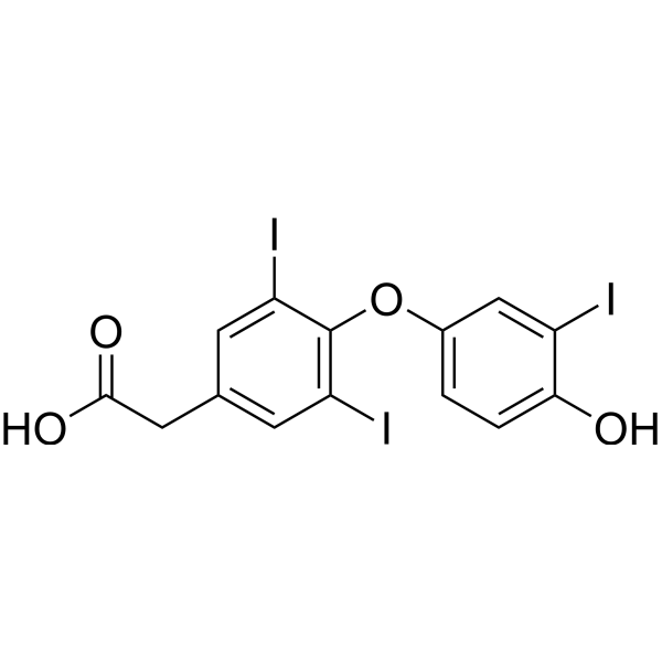
Tiratricol is an orally available thyroid hormone analog that inhibits pituitary thyroid-stimulating hormone secretion. Tiratricol is an intracellular toxin neutralizer that inhibits LPS and lipid A cytotoxicity with IC50s of 20 μM and 32 μM, respectively. Tiratricol reduces TNF production in lipopolysaccharide-stimulated macrophages. Tiratricol also has antiviral activity and is an inhibitor of yellow fever virus (Flavivirus). It can bind to the RdRp domain of the viral NS5 protein to hinder YFV replication..
For research use only. We do not sell to patients.
- Purity : 98.88%
- CAS No.: 51-24-1
- Formula: C14H9I3O4
- Molecular Weight:621.93
-
Storage:Powder -20°C, 3 years ; In solvent -80°C, 6 months , -20°C, 1 month

To place orders, for customer services and technical support, please contact: MedChemExpress USA
Tel: 609-228-6898 E-mail: [email protected] [email protected]


-
 Biological Activity
-
Chemical Information
-
Solvent & Solubility
- Protocols
- Purity & Documentation
- References
-
 Help & FAQs


-
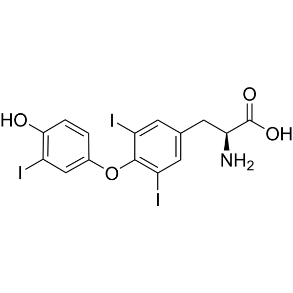
HY-A0070A
LiothyronineTIGIT/PVR Blocker -
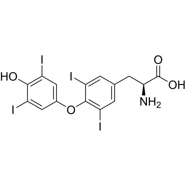
HY-18341
L-ThyroxineThyroid Hormone -
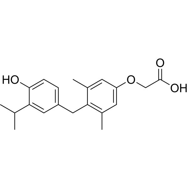
HY-14823
SobetiromeTRβ Agonist -
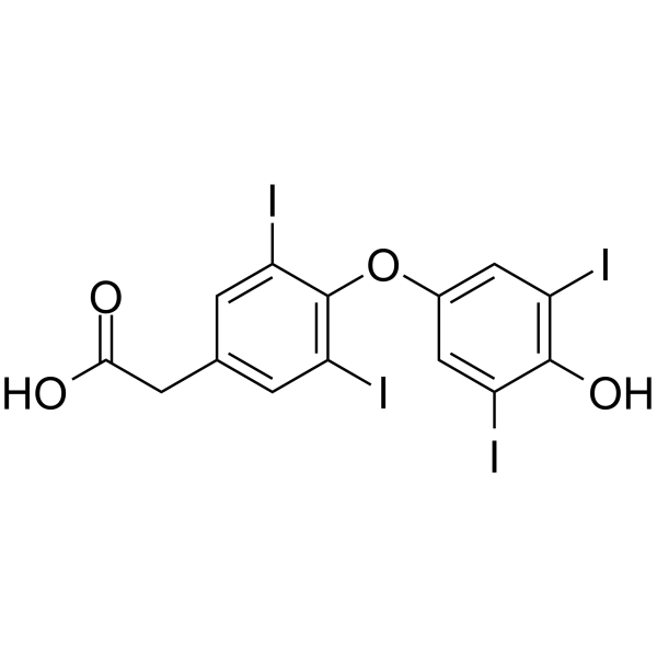
HY-W008859
Tetrac -
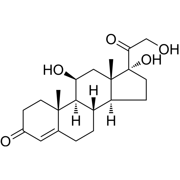
HY-N0583
HydrocortisoneGlucocorticoid Receptor Agonist -
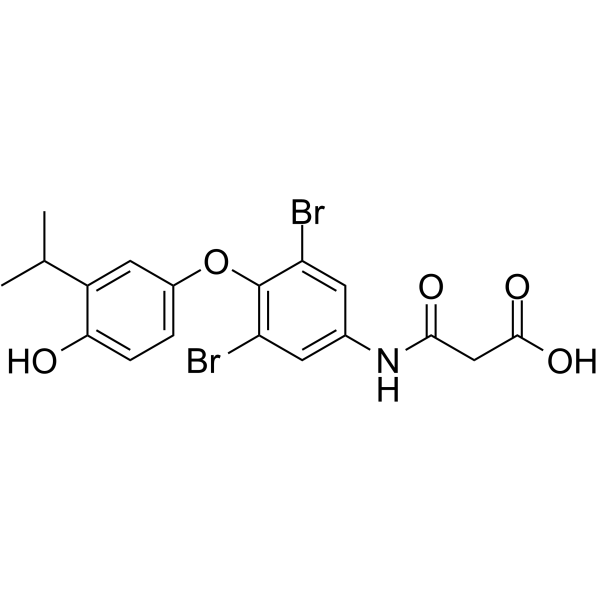
HY-10473
Eprotirome -
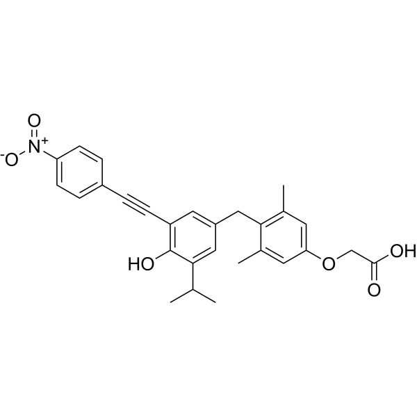
HY-141513
NH-3THR Antagonist -

HY-B1201R
Tiratricol (Standard)Thyroid Hormone Receptor Inhibitor -
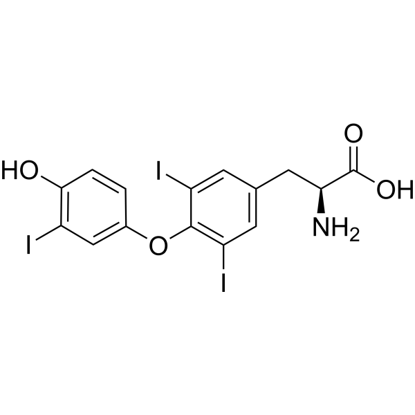
HY-A0070AG
Liothyronine (GMP)TRα/TRβ Agonist (GMP) -
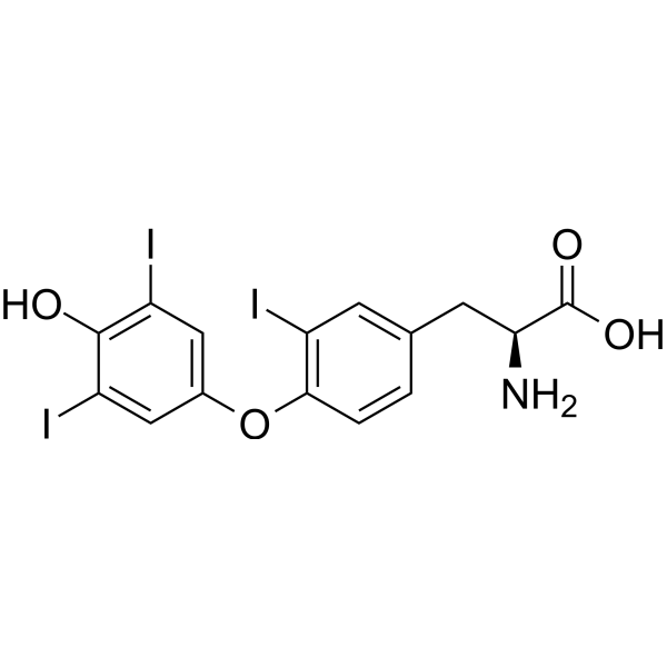
HY-W010696
Reverse T3Thyroid Hormone -
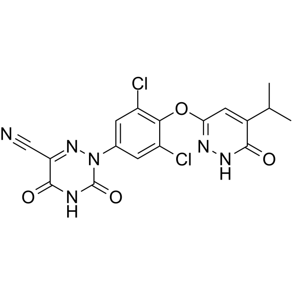
HY-12216
ResmetiromTHR-β Agonist -
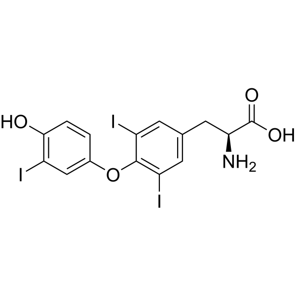
HY-A0070AR
Liothyronine (Standard)TRα/TRβ Agonist
Publications Citing Use of MedChemExpress (MCE) Tiratricol
More

Biological Activity
Description
Cellular Effect
|
Cell Line

|
Type | Value | Description | References |
|---|---|---|---|---|
| HEK-293T | EC50 |
0.37 μM
Compound: TRIAC
|
Agonist activity at recombinant human pFA-CMV fused PPARgamma expressed in HEK293T cells transfected with pFR-luciferase plasmid and pRL-SV40 plasmid incubated for 14 to 16 hrs by dual glo luciferase reporter gene assay
Agonist activity at recombinant human pFA-CMV fused PPARgamma expressed in HEK293T cells transfected with pFR-luciferase plasmid and pRL-SV40 plasmid incubated for 14 to 16 hrs by dual glo luciferase reporter gene assay
|
[PMID: 32356658] |
| HEK-293T | EC50 |
12 μM
Compound: TRIAC
|
Agonist activity at recombinant human pFA-CMV fused RXRalpha LBD expressed in HEK293T cells transfected with pFR-luciferase plasmid and pRL-SV40 plasmid incubated for 14 to 16 hrs by dual glo luciferase reporter gene assay
Agonist activity at recombinant human pFA-CMV fused RXRalpha LBD expressed in HEK293T cells transfected with pFR-luciferase plasmid and pRL-SV40 plasmid incubated for 14 to 16 hrs by dual glo luciferase reporter gene assay
|
[PMID: 32356658] |
| LNCaP | IC50 |
28.3 μM
Compound: 5, TRIAC
|
Binding affinity to human androgen receptor BF3 F673R mutant stably transfected in eGFP-expressing human LNCAP cells after 5 days by fluorometric analysis
Binding affinity to human androgen receptor BF3 F673R mutant stably transfected in eGFP-expressing human LNCAP cells after 5 days by fluorometric analysis
|
[PMID: 22047606] |
| LNCaP | IC50 |
34.8 μM
Compound: 5, TRIAC
|
Transcriptional activity at human androgen receptor BF3 site stably transfected in eGFP-expressing human LNCAP cells after 5 days by fluorometric analysis
Transcriptional activity at human androgen receptor BF3 site stably transfected in eGFP-expressing human LNCAP cells after 5 days by fluorometric analysis
|
[PMID: 22047606] |
| LNCaP | IC50 |
42.8 μM
Compound: 5, TRIAC
|
Binding affinity to human androgen receptor BF3 R840A mutant stably transfected in eGFP-expressing human LNCAP cells after 5 days by fluorometric analysis
Binding affinity to human androgen receptor BF3 R840A mutant stably transfected in eGFP-expressing human LNCAP cells after 5 days by fluorometric analysis
|
[PMID: 22047606] |
| U2OS | IC50 |
6.9 μM
Compound: TRIAC
|
Inhibition of HA-tagged human NTCP expressed in human U2OS cells assessed as reduction in [14C]taurocholate uptake preincubated for 10 mins followed by [14C] taurocholate addition and further incubation for 10 mins by scintillation counting method
Inhibition of HA-tagged human NTCP expressed in human U2OS cells assessed as reduction in [14C]taurocholate uptake preincubated for 10 mins followed by [14C] taurocholate addition and further incubation for 10 mins by scintillation counting method
|
[PMID: 31439379] |
In Vitro
Tiratricol (10, 40 μM; 24 h) dose-dependently inhibits YFV infection of Huh-7 cells and also reduces the expression of viral non-structural protein (NS3) [2].
MedChemExpress (MCE) has not independently confirmed the accuracy of these methods. They are for reference only. Further protocols information, click here.
-
Cell Line:Huh-7 cells
-
Concentration:0.625, 2.5, 10, and 40 μM
-
Incubation Time:24 h
-
Result:Remarkably suppressed the viral proteins of NS1, NS2B, NS3, prM and capsid.
In Vivo
MedChemExpress (MCE) has not independently confirmed the accuracy of these methods. They are for reference only.
-
Animal Model:C57BL/6 mice infected with YFV[2]
-
Dosage:0.2 mg/kg/day
-
Administration:po; day 0–6 post infection (dpi) or from 2 days before infection to 6 dpi
-
Result:Significantly reduce virus titers and protein expression.
Restored body weight and improved the survival rate of YFV-infected mice by 40%.
Clinical Trial
| NCT Number | Sponsor | Condition | Start Date |
Phase
|
|---|---|---|---|---|
| NCT01329991 | Plexxikon| | 2011-05 | PHASE1 |


Chemical Information
-
CAS No. 51-24-1
-
Appearance Solid
-
Molecular Weight 621.93
-
Formula C14H9I3O4
-
Color White to off-white
-
SMILES
O=C(O)CC1=CC(I)=C(OC2=CC=C(O)C(I)=C2)C(I)=C1
-
Synonyms
3,3',5-Triiodothyroacetic acid
-
Shipping
Room temperature in continental US; may vary elsewhere.
-
Storage
Powder -20°C 3 years In solvent -80°C 6 months -20°C 1 month


Publications (2)
-
Journal Impact Factor
-
Most Recent
-
Environ Sci Technol
IodoFinder: Machine Learning-Guided Recognition of Iodinated Chemicals in Nontargeted LC-MS/MS Analysis. [Abstract]2025 Mar 11;59(9):4530-4539. PMID: 40015982 -


Solvent & Solubility
In Vitro:
DMSO : ≥ 42 mg/mL (67.53 mM; Hygroscopic DMSO has a significant impact on the solubility of product, please use newly opened DMSO)
* "≥" means soluble, but saturation unknown.
Please refer to the solubility information to select the appropriate solvent. Once prepared, please aliquot and store the solution to prevent product inactivation from repeated freeze-thaw cycles.
Storage method and period of stock solution: -80°C, 6 months; -20°C, 1 month. When stored at -80°C, please use it within 6 months. When stored at -20°C, please use it within 1 month.
Please refer to the solubility information to select the appropriate solvent. Once prepared, please aliquot and store the solution to prevent product inactivation from repeated freeze-thaw cycles.
Storage method and period of stock solution: -80°C, 6 months; -20°C, 1 month. When stored at -80°C, please use it within 6 months. When stored at -20°C, please use it within 1 month.
Concentration (start) × Volume (start) = Concentration (final) × Volume (final)
In Vivo:
Select the appropriate dissolution method based on your experimental animal and administration route.
- For the following dissolution methods, please ensure to first prepare a clear stock solution using an In Vitro approach and then sequentially add co-solvents:
- To ensure reliable experimental results, the clarified stock solution can be appropriately stored based on storage conditions. As for the working solution for In Vivo experiments, it is recommended to prepare freshly and use it on the same day.
- The percentages shown for the solvents indicate their volumetric ratio in the final prepared solution. If precipitation or phase separation occurs during preparation, heat and/or sonication can be used to aid dissolution.
Add each solvent one by one: 10% DMSO 40% PEG300 5% Tween-80 45% Saline
Solubility: ≥ 2.5 mg/mL (4.02 mM); Clear solution
This protocol yields a clear solution of ≥ 2.5 mg/mL (saturation unknown).
Taking 1 mL working solution as an example, add 100 μL DMSO stock solution (25.0 mg/mL) to 400 μL PEG300, and mix evenly; then add 50 μL Tween-80 and mix evenly; then add 450 μL Saline to adjust the volume to 1 mL.
Preparation of Saline: Dissolve 0.9 g sodium chloride in ddH₂O and dilute to 100 mL to obtain a clear Saline solution.
Add each solvent one by one: 10% DMSO 90% (20% SBE-β-CD in Saline)
Solubility: ≥ 2.5 mg/mL (4.02 mM); Clear solution
This protocol yields a clear solution of ≥ 2.5 mg/mL (saturation unknown).
Taking 1 mL working solution as an example, add 100 μL DMSO stock solution (25.0 mg/mL) to 900 μL 20% SBE-β-CD in Saline, and mix evenly.
Preparation of 20% SBE-β-CD in Saline (4°C, storage for one week): 2 g SBE-β-CD powder is dissolved in 10 mL Saline, completely dissolve until clear.
In Vivo Dissolution Calculator
Please enter the basic information of animal experiments:
-
-
-
-
Recommended: Prepare an additional quantity of animals to account for potential losses during experiments.
Please enter your animal formula composition:
-
%DMSO +
Recommended: Keep the proportion of DMSO in working solution below 2% if your animal is weak.
-
%+
-
+%Tween-80 + +
-
%Saline +
The co-solvents required include: DMSO, . All of co-solvents are available by MedChemExpress (MCE). , Tween 80. All of co-solvents are available by MedChemExpress (MCE).
Working solution concentration: 0.22 mg/mL
Method for preparing stock solution: mg drug dissolved in μL DMSO. Stock solution concentration: mg/mL.
1. Take μL DMSO stock solution;
2. Add μL .
μL , mix evenly;
3. Then add μL Tween 80, mix evenly;
4. Then add μL
Please ensure that the stock solution in the first step is dissolved to a clear state, and add co-solvents in sequence. You can use ultrasonic heating (ultrasonic cleaner, recommended frequency 20-40 kHz), vortexing, etc. to assist dissolution.

Protocols
-
Research Protocol for Infectious Diseases
Infectious-disease experiments test how pathogens interact with host barriers, innate immune receptors, inflammatory signaling, pathogen replication, and tissue injury; pattern-recognition receptors such as TLRs, RIG-I-like receptors, NOD-like receptors, and inflammasomes detect microbial molecules and activate NF-κB, interferon, and cytokine responses. The central hypothesis is that infection severity reflects the balance between pathogen burden and host response: protective inflammation restricts pathogen growth, whereas excessive or mislocalized inflammation contributes to tissue damage and disease phenotype. Unresolved questions include which host pathways are protective versus pathogenic, why some infection models fail to translate to human disease, and which combined readouts best predict clinically relevant infection outcomes.
-
Cell Cytotoxicity Assay
Cytotoxicity assays are usually based on the assessment of cell membrane damage, which can also be indirectly detected by measuring cell viability. Detection methods include MTT assay, CKK-8 assay, LDH assay and ATP assay, etc.
-
Mammalian live/dead viability and cytotoxicity staining
Live/dead viability and cytotoxicity staining assays are based on the simultaneous detection of intracellular esterase activity in metabolically active (viable) cells and membrane integrity loss in non-viable cells. In commonly used dual-staining approaches, membrane-permeant fluorogenic substrates are converted by intracellular esterases into fluorescent products in live cells, while impermeant DNA-binding dyes selectively enter cells with compromised plasma membranes and label nucleic acids in dead or dying cells, enabling discrimination between viable and non-viable populations by fluorescence microscopy or flow cytometry.
-
Research Protocol for Endocrine Diseases
Endocrine diseases often arise from disrupted hormone production, hormone signaling, or target-tissue responsiveness; for diabetes-focused endocrine disease models, insulin signaling regulates glucose uptake, hepatic glucose output, lipid metabolism, and β-cell compensation. Type 2 diabetes develops through interacting defects in insulin resistance, β-cell dysfunction, adipose inflammation, hepatic glucose overproduction, altered incretin signaling, and ectopic lipid metabolism. A major unresolved question is whether endocrine dysfunction is driven primarily by target-tissue insulin resistance, intrinsic β-cell failure, immune/inflammatory stress, or combined multi-organ failure that differs by disease stage.
-
Cell Viability Determination by MTT Colorimetric Assay
The following protocol uses the MTT colorimetric assay as a classic literature-established method for assessing cell viability/metabolic activity in cultured mammalian cells. MTT[3-(4,5-dimethylthiazol-2-yl)-2,5-diphenyltetrazolium bromide] is reduced by metabolically active cells to a colored formazan product; the amount of formazan is quantified spectrophotometrically and provides an indirect measure of metabolically active viable cells. Importantly, MTT reduction reflects cellular oxidoreductase/metabolic activity rather than an absolute direct count of living cells, so changes in cellular metabolism can alter the signal independently of cell number.


Purity & Documentation
-
Data Sheet (273 KB)
-
SDS (393 KB)
- English - EN (393 KB)
- Français - FR (393 KB)
- Deutsch - DE (393 KB)
- Norwegian - NO (393 KB)
- Español - ES (393 KB)
- Swedish - SV (393 KB)
- Italian - IT (393 KB)
- Korean - KR (393 KB)
- Portuguese - PT (393 KB)
-
Handling Instructions (2659 KB)
References
[1]. Ren H, et al. Tiratricol inhibits yellow fever virus replication through targeting viral RNA-dependent RNA polymerase of NS5. Antiviral Res. 2023 Nov;219:105737. [Content Brief]
[2]. Cascales L, et al. Tiratricol neutralizes bacterial endotoxins and reduces lipopolysaccharide-induced TNF-alpha production in the cell. Chem Biol Drug Des. 2008 Oct;72(4):320-8. [Content Brief]


Complete Stock Solution Preparation Table
Please refer to the solubility information to select the appropriate solvent. Once prepared, please aliquot and store the solution to prevent product inactivation from repeated freeze-thaw cycles.
Storage method and period of stock solution: -80°C, 6 months; -20°C, 1 month. When stored at -80°C, please use it within 6 months. When stored at -20°C, please use it within 1 month.
| Optional Solvent | Concentration Solvent Mass | 1 mg | 5 mg | 10 mg | 25 mg |
|---|---|---|---|---|---|
| DMSO | 1 mM | 1.6079 mL | 8.0395 mL | 16.0790 mL | 40.1975 mL |
| 5 mM | 0.3216 mL | 1.6079 mL | 3.2158 mL | 8.0395 mL | |
| 10 mM | 0.1608 mL | 0.8039 mL | 1.6079 mL | 4.0197 mL | |
| 15 mM | 0.1072 mL | 0.5360 mL | 1.0719 mL | 2.6798 mL | |
| 20 mM | 0.0804 mL | 0.4020 mL | 0.8039 mL | 2.0099 mL | |
| 25 mM | 0.0643 mL | 0.3216 mL | 0.6432 mL | 1.6079 mL | |
| 30 mM | 0.0536 mL | 0.2680 mL | 0.5360 mL | 1.3399 mL | |
| 40 mM | 0.0402 mL | 0.2010 mL | 0.4020 mL | 1.0049 mL | |
| 50 mM | 0.0322 mL | 0.1608 mL | 0.3216 mL | 0.8039 mL | |
| 60 mM | 0.0268 mL | 0.1340 mL | 0.2680 mL | 0.6700 mL |


Tiratricol Related Classifications
HY-B1201 Related Classifications
 Powered by Bioz