



Alirocumab
Based on 1 publication(s) in Google Scholar
Alirocumab is an anti-PCSK9 human monoclonal antibody. Alirocumab inhibits PCSK9. Alirocumab reduces NLRP3 inflammasome, regulates Nrf2/HO-1, HMGB1/NF-κB and Fractalkine/CX3CR1. Alirocumab increases the ability of the liver to bind LDL-cholesterol (LDL-C) and reduces levels of LDL-C in blood. Alirocumab improves atherosclerosis and inflammation.
For research use only. We do not sell to patients.
- Purity : 97.00%
- CAS No.: 1245916-14-6
- Molecular Weight:146.24 kDa
-
Storage:
Please store the product under the recommended conditions in the Certificate of Analysis.

To place orders, for customer services and technical support, please contact: MedChemExpress USA
Tel: 609-228-6898 E-mail: [email protected] [email protected]


-
 Biological Activity
-
Chemical Information
- Protocols
- Purity & Documentation
- References
-
 Help & FAQs


-
HY-P9930
EvolocumabPCSK9 Inhibitor -
HY-P9908
AdalimumabTNF-α Inhibitor -
HY-P99187
BococizumabAnti-human PCSK9 Antibody -
HY-148673
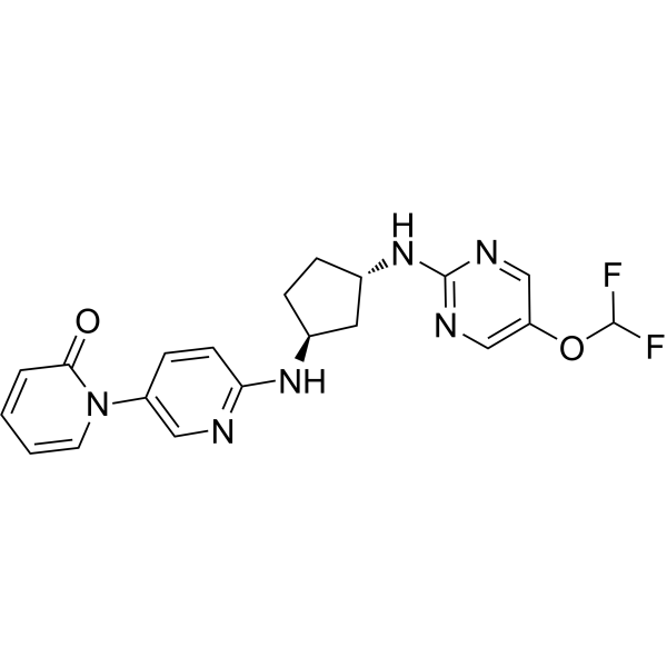
LaroprovstatPCSK9 Inhibitor -
HY-P9928A
Alirocumab (anti-PCSK9)PCSK9 Inhibitor -

HY-P9905
CetuximabEGFR Inhibitor -
HY-P9903
NivolumabPD-1 Inhibitor -
HY-P9917
TocilizumabIL-6R Neutralizing Antibody -
HY-114118
SemaglutideGLP-1R Agonist -
HY-108810
CanakinumabAnti-IL-1β Monoclonal Antibody -
HY-P9906
BevacizumabVEGF-A Inhibitor -

HY-P9927
SecukinumabIL-17A Antagonist
Publications Citing Use of MedChemExpress (MCE) Alirocumab
More

Biological Activity
Description
Isotype
Human IgG1 kappa
Recommend Isotype Controls
Species Reactivity
Human
IC50 & Target
PCSK9
In Vitro
Alirocumab (40 μg/mL, 24 h) alleviates basal PCSK9 overexpression in vascular smooth muscle cells (VSMCs) of obese insulin-resistant Zucker rats (OZR)[3].
Alirocumab (8 μg/mL, 72 h) attenuates Lp(a) secretion in primary human hepatocytes via inhibition of PCSK9[4].
Alirocumab (10 μg/mL, 24 h) inhibits lipid-induced inflammation in HepG2 cells[5].
MedChemExpress (MCE) has not independently confirmed the accuracy of these methods. They are for reference only. Further protocols information, click here.
-
Cell Line:HepG2 incubated with 0.5 mM cis-9-octadecenoic acid and 0.25 mM palmitic acid
-
Concentration:10 μg/mL
-
Incubation Time:24 h
-
Result:Decreased PCSK9 protein levels by 65.3%.
Attenuated increased IL-6, IL-1β, and TNFα protein levels.
Decreased p65-NF-κB phosphorylation.
Reduced the phosphorylation levels of AP-1 by 61.0%.
Decreased the phosphorylation levels of PI3K and AKT.
Decreased the mTOR protein phosphorylation levels by 46.2%.
In Vivo
Alirocumab (16 mg/kg/week, s.c., on day 0, day 7, and day 14) boosts antioxidant status and halts inflammation in rat model of sepsis-induced nephrotoxicity via modulation of Nrf2/HO-1, PCSK9/HMGB1/NF-ᴋB/NLRP3 and Fractalkine/CX3CR1 hubs[7].
Alirocumab (50 mg/kg, s.c., weekly prior to exposure to the liquid diets) attenuates ethanol-induced neuronal injury in the brain and oxidative stress in rats[8].
Alirocumab (1 mg/kg/week, s.c.) activates brown fat, increases hepatic uptake of cholesterol-rich TRL remnants, thereby lowering non-HDL-C, and increases HDL-C levels and cholesterol efflux capacity of HDL, further improving dyslipidemia in APOE*3-Leiden.CETP mice[9].
Alirocumab (10 mg/kg, s.c., 2 weeks) reduces lipoprotein(a) levels in nonhuman primates by lowering apolipoprotein(a) production rate[10].
Alirocumab (3-10 mg/kg, i.p., weekly for 16 weeks) reduces RAS, NLRP3 inflammasome, and cholecystokinin in lung tissue of obese mice[11].
MedChemExpress (MCE) has not independently confirmed the accuracy of these methods. They are for reference only.
-
Animal Model:Male albino Wistar rats model (LPS-intoxicated)[7]
-
Dosage:16 mg/kg/week
-
Administration:Subcutaneous injection (s.c.), on day 0, day 7, and day 14
-
Result:Mitigated LPS-mediated increments in serum creatinine and cystatin C, together with renal contents of both KIM-1 and NGAL.
Restored renal NGAL content to its normal values.
Boosted mRNA expression levels of both Nrf2 and HO-1 and renal TAC content (2.5, 2, and 3.2-folds, respectively).
Produced pronounced hampering in LPS-mediated elevation in mRNA expression levels of PCSK9 and RAGE, along with renal contents of PCSK9 and HMGB1 by 80.9 %, 49.6 %, 53.1 % and 59.8 %, respectively.
Resulted in a marked reduction in the protein expression of TLR4, MYD88, and NLRP3, along with mRNA expression levels of NF-ᴋB by 62.9 %, 58.1 %, 50.9 %, respectively.
Caused remarkable alleviation in LPS-mediated increment in TNF-α, IL-1β, and caspase-1 by 48.5 %, 68.3 % and 58.5 %, respectively.
Produced prominent downregulation in mRNA expression levels of CX3CL1 and CX3CR1 by 88.4 % and 87.5 %, respectively.
Exhibited prominent elevation in mRNA expression level of Bcl-2 (1.7-folds), along with a marked reduction in both mRNA expression level of Bax and renal caspase-3 content (by 66.7 % and 58.5 %, respectively) .
Regressed glomerular and tubular lesions.
Clinical Trial
| NCT Number | Sponsor | Condition | Start Date |
Phase

|
|---|---|---|---|---|
| NCT01329991 | Plexxikon| | 2011-05 | PHASE1 |
Gene ID
Accession
Conjugated
Unconjugated
Reconsititution
The product can be reconstituted/diluted with sterile PBS or saline.
Format
-
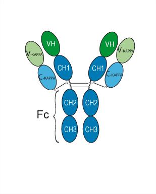
Human IgG1 kappa
Application
ELISA, FACS, Functional assay
Verified Bioactivity
-
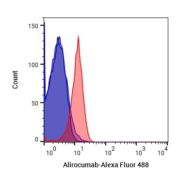 Flow Cytometry analysis of Hela cells labelling PCSK9 (red) with Alirocumab (anti-PCSK9) (HY-P9928). Goat Anti-Human IgG (Alexa Fluor 488) (HY-P83776) at a dilution of 1/1000 was used as the secondary antibody. Blue-Human IgG1 kappa (HY-P99001). Black-Unlabelled control, cells without incubation with primary antibody.


Chemical Information
-
CAS No. 1245916-14-6
-
Appearance Liquid
-
Molecular Weight 146.24 kDa
-
Color Colorless to light yellow
-
SMILES
OC(CO)COP(O)(O)=O.[Na].[Na]
-
Synonyms
REGN 727; SAR 236553
-
Formulation
Please refer to the lot-specific COA for specific buffer information.
-
Storage
Please store the product under the recommended conditions in the Certificate of Analysis.


Publications (1)
-
Journal Impact Factor
-
Most Recent
-
Nat Commun
The Lin28b/Wnt5a axis drives pancreas cancer through crosstalk between cancer associated fibroblasts and tumor epithelium. [Abstract]2023 Oct 28;14(1):6885. PMID: 37898598

Protocols
-
Research Protocol for Cardiovascular Diseases
Cardiovascular disease can be modeled as maladaptive cardiac remodeling, where ischemic injury or pressure overload activates inflammatory signaling, fibroblast activation, extracellular-matrix deposition, cardiomyocyte hypertrophy, vascular remodeling, and progressive ventricular dysfunction. The TGF-β/SMAD axis is a central profibrotic pathway after myocardial injury and pressure overload, while innate immune and cytokine pathways regulate leukocyte recruitment, scar formation, and adverse remodeling. Key unresolved questions include which inflammatory signals are reparative versus harmful, when fibrosis is protective versus maladaptive, and whether pathway inhibition improves function without weakening necessary infarct healing or compensatory remodeling.
-
Research Protocol for Inflammation-related Diseases
The NLRP3 inflammasome is a cytosolic innate immune signaling platform that integrates priming signals and danger-signal activation to promote caspase-1 activation, maturation of IL-1β and IL-18, and gasdermin D-mediated pyroptotic cell death. The core experimental logic is to determine whether inflammatory disease phenotypes are driven by increased NLRP3 expression, ASC-containing inflammasome assembly, caspase-1 cleavage, GSDMD cleavage, and extracellular release of IL-1β/IL-18 rather than by nonspecific cell injury alone. The pathway is strongly linked to inflammation-related disease phenotypes because monosodium urate crystals activate NALP3/NLRP3 inflammasome signaling in gout-like crystal inflammation, cholesterol crystals activate NLRP3 inflammasomes in atherogenesis models, and DSS-induced intestinal inflammation has been reported to involve NLRP3 inflammasome activity. However, experimental colitis studies also show context-dependent protective effects of NLRP3 inflammasome co
-
Pyroptosis Solutions
Pyroptosis is a lytic inflammatory cell-death pathway executed by gasdermin pores, most classically through inflammasome-mediated activation of caspase-1, cleavage of gasdermin D, membrane pore formation, LDH release, and secretion of IL-1β and IL-18. The canonical pathway is commonly modeled by priming cells with an inflammatory signal such as LPS to induce pro-IL-1β and inflammasome components, followed by an activation signal such as ATP or nigericin to activate NLRP3, ASC speck formation, caspase-1 cleavage, GSDMD cleavage, cytokine release, and pyroptotic membrane rupture. The non-canonical pathway is triggered when cytosolic LPS activates mouse caspase-11 or human caspase-4/5, leading to GSDMD cleavage and pyroptosis, and this can secondarily activate NLRP3-dependent IL-1β release. Pyroptosis is linked to inflammatory injury, infection, cancer, liver disease, ocular disease, placental inflammation, and other disease phenotypes, but unresolved questions include which gasdermin fam


Purity & Documentation
-
Data Sheet (269 KB)
-
SDS (251 KB)
- English - EN (251 KB)
- Français - FR (251 KB)
- Deutsch - DE (251 KB)
- Norwegian - NO (251 KB)
- Español - ES (251 KB)
- Swedish - SV (251 KB)
- Italian - IT (251 KB)
- Korean - KR (251 KB)
- Portuguese - PT (251 KB)
-
Inhibitory Antibodies User Guide (603 KB)
References
[1]. Markham A. Alirocumab: First Global Approval. Drugs. 2015;75(14):1699-1705. [Content Brief]
[2]. Tavori H, et al. Alirocumab: PCSK9 inhibitor for LDL cholesterol reduction. Expert Rev Cardiovasc Ther. 2014 Oct;12(10):1137-44. [Content Brief]
[3]. Barale C, et al. PCSK9 Expression in Vascular Smooth Muscle Cells: Role of Insulin Resistance and High Glucose. Int J Mol Sci. 2025 Jan 24;26(3):1003. [Content Brief]
[4]. Villard EF, et al. PCSK9 Modulates the Secretion But Not the Cellular Uptake of Lipoprotein(a) Ex Vivo: An Effect Blunted by Alirocumab. JACC Basic Transl Sci. 2016 Oct;1(6):419-427. [Content Brief]
[6]. Kühnast S, et al. Alirocumab inhibits atherosclerosis, improves the plaque morphology, and enhances the effects of a statin. J Lipid Res. 2014 Oct;55(10):2103-12. [Content Brief]
[7]. Hassan NF, et al. Alirocumab boosts antioxidant status and halts inflammation in rat model of sepsis-induced nephrotoxicity via modulation of Nrf2/HO-1, PCSK9/HMGB1/NF-ᴋB/NLRP3 and Fractalkine/CX3CR1 hubs. Biomed Pharmacother. 2024 Aug;177:116929. [Content Brief]
[8]. Wagner J, et al. PCSK9 inhibition attenuates alcohol-associated neuronal oxidative stress and cellular injury. Brain Behav Immun. 2024 Jul;119:494-506. [Content Brief]
[9]. Zhou E, et al. Beneficial effects of brown fat activation on top of PCSK9 inhibition with alirocumab on dyslipidemia and atherosclerosis development in APOE*3-Leiden.CETP mice. Pharmacol Res. 2021 May;167:105524. [Content Brief]
[10]. Croyal M, et al. PCSK9 inhibition with alirocumab reduces lipoprotein(a) levels in nonhuman primates by lowering apolipoprotein(a) production rate. Clin Sci (Lond). 2018 May 31;132(10):1075-1083. [Content Brief]
[11]. Liang L, et al. Statin administration or blocking PCSK9 alleviates airway hyperresponsiveness and lung fibrosis in high-fat diet-induced obese mice. Respir Res. 2024 May 18;25(1):213. [Content Brief]

Calculators
Concentration (start) × Volume (start) = Concentration (final) × Volume (final)


Alirocumab Related Classifications
HY-P9928 Related Classifications
 Powered by Bioz