



AMPK activator 11
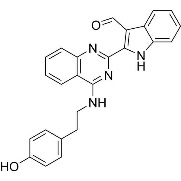
AMPK activator 11 is an AMP-activated protein kinase (AMPK) activator with nanomolelevel antiproliferation activities against several CRCs. AMPK activator 11 selectively inhibits the RKO xenograft growth along by activating AMPK and upregulating oxidative phosphorylation (OXPHOS) ( mitochondrial metabolism ) and can be used for anti-tumor and metabolic disease research.
For research use only. We do not sell to patients.
- CAS No.: 2948304-00-3
- Formula: C25H20N4O2
- Molecular Weight:408.45
-
Storage:
Please store the product under the recommended conditions in the Certificate of Analysis.

To place orders, for customer services and technical support, please contact: MedChemExpress USA
Tel: 609-228-6898 E-mail: [email protected] [email protected]


-
 Biological Activity
-
Chemical Information
- Protocols
- Purity & Documentation
- References
-
 Help & FAQs


All AMPK Isoforms
More

Biological Activity
Description
Cellular Effect
|
Cell Line

|
Type | Value | Description | References |
|---|---|---|---|---|
| DLD-1 | IC50 |
<1 μM
Compound: 18a
|
Antiproliferative activity against human DLD-1 cells assessed as reduction in cell viability incubated for 24 hrs
Antiproliferative activity against human DLD-1 cells assessed as reduction in cell viability incubated for 24 hrs
|
[PMID: 37253101] |
| HCT-116 | IC50 |
<1 μM
Compound: 18a
|
Antiproliferative activity against human HCT-116 cells assessed as reduction in cell viability incubated for 24 hrs
Antiproliferative activity against human HCT-116 cells assessed as reduction in cell viability incubated for 24 hrs
|
[PMID: 37253101] |
| L02 | IC50 |
>15 μM
Compound: 18a
|
Cytotoxicity against human L02 cells assessed as reduction in cell viability incubated for 24 hrs
Cytotoxicity against human L02 cells assessed as reduction in cell viability incubated for 24 hrs
|
[PMID: 37253101] |
| LX-2 | IC50 |
>15 μM
Compound: 18a
|
Cytotoxicity against human LX2 cells assessed as reduction in cell viability incubated for 24 hrs
Cytotoxicity against human LX2 cells assessed as reduction in cell viability incubated for 24 hrs
|
[PMID: 37253101] |
| NCM460 | IC50 |
>15 μM
Compound: 18a
|
Cytotoxicity against human NCM460 cells assessed as reduction in cell viability incubated for 24 hrs
Cytotoxicity against human NCM460 cells assessed as reduction in cell viability incubated for 24 hrs
|
[PMID: 37253101] |
| RKO | IC50 |
>1 μM
Compound: 18a
|
Antiproliferative activity against human RKO cells assessed as reduction in cell viability incubated for 24 hrs in presence of FCCP by propidium iodide staining based high-content screening method
Antiproliferative activity against human RKO cells assessed as reduction in cell viability incubated for 24 hrs in presence of FCCP by propidium iodide staining based high-content screening method
|
[PMID: 37253101] |
| RKO | IC50 |
>1 μM
Compound: 18a
|
Antiproliferative activity against human RKO cells assessed as reduction in cell viability incubated for 24 hrs in presence of oligomycin by propidium iodide staining based high-content screening method
Antiproliferative activity against human RKO cells assessed as reduction in cell viability incubated for 24 hrs in presence of oligomycin by propidium iodide staining based high-content screening method
|
[PMID: 37253101] |
| RKO | IC50 |
0.07 μM
Compound: 18a
|
Antiproliferative activity against human RKO cells assessed as reduction in cell viability incubated for 24 hrs by propidium iodide staining based high-content screening method
Antiproliferative activity against human RKO cells assessed as reduction in cell viability incubated for 24 hrs by propidium iodide staining based high-content screening method
|
[PMID: 37253101] |
| RKO | IC50 |
0.1 μM
Compound: 18a
|
Antiproliferative activity against human RKO cells assessed as reduction in cell viability incubated for 24 hrs
Antiproliferative activity against human RKO cells assessed as reduction in cell viability incubated for 24 hrs
|
[PMID: 37253101] |
| SW480 | IC50 |
<1 μM
Compound: 18a
|
Antiproliferative activity against human SW480 cells assessed as reduction in cell viability incubated for 24 hrs
Antiproliferative activity against human SW480 cells assessed as reduction in cell viability incubated for 24 hrs
|
[PMID: 37253101] |
In Vitro
AMPK activator 11 (Compound 18a) (0-30 μΜ;0-7 days) inhibits the growth and migration of CRC cells with IC50 values below 1 μM[1].
AMPK activator 11 (0-0.1 μΜ;24 hours) dramatically enhances of global oxygen consumption rate (OCR) in RKO cells while the slightly changed extracellular acidification rate (ECAR). AMPK activator 11 upregulates the expression of p-AMPK and mitochondrial complex III and V and has the ability to selectively activate OXPHOS in CRCs[1].
MedChemExpress (MCE) has not independently confirmed the accuracy of these methods. They are for reference only.
-
Cell Line:CRC cells
-
Concentration:0-30 μM
-
Incubation Time:0-7 days
-
Result:Selectively inhibited the growth and migration of CRC cells.
Inhibited the proliferation of different CRCs with IC50 values below 1 μM.
-
Cell Line:RKO cells
-
Concentration:0.1 μM
-
Incubation Time:24 hours
-
Result:Upregulated the expression of p-AMPK and mitochondrial complex III and V and has the ability to selectively activate OXPHOS.
In Vivo
MedChemExpress (MCE) has not independently confirmed the accuracy of these methods. They are for reference only.
-
Animal Model:RKO cells related axenograft model on male BALB/nude mice[1]
-
Dosage:2.5 or 10 mg/kg
-
Administration:Intraperitoneal injection (i.p.) for 25 days
-
Result:Resulted in 58.2% tumor growth inhibition, with no significant weight loss observed.
Resulted in 77.1% inhibition of tumor growth but also caused a 36% weight loss.
Immunohistochemistry results also showed activation of AMPK in tumor tissue.
- Kinase activity and phosphorylation assays
- Western Blot
- Patient-Derived Xenograft (PDX)
- Splenic/Portal-Vein Liver Metastasis Xenograft
- Protocol for Kinase activity and phosphorylation assays
- Subcutaneous Cell-Line-Derived Xenograft
- Research Protocol for Metabolic Diseases
- Orthotopic Cell-Line Xenograft
- Intraperitoneal/Peritoneal Dissemination Xenograft


Chemical Information
-
CAS No. 2948304-00-3
-
Molecular Weight 408.45
-
Formula C25H20N4O2
-
SMILES
OC1=CC=C(CCNC2=NC(C3=C(C4=CC=CC=C4N3)C=O)=NC5=C2C=CC=C5)C=C1
-
Shipping
Room temperature in continental US; may vary elsewhere.
-
Storage
Please store the product under the recommended conditions in the Certificate of Analysis.


Purity & Documentation
References

Calculators
Concentration (start) × Volume (start) = Concentration (final) × Volume (final)


AMPK activator 11 Related Classifications
HY-154973 Related Classifications
 Powered by Bioz