



Anthraquinone
Based on 3 publication(s) in Google Scholar
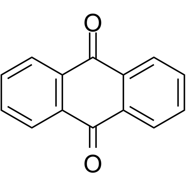
Anthraquinone is used as a precursor for dye formation and agrochemicals, existing in different organisms, such as bacteria, fungi, plants, and some animals. Anthraquinone has biological activities: anticancer, antiinflammatory, diuretic, antiarthritic, antifungal, antibacterial, antimalarial and antioxidant. Anthraquinone also plays an important role in the primary metabolism of plants by acting on the electron transport chain through the inhibition of energy transfer in the photosynthetic process. Anthraquinone can intercalates into DNA and inhibits the topoisomerase II (topo II) enzyme, resulting in cell death via Apoptosis.
For research use only. We do not sell to patients.
- Purity : 99.80%
- CAS No.: 84-65-1
- Formula: C14H8O2
- Molecular Weight:208.22
-
Storage:Powder -20°C, 3 years , 4°C, 2 years ; In solvent -80°C, 6 months , -20°C, 1 month

To place orders, for customer services and technical support, please contact: MedChemExpress USA
Tel: 609-228-6898 E-mail: [email protected] [email protected]


-
 Biological Activity
-
Chemical Information
-
Solvent & Solubility
- Protocols
- Purity & Documentation
- References
-
 Help & FAQs


-
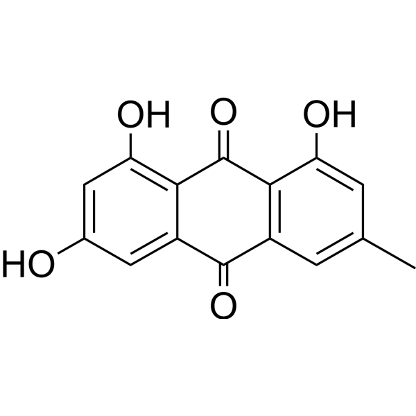
HY-14393
EmodinNatural Anthraquinone -
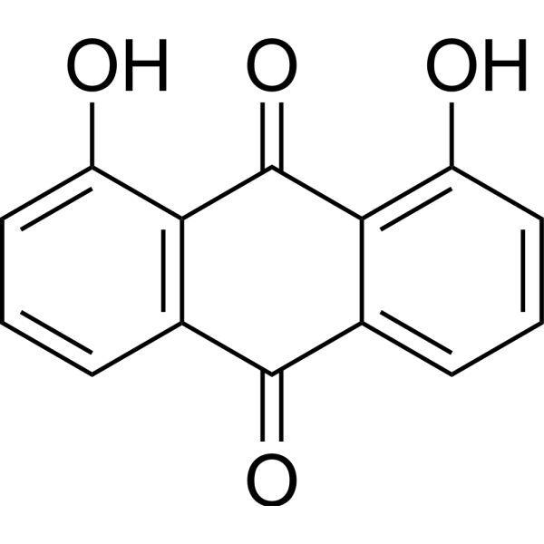
HY-B0923
DanthronAMPK Activator -
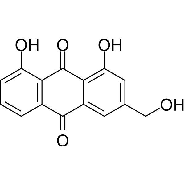
HY-N0189
Aloe emodinmTORC2 Inhibitor -
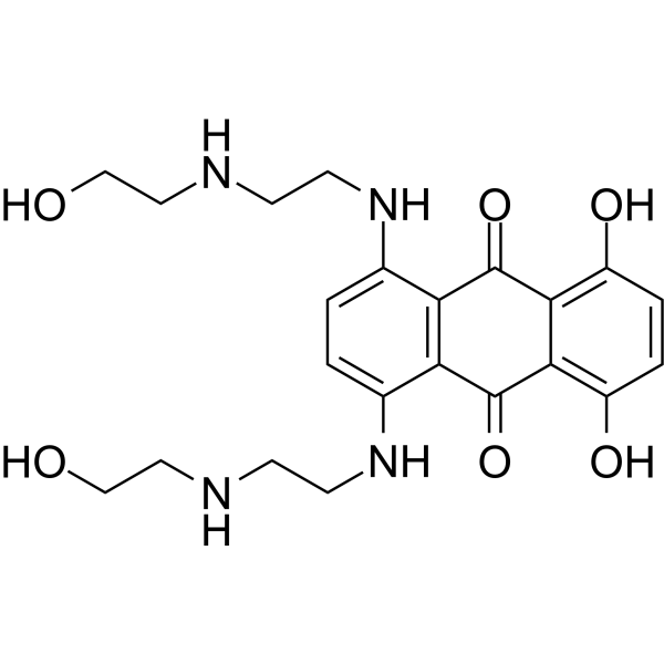
HY-13502
MitoxantroneTop II/PKC Inhibitor -
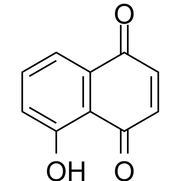
HY-N6949
JugloneAntimicrobial Drug -
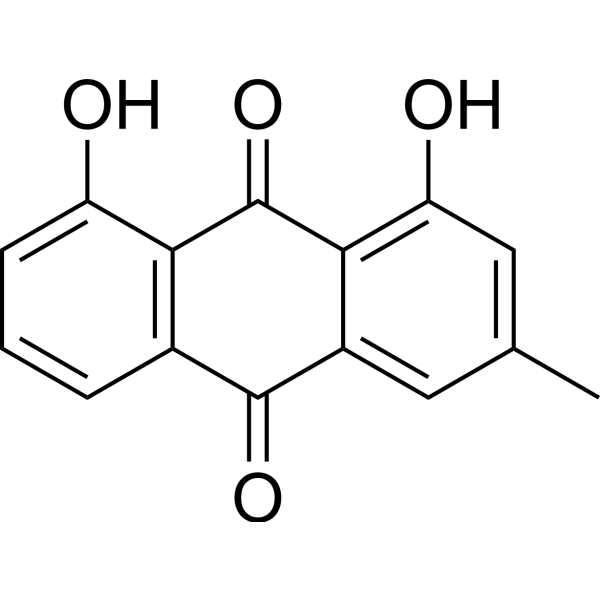
HY-13595
ChrysophanolEGFR Inhibitor -
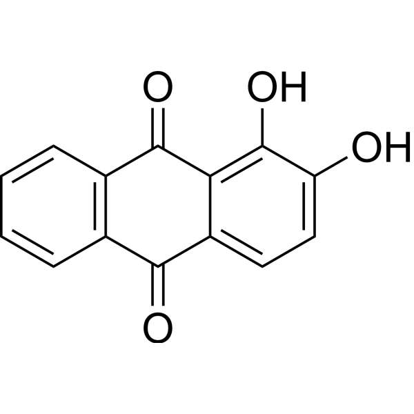
HY-N0563
AlizarinNatural Dye -

HY-N0354R
Anthraquinone (Standard)Virus Protease Inhibitor -
HY-N0105
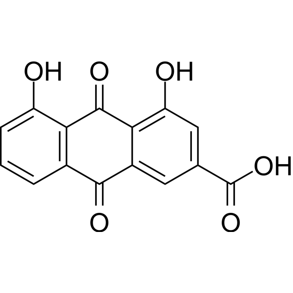
RheinAntioxidant -
HY-D0908
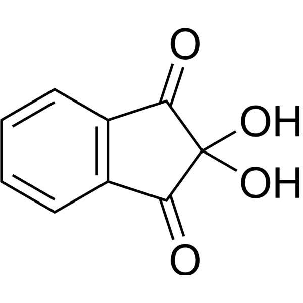
NinhydrinChromogenic Probe -

HY-B0923R
Danthron (Standard)AMPK Activator -
HY-14393R
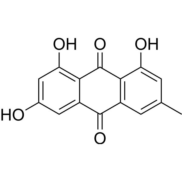
Emodin (Standard)
Publications Citing Use of MedChemExpress (MCE) Anthraquinone
More

Biological Activity
Description
Cellular Effect
|
Cell Line

|
Type | Value | Description | References |
|---|---|---|---|---|
| A2780 | IC50 |
0.58 μM
Compound: 12
|
Antiproliferative activity against human A2780 cells after 2 days by alamar-blue assay
Antiproliferative activity against human A2780 cells after 2 days by alamar-blue assay
|
[PMID: 19028102] |
| AGS | IC50 |
>100 μM
Compound: 1B
|
Cytotoxicity against human AGS cells assessed as cell viability after 72 hrs by MTT assay
Cytotoxicity against human AGS cells assessed as cell viability after 72 hrs by MTT assay
|
[PMID: 23454511] |
| HeLa | IC50 |
>50 μM
Compound: 12
|
Inhibition of histidine-tagged mouse MKP1 catalytic domain expressed in human Hela cells
Inhibition of histidine-tagged mouse MKP1 catalytic domain expressed in human Hela cells
|
[PMID: 19028102] |
| HL-60 | IC50 |
>100 μM
Compound: 1B
|
Cytotoxicity against human HL60 cells assessed as cell viability after 72 hrs by MTT assay
Cytotoxicity against human HL60 cells assessed as cell viability after 72 hrs by MTT assay
|
[PMID: 23454511] |
| J82 | IC50 |
>100 μM
Compound: 1B
|
Cytotoxicity against human J82 cells assessed as cell viability after 72 hrs by MTT assay
Cytotoxicity against human J82 cells assessed as cell viability after 72 hrs by MTT assay
|
[PMID: 23454511] |
| MRC5 | IC50 |
>100 μM
Compound: 1B
|
Cytotoxicity against human MRC5 cells assessed as cell viability after 72 hrs by MTT assay
Cytotoxicity against human MRC5 cells assessed as cell viability after 72 hrs by MTT assay
|
[PMID: 23454511] |
| SK-MES-1 | IC50 |
>100 μM
Compound: 1B
|
Cytotoxicity against human SKMES1 cells assessed as cell viability after 72 hrs by MTT assay
Cytotoxicity against human SKMES1 cells assessed as cell viability after 72 hrs by MTT assay
|
[PMID: 23454511] |
In Vitro
Anthraquinone (0.2 mg/mL, 3 min) shows a strong antioxidant activity in vitro[2].
MedChemExpress (MCE) has not independently confirmed the accuracy of these methods. They are for reference only. Further protocols information, click here.


Chemical Information
-
CAS No. 84-65-1
-
Appearance Solid
-
Molecular Weight 208.22
-
Formula C14H8O2
-
Color Light yellow to yellow
-
SMILES
O=C1C2=C(C=CC=C2)C(C3=CC=CC=C13)=O
-
Structure Classification
-
Shipping
Room temperature in continental US; may vary elsewhere.
-
Storage
Powder -20°C 3 years 4°C 2 years In solvent -80°C 6 months -20°C 1 month


Publications (3)
-
Journal Impact Factor
-
Most Recent
-
Vet Microbiol
The Chinese medicine monomer Schisandrin C inhibits PRRSV infection by regulating the OGT-PI3K/AKT/mTOR signaling pathway. [Abstract]2026 May:316:110992. PMID: 41865607 -
Cancer Manag Res
Dihydrocapsaicin Secreted by RYK Silenced Bone Marrow-Derived Mesenchymal Stem Cells Triggers Apoptosis of Gastric Cancer Cells. [Abstract]2026 Mar 21:18:579598. PMID: 41890221 -


Solvent & Solubility
In Vitro:
DMSO : 2 mg/mL (9.61 mM; ultrasonic and warming and heat to 60°C; Hygroscopic DMSO has a significant impact on the solubility of product, please use newly opened DMSO)
Please refer to the solubility information to select the appropriate solvent. Once prepared, please aliquot and store the solution to prevent product inactivation from repeated freeze-thaw cycles.
Storage method and period of stock solution: -80°C, 6 months; -20°C, 1 month. When stored at -80°C, please use it within 6 months. When stored at -20°C, please use it within 1 month.
Please refer to the solubility information to select the appropriate solvent. Once prepared, please aliquot and store the solution to prevent product inactivation from repeated freeze-thaw cycles.
Storage method and period of stock solution: -80°C, 6 months; -20°C, 1 month. When stored at -80°C, please use it within 6 months. When stored at -20°C, please use it within 1 month.
Concentration (start) × Volume (start) = Concentration (final) × Volume (final)

Protocols
-
Research Protocol for Infectious Diseases
Infectious-disease experiments test how pathogens interact with host barriers, innate immune receptors, inflammatory signaling, pathogen replication, and tissue injury; pattern-recognition receptors such as TLRs, RIG-I-like receptors, NOD-like receptors, and inflammasomes detect microbial molecules and activate NF-κB, interferon, and cytokine responses. The central hypothesis is that infection severity reflects the balance between pathogen burden and host response: protective inflammation restricts pathogen growth, whereas excessive or mislocalized inflammation contributes to tissue damage and disease phenotype. Unresolved questions include which host pathways are protective versus pathogenic, why some infection models fail to translate to human disease, and which combined readouts best predict clinically relevant infection outcomes.
-
Apoptosis
Apoptosis, also called programmed cell death, is generally characterized by distinct morphological characteristics.
-
TUNEL staining for apoptotic DNA fragmentation
TUNEL staining detects DNA strand breaks by using terminal deoxynucleotidyl transferase to add labeled nucleotides to exposed 3′-OH DNA termini, generating either microscopic staining in fixed cells or tissue sections, or fluorescence/cytometric signal in cell suspensions. TUNEL positivity reflects DNA fragmentation but should not be interpreted alone as definitive apoptosis, because TUNEL can also label necrotic, autolytic, mechanically damaged, or DNA-repair-associated DNA breaks.
-
Cell Cytotoxicity Assay
Cytotoxicity assays are usually based on the assessment of cell membrane damage, which can also be indirectly detected by measuring cell viability. Detection methods include MTT assay, CKK-8 assay, LDH assay and ATP assay, etc.
-
Mammalian live/dead viability and cytotoxicity staining
Live/dead viability and cytotoxicity staining assays are based on the simultaneous detection of intracellular esterase activity in metabolically active (viable) cells and membrane integrity loss in non-viable cells. In commonly used dual-staining approaches, membrane-permeant fluorogenic substrates are converted by intracellular esterases into fluorescent products in live cells, while impermeant DNA-binding dyes selectively enter cells with compromised plasma membranes and label nucleic acids in dead or dying cells, enabling discrimination between viable and non-viable populations by fluorescence microscopy or flow cytometry.
-
Annexin V plus membrane-impermeant dye apoptosis staining
Annexin V-based apoptosis assays rely on the detection of phosphatidylserine (PS) externalization from the inner leaflet of the plasma membrane to the outer leaflet, an early biochemical hallmark of apoptosis. Fluorescently labeled Annexin V binds PS in a calcium-dependent manner, enabling identification of early apoptotic cells by flow cytometry or fluorescence microscopy. When combined with a membrane-impermeant DNA-binding dye (e. g. , propidium iodide), this approach allows discrimination between viable (Annexin V−/dye−), early apoptotic (Annexin V+/dye−), and late apoptotic or necrotic (Annexin V+/dye+) cell populations by assessing membrane integrity and PS exposure.
-
Apoptosis Solutions
Apoptosis is a regulated, generally non-lytic cell-death pathway that removes unwanted, damaged, infected, or abnormal cells through coordinated morphological changes, caspase activation, DNA fragmentation, and membrane remodeling. The intrinsic apoptosis pathway is controlled mainly by mitochondrial outer membrane permeabilization, BCL-2 family proteins, cytochrome c release, apoptosome formation, caspase-9 activation, and downstream executioner caspase-3/7 activation. The extrinsic apoptosis pathway is initiated by death receptors such as Fas, TNFR, and TRAIL receptors, which recruit adaptor proteins and activate caspase-8 before engaging executioner caspases or mitochondrial amplification through BID cleavage. Apoptosis is linked to many phenotypes, including cancer cell killing, tissue homeostasis, immune regulation, neurodegeneration, infection response, and treatment-induced cytotoxicity; unresolved questions include how apoptosis interacts with necroptosis, pyroptosis, ferroptos
-
Directly Induced Neuron Culture
Directly induced neuron culture converts somatic cells, most commonly fibroblasts, into induced neurons without passing through a pluripotent or neural progenitor stage; classic evidence shows that mouse fibroblasts can be converted by Ascl1, Brn2/Pou3f2, and Myt1l, human fibroblasts can be converted by defined neuronal transcription factors, and human fibroblasts can also be converted by miR-9/9-124 with neurogenic or subtype-specifying transcription factors. The readout is acquisition of neuronal identity and function, assessed by neuronal morphology, neuronal markers such as Tuj1/βIII-tubulin, MAP2, synapsin, and subtype markers when relevant, together with functional assays such as action-potential firing, synaptic activity, and electrophysiology.


Purity & Documentation
-
Data Sheet (277 KB)
-
SDS (557 KB)
- English - EN (557 KB)
- Français - FR (557 KB)
- Deutsch - DE (557 KB)
- Norwegian - NO (557 KB)
- Español - ES (557 KB)
- Swedish - SV (557 KB)
- Italian - IT (557 KB)
- Korean - KR (557 KB)
- Portuguese - PT (557 KB)
-
Handling Instructions (2659 KB)
References


Complete Stock Solution Preparation Table
Please refer to the solubility information to select the appropriate solvent. Once prepared, please aliquot and store the solution to prevent product inactivation from repeated freeze-thaw cycles.
Storage method and period of stock solution: -80°C, 6 months; -20°C, 1 month. When stored at -80°C, please use it within 6 months. When stored at -20°C, please use it within 1 month.
| Optional Solvent | Concentration Solvent Mass | 1 mg | 5 mg | 10 mg | 25 mg |
|---|---|---|---|---|---|
| DMSO | 1 mM | 4.8026 mL | 24.0131 mL | 48.0261 mL | 120.0653 mL |
| 5 mM | 0.9605 mL | 4.8026 mL | 9.6052 mL | 24.0131 mL |


Anthraquinone Related Classifications
HY-N0354 Related Classifications
 Powered by Bioz