



Anti-inflammatory agent 21
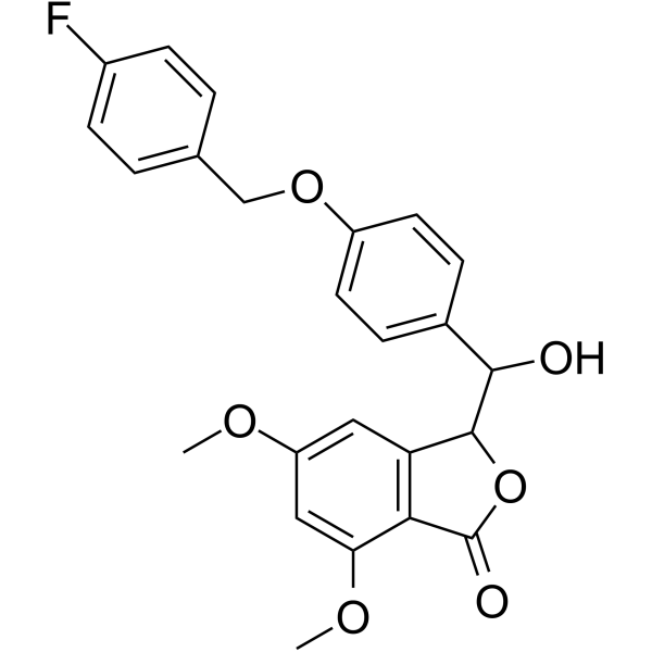
Anti-inflammatory agent 21 (compound 9o) is an orally active and low cytotoxic anti-inflammatory agent, with an IC50 value of 0.76 μM for NO. Anti-inflammatory agent 21 acts via accumulation ROS and blocks the NF-κB/MAPK signaling pathway. Anti-inflammatory agent 21 can ameliorate cartilage destruction and inflammatory cell infiltration in arthritis rats model.
For research use only. We do not sell to patients.
- CAS No.: 2408836-40-6
- Formula: C24H21FO6
- Molecular Weight:424.42
-
Storage:
Please store the product under the recommended conditions in the Certificate of Analysis.

To place orders, for customer services and technical support, please contact: MedChemExpress USA
Tel: 609-228-6898 E-mail: [email protected] [email protected]


-
 Biological Activity
-
Chemical Information
- Protocols
- Purity & Documentation
- References
-
 Help & FAQs




Biological Activity
Description
IC50 & Target
IC50: 0.76 μM (NO)[1]
Cellular Effect
|
Cell Line

|
Type | Value | Description | References |
|---|---|---|---|---|
| RAW264.7 | IC50 |
>40 μM
Compound: 9o
|
Cytotoxicity against mouse RAW264.7 cells assessed as reduction in cell viability incubated for 24 hrs in absence of LPS by MTT assay
Cytotoxicity against mouse RAW264.7 cells assessed as reduction in cell viability incubated for 24 hrs in absence of LPS by MTT assay
|
[PMID: 32823004] |
| RAW264.7 | IC50 |
>40 μM
Compound: 9o
|
Cytotoxicity against mouse RAW264.7 cells assessed as reduction in cell viability incubated for 24 hrs in presence of LPS by MTT assay
Cytotoxicity against mouse RAW264.7 cells assessed as reduction in cell viability incubated for 24 hrs in presence of LPS by MTT assay
|
[PMID: 32823004] |
| RAW264.7 | IC50 |
0.76 μM
Compound: 9o
|
Antiinflammatory activity in mouse RAW264.7 cells assessed as inhibition of LPS-induced nitric oxide production preincubated for 1 hr followed by LPS stimulation and measured after 24 hrs by Griess reagent-based assay
Antiinflammatory activity in mouse RAW264.7 cells assessed as inhibition of LPS-induced nitric oxide production preincubated for 1 hr followed by LPS stimulation and measured after 24 hrs by Griess reagent-based assay
|
[PMID: 32823004] |


Chemical Information
-
CAS No. 2408836-40-6
-
Molecular Weight 424.42
-
Formula C24H21FO6
-
SMILES
COC1=CC2=C(C(OC)=C1)C(OC2C(C3=CC=C(C=C3)OCC4=CC=C(C=C4)F)O)=O
-
Shipping
Room temperature in continental US; may vary elsewhere.
-
Storage
Please store the product under the recommended conditions in the Certificate of Analysis.

Protocols
-
Collagen-Induced Arthritis
Collagen-induced arthritis (CIA) is an autoimmune murine model of rheumatoid arthritis in which immunization with type II collagen (CII) emulsified in an adjuvant induces a T cell- and autoantibody-driven inflammatory arthritis characterized by synovial hyperplasia, immune cell infiltration, and joint destruction. The model typically relies on genetically susceptible mouse strains (e. g. , DBA/1) and reproduces key features of human rheumatoid arthritis, including anti-collagen immune responses and progressive joint inflammation. Disease onset generally occurs within ~3-4 weeks after immunization, depending on antigen/adjuvant combinations and protocol variation. The immunopathology is driven by adaptive immune activation against CII, leading to systemic and local joint inflammation mediated by pro-inflammatory cytokines and effector immune cells, making CIA a standard preclinical platform for evaluating immunomodulatory and anti-arthritic interventions.
-
Cell Cytotoxicity Assay
Cytotoxicity assays are usually based on the assessment of cell membrane damage, which can also be indirectly detected by measuring cell viability. Detection methods include MTT assay, CKK-8 assay, LDH assay and ATP assay, etc.
-
ROS/oxidative-stress fluorescent staining
ROS/oxidative-stress fluorescent staining uses cell-permeant fluorogenic probes that become fluorescent after oxidation inside cells or tissues; commonly used examples include DCFH-DA/DCFDA for broad cellular oxidant detection, DHE for superoxide-related signal detection, MitoSOX for mitochondrial superoxide-related signal detection, and CellROX probes for oxidative-stress-associated fluorescence readouts. The assay detects probe oxidation rather than a single ROS species unless the probe and analysis method have been chemically validated for that species. DCFH-DA enters cells, is deacetylated by intracellular esterases to DCFH, and produces fluorescent DCF after oxidation, so the readout is used as an operational measure of total cellular oxidative stress rather than a species-specific ROS measurement. DHE and MitoSOX can report superoxide-related oxidation, but red fluorescence alone can include non-specific ethidium-like oxidation products; HPLC or optimized spectral approaches are
-
Research Protocol for Inflammation-related Diseases
The NLRP3 inflammasome is a cytosolic innate immune signaling platform that integrates priming signals and danger-signal activation to promote caspase-1 activation, maturation of IL-1β and IL-18, and gasdermin D-mediated pyroptotic cell death. The core experimental logic is to determine whether inflammatory disease phenotypes are driven by increased NLRP3 expression, ASC-containing inflammasome assembly, caspase-1 cleavage, GSDMD cleavage, and extracellular release of IL-1β/IL-18 rather than by nonspecific cell injury alone. The pathway is strongly linked to inflammation-related disease phenotypes because monosodium urate crystals activate NALP3/NLRP3 inflammasome signaling in gout-like crystal inflammation, cholesterol crystals activate NLRP3 inflammasomes in atherogenesis models, and DSS-induced intestinal inflammation has been reported to involve NLRP3 inflammasome activity. However, experimental colitis studies also show context-dependent protective effects of NLRP3 inflammasome co
-
Cell Viability Determination by MTT Colorimetric Assay
The following protocol uses the MTT colorimetric assay as a classic literature-established method for assessing cell viability/metabolic activity in cultured mammalian cells. MTT[3-(4,5-dimethylthiazol-2-yl)-2,5-diphenyltetrazolium bromide] is reduced by metabolically active cells to a colored formazan product; the amount of formazan is quantified spectrophotometrically and provides an indirect measure of metabolically active viable cells. Importantly, MTT reduction reflects cellular oxidoreductase/metabolic activity rather than an absolute direct count of living cells, so changes in cellular metabolism can alter the signal independently of cell number.


Purity & Documentation
References

Calculators
Concentration (start) × Volume (start) = Concentration (final) × Volume (final)


Anti-inflammatory agent 21 Related Classifications
HY-146421 Related Classifications
 Powered by Bioz