



Anti-inflammatory agent 22
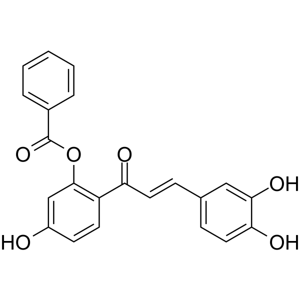
Anti-inflammatory agent 22 (compound 14a) is an orally active anti-inflammatory agent. Anti-inflammatory agent 22 inhibits LPS-induced TNF-α production with an IC50 value of 14.6 μM. Anti-inflammatory agent 22 has preventive effects on lymphedematous tissue via suppression of adipogenesis. Anti-inflammatory agent 22 suppresses limb lymphedema volume in mice.
For research use only. We do not sell to patients.
- CAS No.: 2721484-76-8
- Formula: C22H16O6
- Molecular Weight:376.36
-
Storage:
Please store the product under the recommended conditions in the Certificate of Analysis.

To place orders, for customer services and technical support, please contact: MedChemExpress USA
Tel: 609-228-6898 E-mail: [email protected] [email protected]


-
 Biological Activity
-
Chemical Information
- Protocols
- Purity & Documentation
- References
-
 Help & FAQs




Biological Activity
Description
Cellular Effect
|
Cell Line

|
Type | Value | Description | References |
|---|---|---|---|---|
| Macrophage | IC50 |
12.8 μM
Compound: 14a
|
Inhibition of LPS stimulated TNF alpha production in Balb/c mouse peritoneal macrophages measured by ELISA plate reader method
Inhibition of LPS stimulated TNF alpha production in Balb/c mouse peritoneal macrophages measured by ELISA plate reader method
|
[PMID: 32361286] |


Chemical Information
-
CAS No. 2721484-76-8
-
Molecular Weight 376.36
-
Formula C22H16O6
-
SMILES
O=C(C1=C(C=C(C=C1)O)OC(C2=CC=CC=C2)=O)/C=C/C3=CC=C(C(O)=C3)O
-
Shipping
Room temperature in continental US; may vary elsewhere.
-
Storage
Please store the product under the recommended conditions in the Certificate of Analysis.

Protocols
-
3T3-L1 preadipocyte-to-adipocyte differentiation
3T3-L1 preadipocytes are induced to differentiate after growth arrest using adipogenic media containing insulin, dexamethasone, and IBMX; differentiation is assessed by lipid-droplet accumulation, triglyceride increase, Oil Red O staining, and adipocyte-marker induction such as PPARγ and C/EBPα.
-
Cell Cytotoxicity Assay
Cytotoxicity assays are usually based on the assessment of cell membrane damage, which can also be indirectly detected by measuring cell viability. Detection methods include MTT assay, CKK-8 assay, LDH assay and ATP assay, etc.
-
Research Protocol for Inflammation-related Diseases
The NLRP3 inflammasome is a cytosolic innate immune signaling platform that integrates priming signals and danger-signal activation to promote caspase-1 activation, maturation of IL-1β and IL-18, and gasdermin D-mediated pyroptotic cell death. The core experimental logic is to determine whether inflammatory disease phenotypes are driven by increased NLRP3 expression, ASC-containing inflammasome assembly, caspase-1 cleavage, GSDMD cleavage, and extracellular release of IL-1β/IL-18 rather than by nonspecific cell injury alone. The pathway is strongly linked to inflammation-related disease phenotypes because monosodium urate crystals activate NALP3/NLRP3 inflammasome signaling in gout-like crystal inflammation, cholesterol crystals activate NLRP3 inflammasomes in atherogenesis models, and DSS-induced intestinal inflammation has been reported to involve NLRP3 inflammasome activity. However, experimental colitis studies also show context-dependent protective effects of NLRP3 inflammasome co
-
Lipid Droplets: Oil Red O/Sudan Dye Lipid Staining
Lipid droplets are intracellular organelles with a neutral-lipid core that stores triacylglycerols and sterol esters, and Oil Red O or Sudan dyes detect these hydrophobic lipid deposits by partitioning into retained lipids in fresh or frozen specimens. Oil Red O stains neutral triglycerides and lipids in frozen tissue sections or air-dried cytologic preparations, while Sudan Black B has also been used as a histochemical fat stain for lipid-rich tissue structures.


Purity & Documentation
References

Calculators
Concentration (start) × Volume (start) = Concentration (final) × Volume (final)


Anti-inflammatory agent 22 Related Classifications
HY-146435 Related Classifications
 Powered by Bioz