



Anti-Mouse CD25/IL-2Rα Antibody (PC-61.5.3)

Based on 1 Customer Validation
Anti-Mouse CD25/IL-2Rα Antibody (PC-61.5.3) is a rat-derived IgG1 λ type antibody inhibitor, targeting to mouse CD25/IL-2Rα. Anti-Mouse CD25/IL-2Rα Antibody (PC-61.5.3) can deplete CD25+ T cells. Anti-Mouse CD25/IL-2Rα Antibody (PC-61.5.3) can be used for the researches of immunology and inflammation, such as atopic dermatitis.
For research use only. We do not sell to patients.
- Purity : 99%
- Molecular Weight:150 kDa
-
Storage:
Please store the product under the recommended conditions in the Certificate of Analysis.

To place orders, for customer services and technical support, please contact: MedChemExpress USA
Tel: 609-228-6898 E-mail: [email protected] [email protected]


-
 Biological Activity
-
Chemical Information
- Protocols
- Purity & Documentation
- References
-
 Help & FAQs




Biological Activity
Description
Isotype
Rat IgG1 lambda
Recommend Isotype Controls
Species Reactivity
Mouse
IC50 & Target
CD25/IL-2Rα
In Vitro
The antibody framework is stable, specific and adaptable, and has the ability to bind both antigens and endogenous immune receptors. Monoclonal antibodies have several derivatives, including bispecific antibodies, antibody-drug conjugates, and antibody fragments, and have significant effects in fields such as immunology and oncology. When designing inhibitory antibodies, considerations include identification of antigen-specific variable regions, choice of expression system, use of multispecific formats, and antibody derivatives based on fragmentation, oligomerization, or conjugation with other functional moieties[1].
MedChemExpress (MCE) has not independently confirmed the accuracy of these methods. They are for reference only. Further protocols information, click here.
In Vivo
Anti-Mouse CD25/IL-2Rα Antibody (PC-61.5.3) (300 μg, i.p., every 2 days starting 2 days prior to treatment with MC903 (HY-10001)) attenuates atopic dermatitis in mice[2].
MedChemExpress (MCE) has not independently confirmed the accuracy of these methods. They are for reference only.
-
Animal Model:Atopic dermatitis-like dermatitis mice models[2]
-
Dosage:300 μg
-
Administration:Intraperitoneally injection, every 2 days two days prior to treatment with MC903 (HY-10001)
-
Result:Effectively depleted CD25+ IL-33R+ ILCs in the skin dLNs.
Reduced IL-5 and IL-13 production in the ear skin.
Significantly diminished ear thickening.
Reduced orthokeratosis and acanthosis and reduced dermal inflammation consisting ofmononuclear leukocytes and granulocytes.
Gene ID
Accession
P01590 / NP_032393.3
Conjugated
Unconjugated
Reconsititution
The product can be reconstituted/diluted with sterile PBS or saline.
Format
-
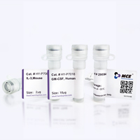
Product Image
Application
ELISA, FACS, Functional assay, Research in vivo


Chemical Information
-
Appearance Liquid
-
Molecular Weight 150 kDa
-
Color Colorless to light yellow
-
SMILES
[Anti-Mouse CD25/IL-2Rα Antibody (PC-61.5.3)]
-
Formulation
Please refer to the lot-specific COA for specific buffer information.
-
Storage
Please store the product under the recommended conditions in the Certificate of Analysis.

Protocols
-
TPA/Croton Oil Ear Edema and Dermatitis
The TPA (12-O-tetradecanoylphorbol-13-acetate) and croton oil-induced mouse ear edema model is a well-established acute cutaneous inflammation system used to evaluate topical anti-inflammatory activity by measuring edema formation, neutrophil infiltration, vascular permeability, and cytokine-mediated skin responses in vivo. The inflammatory response is triggered by topical application of phorbol esters (TPA) or croton oil constituents, leading to rapid activation of protein kinase C signaling, leukocyte recruitment, and increased vascular permeability, which can be quantified by ear thickness, weight, dye extravasation, and biochemical markers such as myeloperoxidase (MPO) activity and pro-inflammatory mediators in ear tissue homogenates. This model is widely used for screening anti-inflammatory agents, where reductions in edema and inflammatory biomarkers reflect suppression of acute dermal inflammation and immune cell infiltration. Histological evaluation typically confirms epidermal
-
Research Protocol for Inflammation-related Diseases
The NLRP3 inflammasome is a cytosolic innate immune signaling platform that integrates priming signals and danger-signal activation to promote caspase-1 activation, maturation of IL-1β and IL-18, and gasdermin D-mediated pyroptotic cell death. The core experimental logic is to determine whether inflammatory disease phenotypes are driven by increased NLRP3 expression, ASC-containing inflammasome assembly, caspase-1 cleavage, GSDMD cleavage, and extracellular release of IL-1β/IL-18 rather than by nonspecific cell injury alone. The pathway is strongly linked to inflammation-related disease phenotypes because monosodium urate crystals activate NALP3/NLRP3 inflammasome signaling in gout-like crystal inflammation, cholesterol crystals activate NLRP3 inflammasomes in atherogenesis models, and DSS-induced intestinal inflammation has been reported to involve NLRP3 inflammasome activity. However, experimental colitis studies also show context-dependent protective effects of NLRP3 inflammasome co


Purity & Documentation
-
Data Sheet (261 KB)
-
SDS (251 KB)
- English - EN (251 KB)
- Français - FR (251 KB)
- Deutsch - DE (251 KB)
- Norwegian - NO (251 KB)
- Español - ES (251 KB)
- Swedish - SV (251 KB)
- Italian - IT (251 KB)
- Korean - KR (251 KB)
- Portuguese - PT (251 KB)
-
Inhibitory Antibodies User Guide (603 KB)
References
[1]. Han P, et al. Low-dose decitabine modulates T-cell homeostasis and restores immune tolerance in immune thrombocytopenia. Blood. 2021 Aug 26;138(8):674-688. [Content Brief]
[2]. Kim BS, et al. TSLP elicits IL-33-independent innate lymphoid cell responses to promote skin inflammation. Sci Transl Med. 2013 Jan 30;5(170):170ra16. [Content Brief]

Calculators
Concentration (start) × Volume (start) = Concentration (final) × Volume (final)


Anti-Mouse CD25/IL-2Rα Antibody (PC-61.5.3) Related Classifications
HY-P990281 Related Classifications
 Powered by Bioz