



CD147 Antibody
(Synonyms: HT-7; CD147; EMMPRIN; AI115436; AI325119)
Based on 1 Customer Validation
CD147 Antibody is a Rabbit-derived and non-conjugated IgG polyclonal antibody, targeting to CD147.
-
Host:
Rabbit
-
Isotype:
IgG
-
Application:
WB, IHC-P, FC, IP
-
Reactivity :
Mouse
-
Formulation:
Supplied in 50 mM Tris-Glycine (pH 7.4), 0.15 M NaCl, 40% Glycerol and 0.05% BSA. Preservative: 0.01% Sodium azide
-
Conjugation:
Non-conjugated


To place orders, for customer services and technical support, please contact: MedChemExpress USA
Tel: 609-228-6898 E-mail: [email protected] [email protected]





Applications
| Application |
WB

WB: Western Blot
|
IHC-P
IHC-P: Immunohistochemistry-Paraffin
|
IP
IP: Immunoprecipitation
|
FC
FC: Flow Cytometry
|
|---|---|---|---|---|
| Dilution Ratio | 1:500-1:1000 | 1:50-1:100 | 1:20 | 1:50-1:100 |


Product Details
CD147 Antibody is a Rabbit-derived and non-conjugated IgG polyclonal antibody, targeting to CD147.
-
Host Rabbit

-
Clonality Polyclonal

-
Species ReactivityMouse
-
Observed Molecular WeightObserved band size: 38-58 kDa
Note: Due to possible protein modifications or aggregation, the molecular weight should be confirmed by actual measurement, and the predicted value is for reference only.

-
Calculated Molecular Weight Predicted band size: 42 kDa

Entrez Gene: 12215 Mouse
SwissProt: P18572 Mouse
Synthetic peptide corresponding to Mouse CD147.The exact sequence is proprietary to MCE.
Endogenous
affinity purified
Non-conjugated
Unmodified
IgG


Product Properties
-
Appearance
Solution
-
Formulation
Supplied in 50 mM Tris-Glycine (pH 7.4), 0.15 M NaCl, 40% Glycerol and 0.05% BSA. Preservative: 0.01% Sodium azide
-
Concentration
Batch-dependent, Please check the COA for the concentration of each lot. Check Lot Concentration
-
Storage & Stability
Stored at -20°C for 1 year. Avoid repeated freeze / thaw cycles.
-
Shipping
Shipping with blue ice.


Verification Images
-
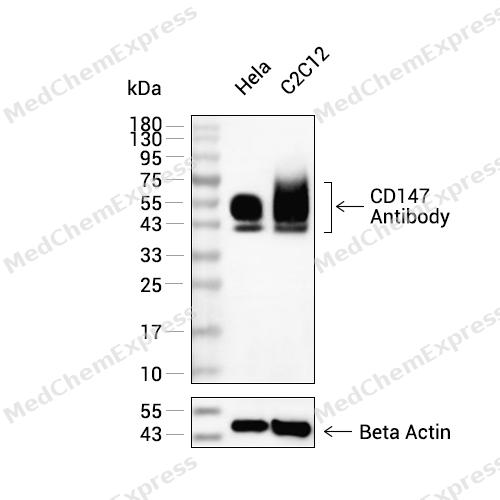 Western blot analysis of extracts from Hela(lane 2(20ug) , C2C12(lane 3(20ug) using CD147 Antibody (HY-P80596) Rabbit mAb. Proteins were transferred to a PVDF membrane and blocked with 5% non-fat milk in TBST for 2 hour at room temperature. The primary antibody (1/1000) and Loading control antibody (Beta Actin, HY-P83730, 1/10000) was used in 5% non-fat milk in TBST at 4°C overnight. Goat Anti-Mouse/Rabbit IgG-HRP Secondary Antibody (1/10000) was used for 1 hour at room temperature.
 -
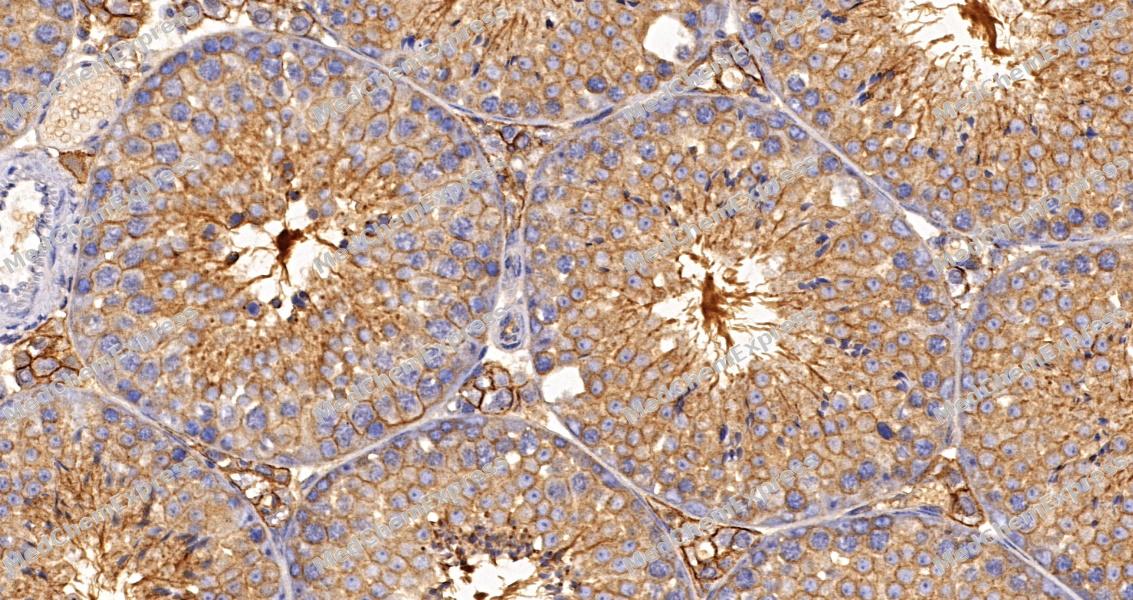 Immunohistochemical analysis of paraffin-embedded mouse testis tissue using CD147 Antibody. The section was pre-treated using heat mediated antigen retrieval with sodium citrate buffer (pH 6.0) for 8 minutes. The tissues were blocked in QuickBlock for 20 minutes at room temperature, washed with ddH2O and PBS, and then probed with the primary antibody at 1/100 dilution in 4℃ overnight. The detection was performed using an HRP conjugated compact polymer system. DAB was used as the chromogen. Tissues were counterstained with hematoxylin and mounted with DPX.
 -
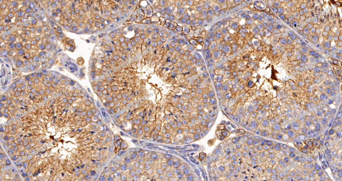 Immunohistochemical analysis of paraffin-embedded mouse testis tissue using CD147 Antibody. The section was pre-treated using heat mediated antigen retrieval with sodium citrate buffer (pH 6.0) for 8 minutes. The tissues were blocked in QuickBlock for 20 minutes at room temperature, washed with ddH2O and PBS, and then probed with the primary antibody at 1/100 dilution in 4℃ overnight. The detection was performed using an HRP conjugated compact polymer system. DAB was used as the chromogen. Tissues were counterstained with hematoxylin and mounted with DPX.
 -
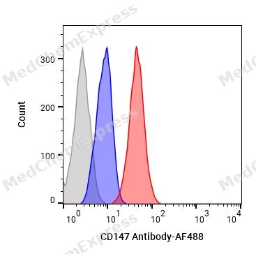 Flow cytometric analysis of 1X10⁶ NIH-3T3 cells labeling CD147 Antibody(HY-P80596, red). Cells were fixed with 4% paraformaldehyde and permeabilised with 90% methanol. Then stained with the primary antibody at 1/100 dilution for an hour at 4℃. AF488-conjugated Goat Anti-Rabbit IgG H&L (HY-P8002) was used as the secondary antibody at 1/1,000 dilution for 30 minutes at 4℃. Rabbit IgG Isotype Control (HY-P80879, blue) was used as the isotype control, cells without incubation with primary antibody were used as the unlabeled control (black).


Background
-
Function
CD147 is essential for normal retinal maturation and development. Acts as a retinal cell surface receptor for NXNL1 and plays an important role in NXNL1-mediated survival of retinal cone photoreceptors. In association with glucose transporter SLC16A1/GLUT1 and NXNL1, promotes retinal cone survival by enhancing aerobic glycolysis and accelerating the entry of glucose into photoreceptors; Signaling receptor for cyclophilins, essential for PPIA/CYPA and PPIB/CYPB-dependent signaling related to chemotaxis and adhesion of immune cells. Plays an important role in targeting the monocarboxylate transporters SLC16A1, SLC16A3 and SLC16A8 to the plasma membrane. Acts as a coreceptor for vascular endothelial growth factor receptor 2 (KDR/VEGFR2) in endothelial cells enhancing its VEGFA-mediated activation and downstream signaling. Promotes angiogenesis through EPAS1/HIF2A-mediated up-regulation of VEGFA and KDR/VEGFR2 in endothelial cells. Plays an important role in spermatogenesis; mediates interactions between germ cells and Sertoli cell and is essential for the development/differentiation of germ cells to round spermatids[1][2][3][4][5][6][7].
-
Subcellular Localization
Cell membrane; Single-pass type I membrane protein; Photoreceptor inner segment; Cell projection, cilium, photoreceptor outer segment; Cell membrane; Single-pass type I membrane protein; Endoplasmic reticulum membrane; Single-pass type I membrane protein; Basolateral cell membrane; Single-pass type I membrane protein
-
Expression
Tissue_specificity:Retina-specific (PubMed:11273674, PubMed:11853760, PubMed:12939332, PubMed:25957687) . Expressed in both rods and cones (at protein level) (PubMed:25957687) ; Testis and caput, corpus and cauda epididymides (at protein level) (PubMed:11882021, PubMed:21792931, PubMed:23727514) . Expressed in the brain, lung, liver, kidney, heart, spleen, uterus, retina and skeletal muscle (PubMed:12601063, PubMed:12939332, PubMed:2361961)
Induction:By estrogen in the uterine epithelium of ovariectomized animals. By eCG in ovary -
Isoforms & Post-Translational Modification
P18572 has 2 isomers: P18572-1: 42445 Da (predicted); P18572-2: 29674 Da (predicted).
N-glycosylated;N-glycosylated (PubMed:12558975, PubMed:23727514). During spermatogenesis, probably deglycosylated during epididymal transit (PubMed:11882021) -
Subunit
Interacts with NXNL1 (PubMed:25957687). Interacts with SLC2A1 and SLC16A1/GLUT1 (By similarity). Interacts with XKR8; promoting its localization at the cell membrane (PubMed:27503893)
-
SwissProt ID
-
Synonyms
HT-7; CD147; EMMPRIN; AI115436; AI325119
-
Research Field
Immunology


Documentation
-
Data Sheet (261 KB)
-
SDS (251 KB)
- English - EN (251 KB)
- Français - FR (251 KB)
- Deutsch - DE (251 KB)
- Norwegian - NO (251 KB)
- Español - ES (251 KB)
- Swedish - SV (251 KB)
- Italian - IT (251 KB)
- Korean - KR (251 KB)
- Portuguese - PT (251 KB)
-
User Guide for Antibodies (1077 KB)
References
[1]. Hori K, et al. Retinal dysfunction in basigin deficiency. Invest Ophthalmol Vis Sci. 2000 Sep;41(10):3128-33. [Content Brief]
[2]. Ochrietor JD, et al. Retinal degeneration following failed photoreceptor maturation in 5A11/basigin null mice. Exp Eye Res. 2001 Apr;72(4):467-77. [Content Brief]
[3]. Ochrietor JD, et al. Inactivation of the Basigin gene impairs normal retinal development and maturation. Vision Res. 2002 Feb;42(4):447-53. [Content Brief]
[4]. Aït-Ali N, et al. Rod-derived cone viability factor promotes cone survival by stimulating aerobic glycolysis. Cell. 2015 May 7;161(4):817-32. [Content Brief]
[5]. Philp NJ, et al. Loss of MCT1, MCT3, and MCT4 expression in the retinal pigment epithelium and neural retina of the 5A11/basigin-null mouse. Invest Ophthalmol Vis Sci. 2003 Mar;44(3):1305-11. [Content Brief]
[6]. Saxena DK, et al. Behaviour of a sperm surface transmembrane glycoprotein basigin during epididymal maturation and its role in fertilization in mice. Reproduction. 2002 Mar;123(3):435-44. [Content Brief]
[7]. Bi J, et al. Basigin null mutant male mice are sterile and exhibit impaired interactions between germ cells and Sertoli cells. Dev Biol. 2013 Aug 15;380(2):145-56. [Content Brief]

 Powered by Bioz
