



CDC37 Antibody
(別名: CDC37A, CDC37, Hsp90 co-chaperone Cdc37, Hsp90 chaperone protein kinase-targeting subunit, p50Cdc37)
Based on 1 Customer Validation
CDC37 Antibody is a Rabbit-derived and non-conjugated IgG polyclonal antibody, targeting to CDC37.
-
Host:
Rabbit
-
Isotype:
IgG
-
Application:
WB, IHC-P, IP, FC
-
Reactivity :
Human, Mouse, Rat
-
Formulation:
Supplied in 50mM Tris-Glycine(pH 7.4), 0.15M NaCl, 40%Glycerol, 0.01% sodium azide and 0.05% BSA.
-
Conjugation:
Non-conjugated


To place orders, for customer services and technical support, please contact: MedChemExpress USA
Tel: 609-228-6898 E-mail: [email protected] [email protected]





Applications
| Application |
WB

WB: Western Blot
|
IHC-P
IHC-P: Immunohistochemistry-Paraffin
|
IP
IP: Immunoprecipitation
|
FC
FC: Flow Cytometry
|
|---|---|---|---|---|
| Dilution Ratio | 1:500-1:1000 | 1:50-1:100 | 1:20 | 1:50-1:100 |


Product Details
CDC37 Antibody is a Rabbit-derived and non-conjugated IgG polyclonal antibody, targeting to CDC37.
-
Host Rabbit

-
Clonality Polyclonal

-
Species ReactivityHuman, Mouse, Rat
-
Observed Molecular WeightObserved band size: 50 kDa
Note: Due to possible protein modifications or aggregation, the molecular weight should be confirmed by actual measurement, and the predicted value is for reference only.

-
Calculated Molecular Weight Predicted band size: 44 kDa

Entrez Gene: 11140 Human ; 12539 Mouse ; 114562 Rat
SwissProt: Q16543 Human ; Q61081 Mouse ; Q63692 Rat
OMIM: 605065 Human
Synthetic peptide corresponding to Human Cdc37.The exact sequence is proprietary to MCE.
Endogenous
Non-conjugated
Unmodified
IgG


Product Properties
-
Appearance
Solution
-
Formulation
Supplied in 50mM Tris-Glycine(pH 7.4), 0.15M NaCl, 40%Glycerol, 0.01% sodium azide and 0.05% BSA.
-
濃度
Batch-dependent, Please check the COA for the concentration of each lot. Check Lot Concentration
-
Storage & Stability
Stored at -20°C for 1 year. Avoid repeated freeze / thaw cycles.
-
輸送条件
Shipping with blue ice.


Verification Images
-
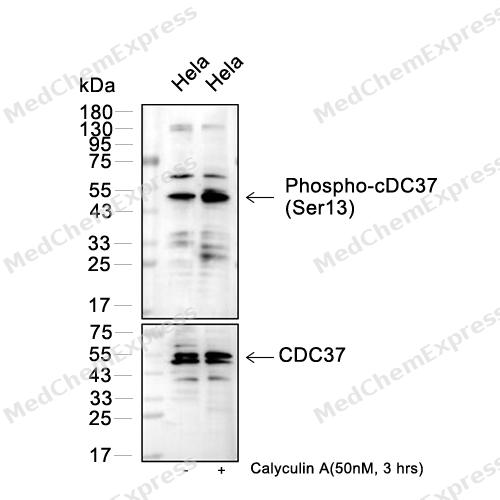 Western blot analysis of extracts from Hela(lane 2(20μg) ) and Hela(lane 3(20μg), Calyculin A treated (HY-18983, 50nm, 3hrs) as indicated, using Phospho-CDC37 (Ser13) (HY-P80962) Rabbit mAb. Proteins were transferred to a PVDF membrane and blocked with 5% BSA in TBST for 2 hour at room temperature. The primary antibody (HY-P80962, 1/1000) and Loading control antibody (CDC37, HY-P80960, 1/1000) was used in 5% BSA in TBST at 4°C overnight. Goat Anti-Rabbit/Mouse IgG-HRP Secondary Antibody (HY-P8001/HY-P8004, 1/10,000) was used for 1 hour at room temperature.
 -
 Western blot analysis was performed on protein extracts (25 μg) from HeLa (lane 2), 293T (lane 3), MCF-7 (lane 4) and RAW264.7 (lane 5) using CDC37 antibody. Proteins were transferred onto a 0.45 μm PVDF membrane using the Trans-Blot® Turbo™ system for 13 min. The membrane was then blocked with 5% nonfat milk in TBST (HY-K1025) for 1 h at room temperature. The primary antibody (1:1000) and loading control antibody GAPDH Antibody (HRP) (HY-P80954A) (1:5000) were diluted in 5% nonfat milk in TBST and incubated with the membrane overnight at 4°C. After washing, the membrane of primary antibody was incubated with HRP-conjugated goat anti-rabbit/mouse IgG secondary antibody (HY-P8001/HY-P8004) (1:5000) diluted in 5% nonfat milk in TBST for 1 h at room temperature. Protein bands were visualized using an Ultra High Sensitivity ECL detection kit (HY-K1005).
 -
 Flow cytometric analysis of 1X10⁶ HeLa cells labeling CDC37 Antibody (HY-P80960, red). Cells were fixed with 4% paraformaldehyde and permeabilised with 90% methanol. Then stained with the primary antibody at 1/100 dilution for an hour at 4℃. AF488-conjugated Goat Anti-Rabbit IgG H&L (HY-P8002) was used as the secondary antibody at 1/1,000 dilution for 30 minutes at 4℃. Rabbit IgG Isotype Control (HY-P80879, blue) was used as the isotype control, cells without incubation with primary antibody were used as the unlabeled control (black).


Background
-
Function
CDC37 is a Co-chaperone that binds to numerous kinases and promotes their interaction with the Hsp90 complex, resulting in stabilization and promotion of their activity. Inhibits HSP90AA1 ATPase activity[1][2].
-
Subcellular Localization
Cytoplasm
-
Subunit
Probably forms a complex composed of chaperones HSP90 and HSP70, co-chaperones STIP1/HOP, CDC37, PPP5C, PTGES3/p23, TSC1 and client protein TSC2 (PubMed:29127155). Probably forms a complex composed of chaperones HSP90 and HSP70, co-chaperones CDC37, PPP5C, TSC1 and client protein TSC2, CDK4, AKT, RAF1 and NR3C1; this complex does not contain co-chaperones STIP1/HOP and PTGES3/p23 (PubMed:29127155). Forms a complex with Hsp90/HSP90AB1 and CDK6 (PubMed:9482106). Interacts with HSP90AA1 (PubMed:23569206, PubMed:27353360). Interacts with AR, CDK4, CDK6 and EIF2AK1 (PubMed:11036079, PubMed:11085988, PubMed:9150368, PubMed:9482106). Interacts with RB1 (By similarity). Interacts with KSR1 (PubMed:10409742). Interacts with FLCN, FNIP1 and FNIP2 (PubMed:27353360)
-
SwissProt ID
-
別名
CDC37A, CDC37, Hsp90 co-chaperone Cdc37, Hsp90 chaperone protein kinase-targeting subunit, p50Cdc37


ドキュメント
-
データシート (234 KB)
-
SDS (252 KB)
- English - EN (252 KB)
- Français - FR (252 KB)
- Deutsch - DE (252 KB)
- Norwegian - NO (252 KB)
- Español - ES (252 KB)
- Swedish - SV (252 KB)
- Italian - IT (252 KB)
- Korean - KR (252 KB)
- Portuguese - PT (252 KB)
-
抗体ユーザーガイドラインです (1077 KB)

 Powered by Bioz

