



HSF1 Antibody (YA896)
(Synonyms: HSF1; HSTF1; Heat shock factor protein 1; HSF 1; Heat shock transcription factor 1; HSTF 1)
Based on 1 Customer Validation
HSF1 Antibody (YA896) is a Rabbit-derived and non-conjugated IgG monoclonal antibody, targeting to HSF1.
-
Host:
Rabbit
-
Isotype:
IgG
-
Application:
WB, IHC-P, ICC/IF, IP, FC
-
Reactivity :
Human
-
Formulation:
Supplied in 50mM Tris-Glycine(pH 7.4), 0.15M NaCl, 40%Glycerol, 0.01% sodium azide and 0.05% BSA.
-
Conjugation:
Non-conjugated


To place orders, for customer services and technical support, please contact: MedChemExpress USA
Tel: 609-228-6898 E-mail: [email protected] [email protected]





Applications
| Application |
WB

WB: Western Blot
|
IHC-P
IHC-P: Immunohistochemistry-Paraffin
|
ICC/IF
ICC/IF: Immunocytochemistry/
Immunofluorescence |
IP
IP: Immunoprecipitation
|
FC
FC: Flow Cytometry
|
|---|---|---|---|---|---|
| Dilution Ratio | 1:500-1:1000 | 1:50-1:100 | 1:50-1:200 | 1:50 | 1:50-1:100 |


Product Details
HSF1 Antibody (YA896) is a Rabbit-derived and non-conjugated IgG monoclonal antibody, targeting to HSF1.
-
Host Rabbit

-
Clonality Recombinant,Monoclonal

-
Species ReactivityHuman
-
Observed Molecular WeightObserved band size: 80 kDa
Note: Due to possible protein modifications or aggregation, the molecular weight should be confirmed by actual measurement, and the predicted value is for reference only.

-
Calculated Molecular Weight Predicted band size: 57 kDa

A synthesized peptide derived from human HSF1 aa485-529.
Endogenous
Affinity Chromatography
Non-conjugated
Unmodified
IgG


Product Properties
-
Appearance
Solution
-
Formulation
Supplied in 50mM Tris-Glycine(pH 7.4), 0.15M NaCl, 40%Glycerol, 0.01% sodium azide and 0.05% BSA.
-
Concentration
Batch-dependent, Please check the COA for the concentration of each lot. Check Lot Concentration
-
Storage & Stability
Stored at -20°C for 1 year. Avoid repeated freeze / thaw cycles.
-
Shipping
Shipping with blue ice.


Verification Images
-
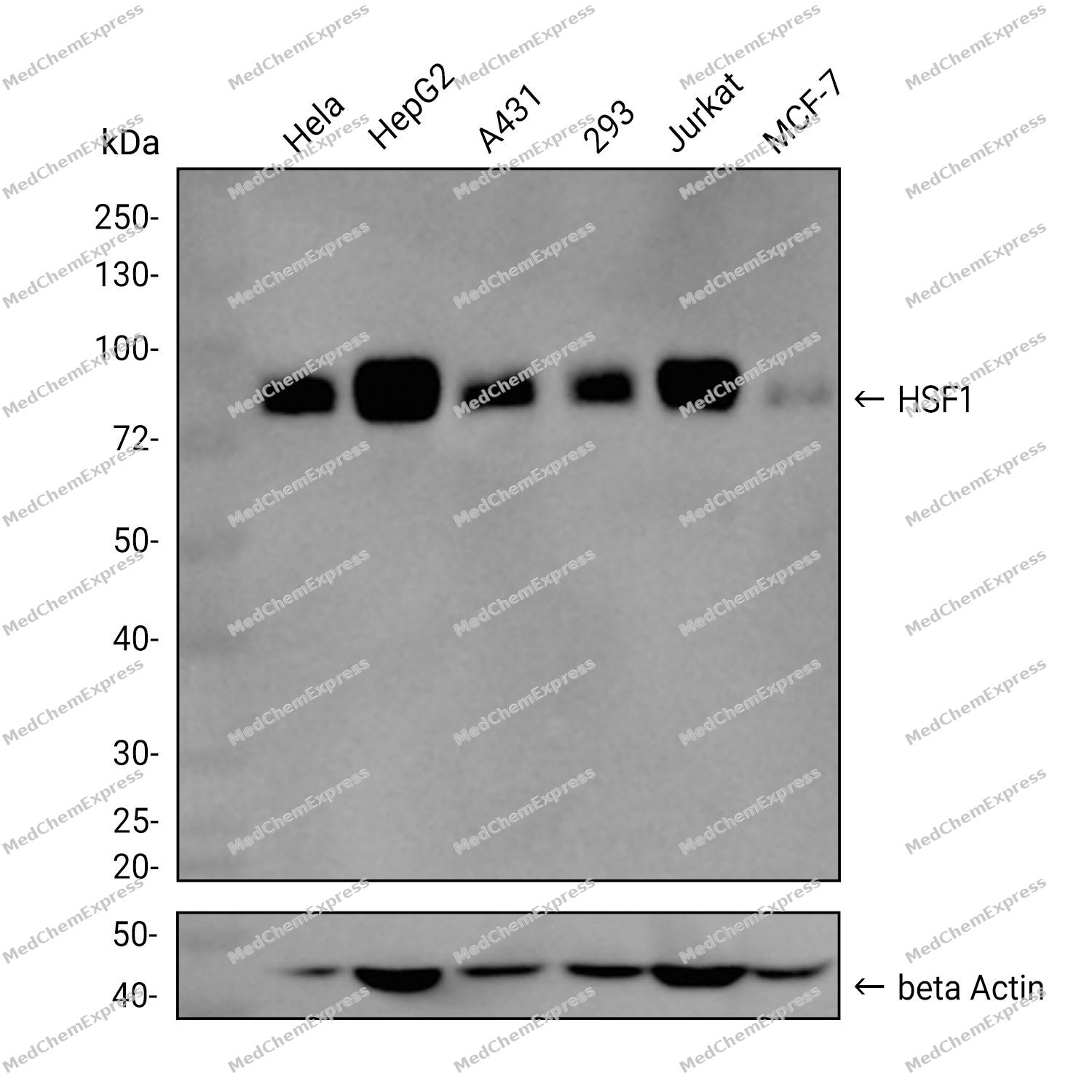
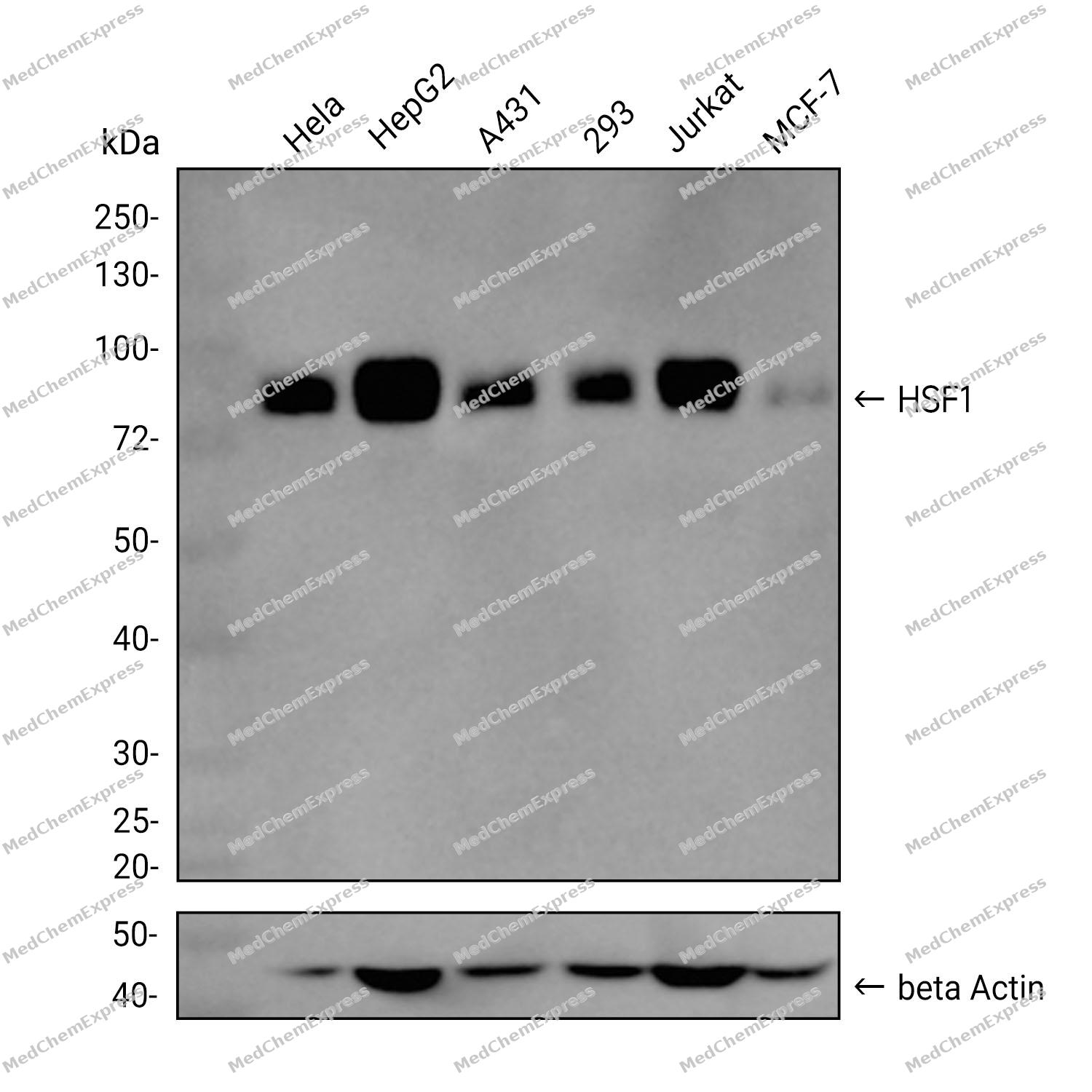 Western blot analysis was performed on extracts from Hela (lane 1, 15 μg), HepG2 (lane 2, 15 μg), A431 (lane 3, 15 μg), 293 (lane 4, 15 μg), Jurkat (lane 5, 15 μg), and MCF-7 (lane 6, 15 μg) using HSF1 Rabbit mAb.Proteins were transferred to a PVDF membrane and blocked with 5% non-fat milk in TBST at 4°C overnight.The primary antibody (1:1000 dilution) and the loading control antibody (beta-Actin, HY-P80438, 1:20000 dilution) were incubated in 5% non-fat milk in TBST for 1 hour at 37°C.Goat Anti-Rabbit IgG-HRP Secondary Antibody (1:20000 dilution) was then applied for 40 minutes at 37°C.
 -
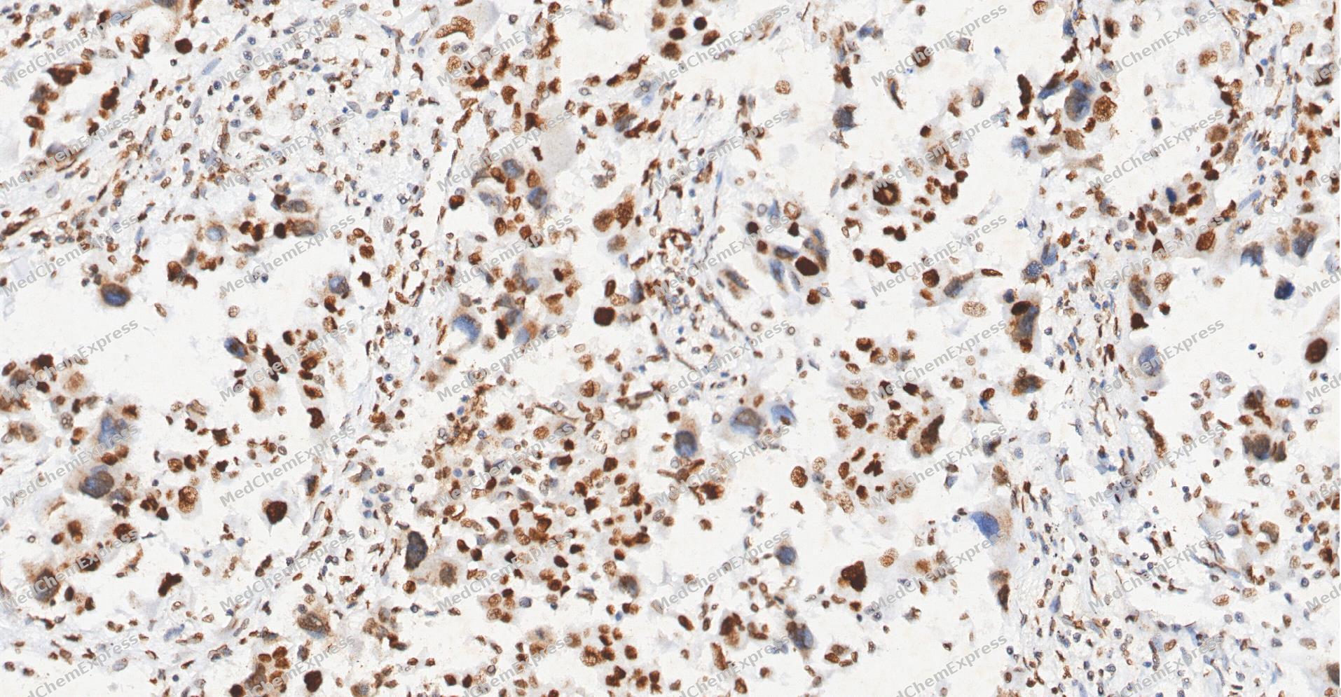 Immunohistochemical analysis of paraffin-embedded human Lung Adenocarcinoma tissue using HSF1 antibody was performed. The section was pretreated using high-temperature and high-pressure mediated EDTA antigen retrieval buffer (pH 9.0), for 5 minutes. The tissues were incubated with primary antibody (HY-P81052, 1:100 dilution) at room temperature for 60 minutes. Detection was performed using an HRP conjugated compact polymer system. DAB was used as the chromogen. The tissues were counterstained with hematoxylin and mounted with neutral balsam mounting medium.
 -
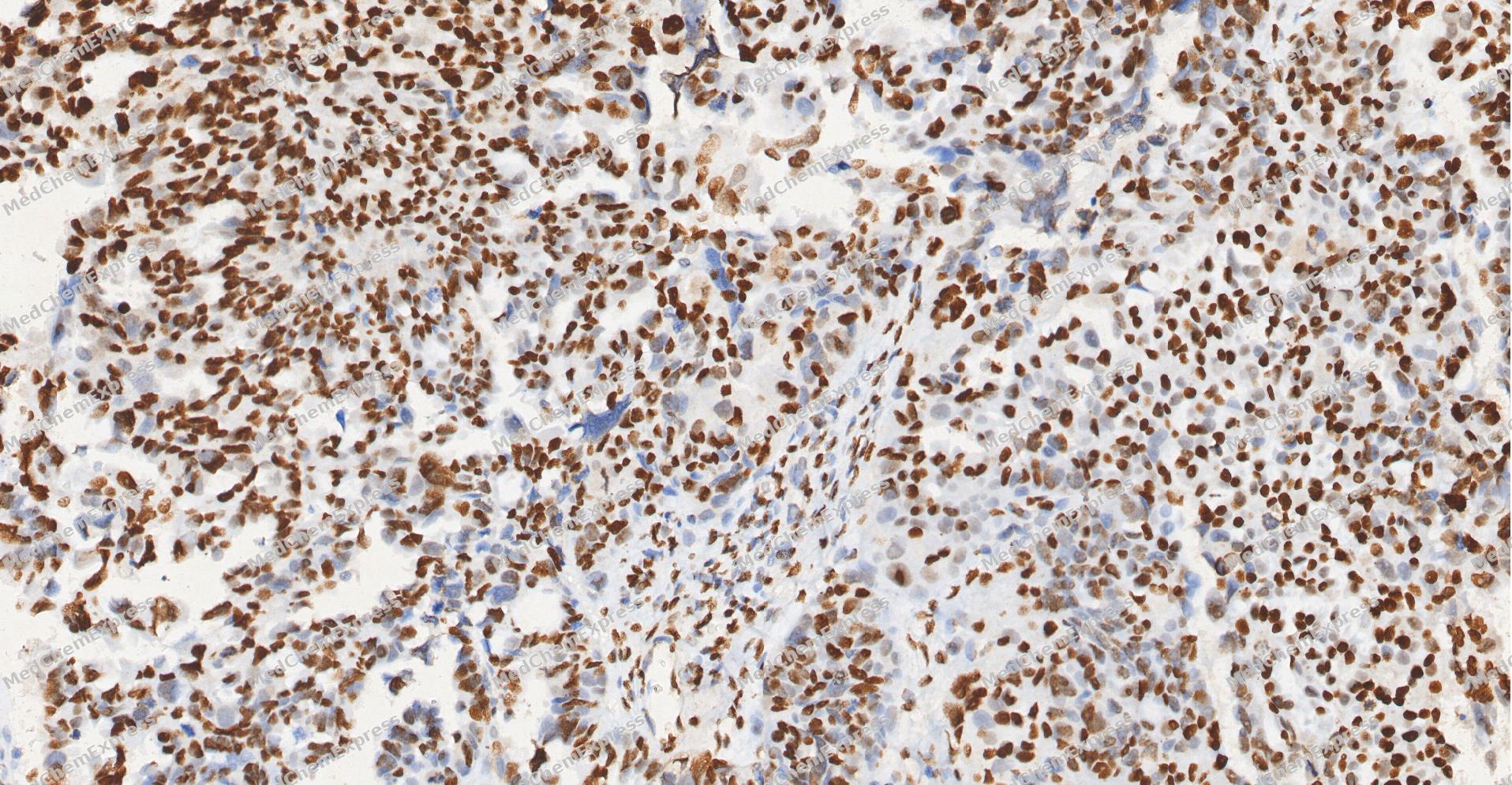 Immunohistochemical analysis of paraffin-embedded human Ovarian Cancer tissue using HSF1 antibody was performed. The section was pretreated using high-temperature and high-pressure mediated EDTA antigen retrieval buffer (pH 9.0), for 5 minutes. The tissues were incubated with primary antibody (HY-P81052, 1:100 dilution) at room temperature for 60 minutes. Detection was performed using an HRP conjugated compact polymer system. DAB was used as the chromogen. The tissues were counterstained with hematoxylin and mounted with neutral balsam mounting medium.
 -
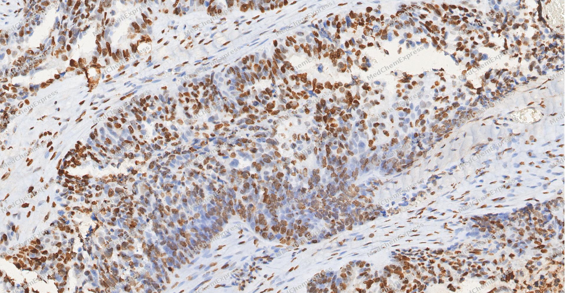 Immunohistochemical analysis of paraffin-embedded human Ovarian Cancer tissue using HSF1 antibody was performed. The section was pretreated using high-temperature and high-pressure mediated EDTA antigen retrieval buffer (pH 9.0), for 5 minutes. The tissues were incubated with primary antibody (HY-P81052, 1:100 dilution) at room temperature for 60 minutes. Detection was performed using an HRP conjugated compact polymer system. DAB was used as the chromogen. The tissues were counterstained with hematoxylin and mounted with neutral balsam mounting medium.
 -
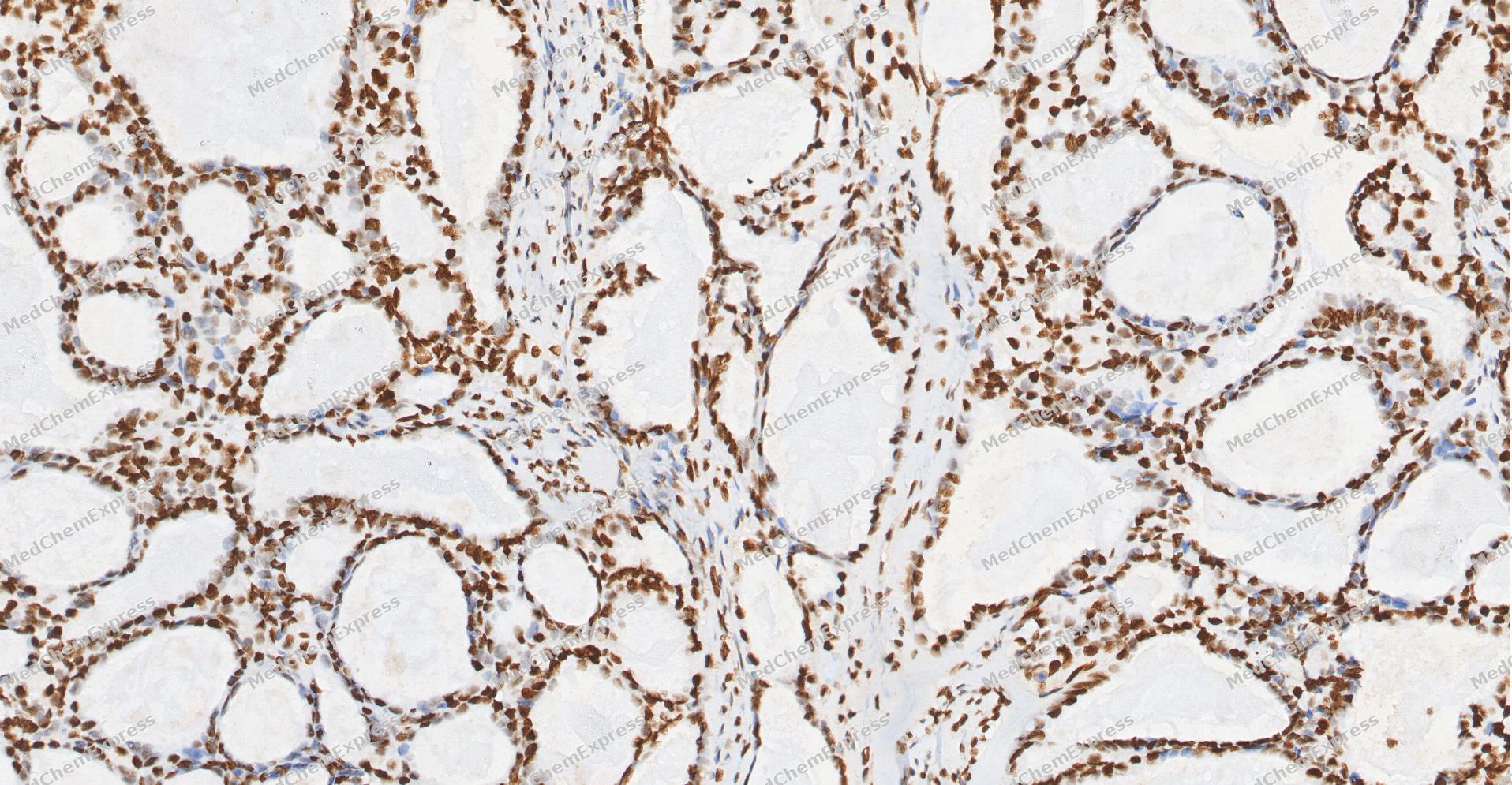 Immunohistochemical analysis of paraffin-embedded human Ovarian Cancer tissue using HSF1 antibody was performed. The section was pretreated using high-temperature and high-pressure mediated EDTA antigen retrieval buffer (pH 9.0), for 5 minutes. The tissues were incubated with primary antibody (HY-P81052, 1:100 dilution) at room temperature for 60 minutes. Detection was performed using an HRP conjugated compact polymer system. DAB was used as the chromogen. The tissues were counterstained with hematoxylin and mounted with neutral balsam mounting medium.
 -
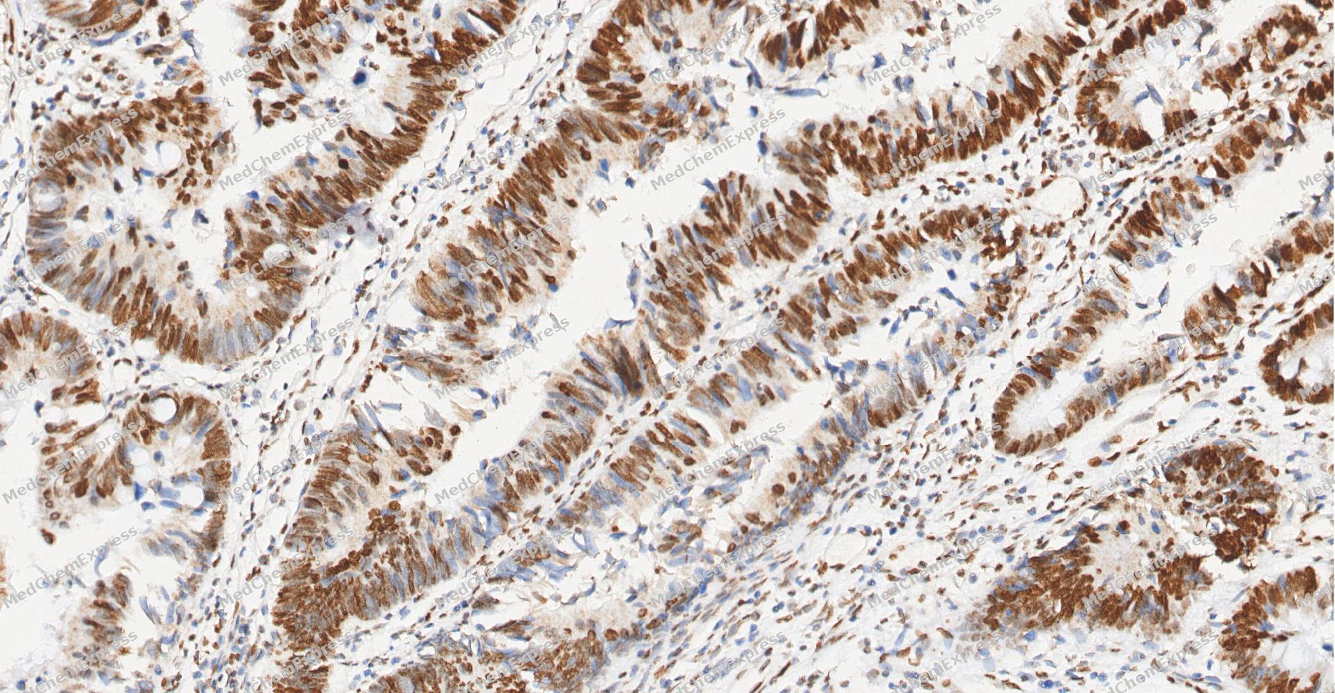 Immunohistochemical analysis of paraffin-embedded human Colon cancer tissue using HSF1 antibody was performed. The section was pretreated using high-temperature and high-pressure mediated EDTA antigen retrieval buffer (pH 9.0), for 5 minutes. The tissues were incubated with primary antibody (HY-P81052, 1:100 dilution) at room temperature for 60 minutes. Detection was performed using an HRP conjugated compact polymer system. DAB was used as the chromogen. The tissues were counterstained with hematoxylin and mounted with neutral balsam mounting medium.
 -
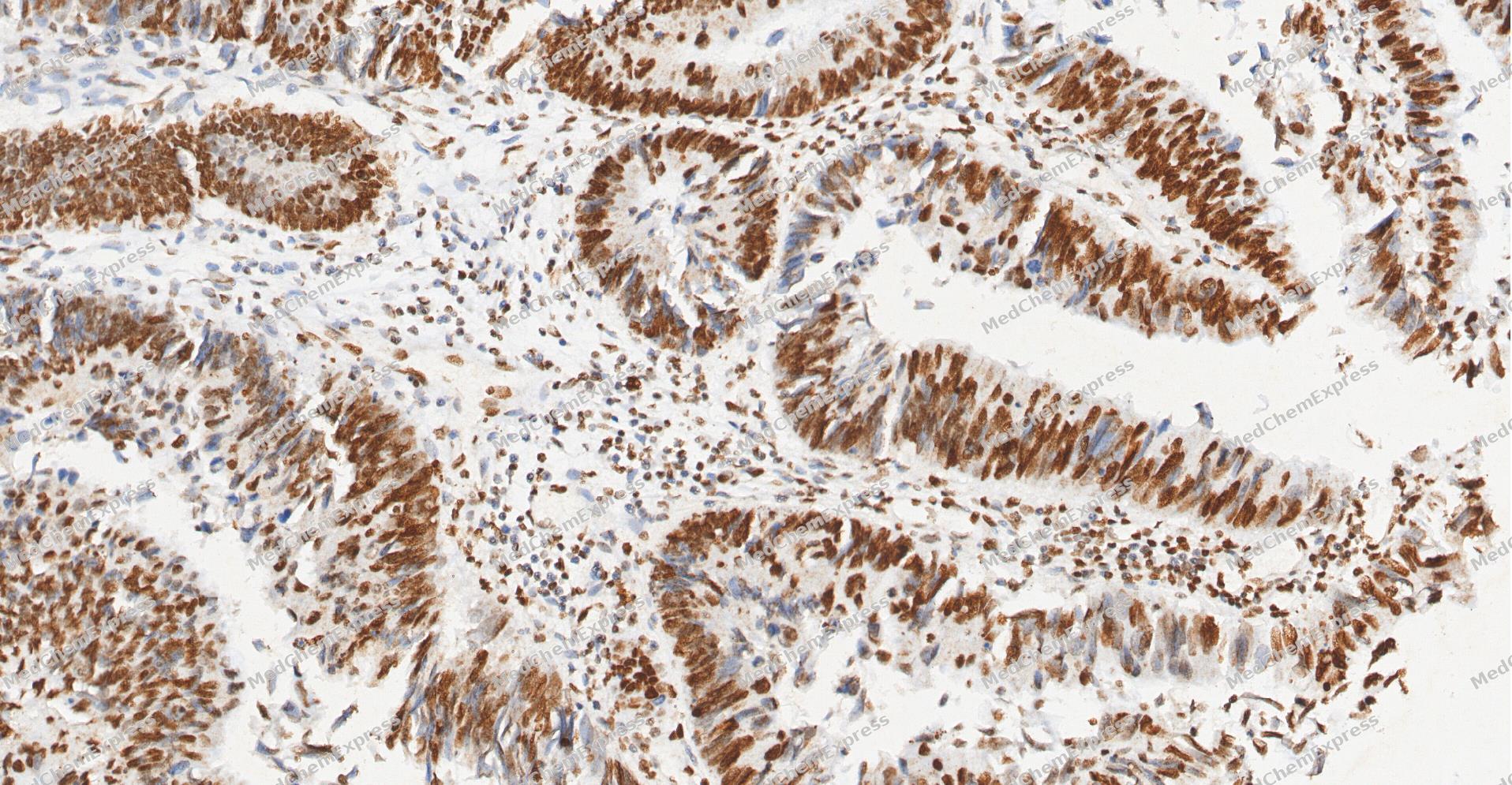 Immunohistochemical analysis of paraffin-embedded human Colon cancer tissue using HSF1 antibody was performed. The section was pretreated using high-temperature and high-pressure mediated EDTA antigen retrieval buffer (pH 9.0), for 5 minutes. The tissues were incubated with primary antibody (HY-P81052, 1:100 dilution) at room temperature for 60 minutes. Detection was performed using an HRP conjugated compact polymer system. DAB was used as the chromogen. The tissues were counterstained with hematoxylin and mounted with neutral balsam mounting medium.
 -
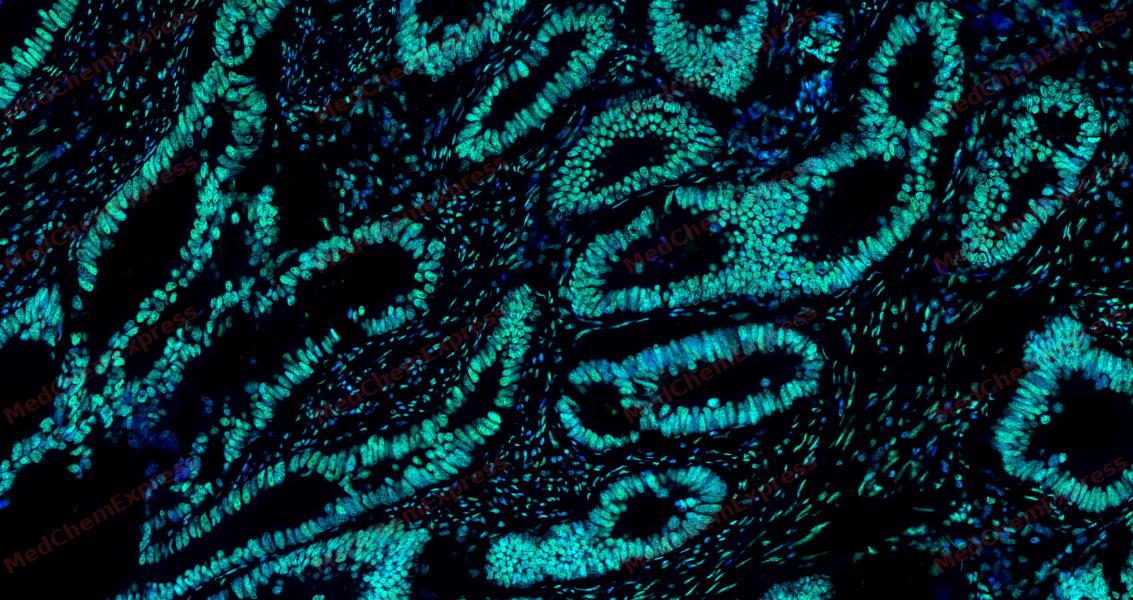 Tyramide signaling amplification based immunofluorescence was performed on paraffin-embedded human Colon cancer tissue using HSF1 antibody. Antigen retrieval was performed in EDTA buffer pH 9.0 (95 °C, 20 min) followed by cooling to RT. Then incubated with primary antibody (HY-P81052, 1:300 dilution) at room temperature for 60 minutes and HRP conjugated secondary antibody for 10 minutes. Fluorescence was then developed with Vari Fluor 532 TSA (200×)(HY-D1832). The tissues were counterstained with DAPI and mounted with Anti-fade mounting medium.
 -
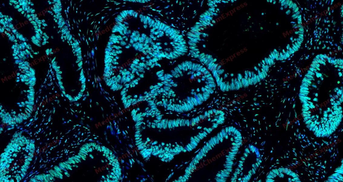 Tyramide signaling amplification based immunofluorescence was performed on paraffin-embedded human Colon cancer tissue using HSF1 antibody. Antigen retrieval was performed in EDTA buffer pH 9.0 (95 °C, 20 min) followed by cooling to RT. Then incubated with primary antibody (HY-P81052, 1:300 dilution) at room temperature for 60 minutes and HRP conjugated secondary antibody for 10 minutes. Fluorescence was then developed with Vari Fluor 532 TSA (200×)(HY-D1832). The tissues were counterstained with DAPI and mounted with Anti-fade mounting medium.
 -
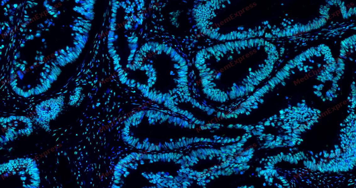 Tyramide signaling amplification based immunofluorescence was performed on paraffin-embedded human Colon cancer tissue using HSF1 antibody. Antigen retrieval was performed in EDTA buffer pH 9.0 (95 °C, 20 min) followed by cooling to RT. Then incubated with primary antibody (HY-P81052, 1:300 dilution) at room temperature for 60 minutes and HRP conjugated secondary antibody for 10 minutes. Fluorescence was then developed with Vari Fluor 532 TSA (200×)(HY-D1832). The tissues were counterstained with DAPI and mounted with Anti-fade mounting medium.
 -
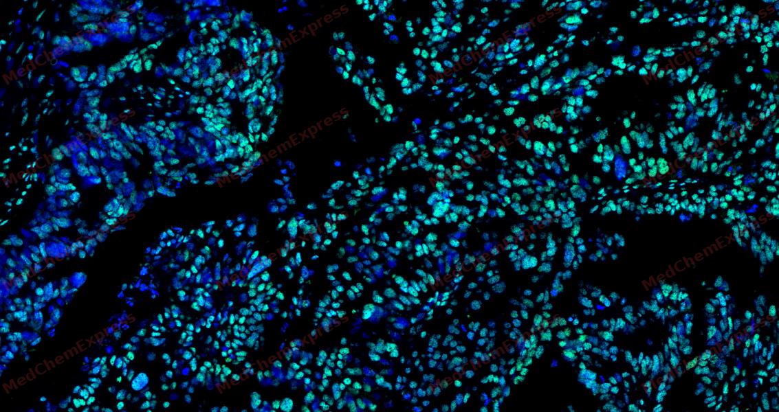 Tyramide signaling amplification based immunofluorescence was performed on paraffin-embedded human ovarian carcinoma tissue using HSF1 antibody. Antigen retrieval was performed in EDTA buffer pH 9.0 (95 °C, 20 min) followed by cooling to RT. Then incubated with primary antibody (HY-P81052, 1:300 dilution) at room temperature for 60 minutes and HRP conjugated secondary antibody for 10 minutes. Fluorescence was then developed with Vari Fluor 532 TSA (200×)(HY-D1832). The tissues were counterstained with DAPI and mounted with Anti-fade mounting medium.
 -
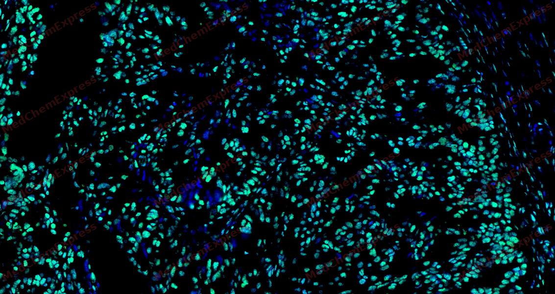 Tyramide signaling amplification based immunofluorescence was performed on paraffin-embedded human ovarian carcinoma tissue using HSF1 antibody. Antigen retrieval was performed in EDTA buffer pH 9.0 (95 °C, 20 min) followed by cooling to RT. Then incubated with primary antibody (HY-P81052, 1:300 dilution) at room temperature for 60 minutes and HRP conjugated secondary antibody for 10 minutes. Fluorescence was then developed with Vari Fluor 532 TSA (200×)(HY-D1832). The tissues were counterstained with DAPI and mounted with Anti-fade mounting medium.
 -
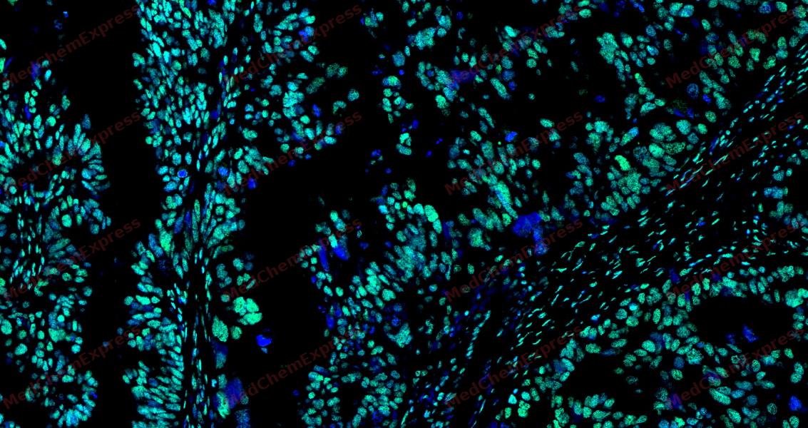 Tyramide signaling amplification based immunofluorescence was performed on paraffin-embedded human ovarian carcinoma tissue using HSF1 antibody. Antigen retrieval was performed in EDTA buffer pH 9.0 (95 °C, 20 min) followed by cooling to RT. Then incubated with primary antibody (HY-P81052, 1:300 dilution) at room temperature for 60 minutes and HRP conjugated secondary antibody for 10 minutes. Fluorescence was then developed with Vari Fluor 532 TSA (200×)(HY-D1832). The tissues were counterstained with DAPI and mounted with Anti-fade mounting medium.


Background
-
Function
Heat Shock Factor 1 (HSF1) functions as a master transcriptional regulator, maintaining cellular proteostasis under stress conditions[1][2]. Mechanistically, HSF1 activation induces heat shock proteins (HSPs), which assist in protein folding, prevent misfolding, and modulate apoptosis pathways[3][2][1]. In metabolic contexts, HSF1 regulates lipid homeostasis through the HSF1-CPT1a axis, influencing hepatic fat accumulation in non-alcoholic fatty liver disease (NAFLD) models[4]. Compared with other isoforms, HSF1 specifically activates transcriptional networks distinct from HSP-independent stress responses, including the FOXO3a-ΔNp63α-CDK4 axis in head and neck squamous cell carcinoma (HNSCC) [5]. In pediatric hepatoblastoma, elevated HSF1 correlates with tumor aggressiveness and decreased differentiation, while its inhibition induces apoptosis and reduces tumor growth[6]. Pharmacological modulation of HSF1, either through activators or inhibitors, demonstrates potential for experimental applications: inhibitors such as KRIBB11 or PU-H71 can suppress HSF1-dependent pathways, decreasing proliferation in cancer models, whereas HSF1 activation enhances inducible HSP expression in neurodegeneration studies[2][1][5][6]. The differential roles of HSF1 in disease versus normal physiology underscore its value as a target for isoform-specific drug development, enabling selective modulation of stress response pathways without broadly affecting related transcription factors[1][2][7]. Therefore, HSF1 serves as both a critical mechanistic node in cellular stress adaptation and a strategic intervention point for disease modeling and therapeutic exploration[1][5][6].
-
Subcellular Localization
Nucleus; Cytoplasm; Nucleus, nucleoplasm; Cytoplasm, perinuclear region; Cytoplasm, cytoskeleton, spindle pole; Cytoplasm, cytoskeleton, microtubule organizing center, centrosome; Chromosome, centromere, kinetochore
-
Isoforms & Post-Translational Modification
Q00613 has 2 isomers: Q00613-1: 57260 Da (predicted); Q00613-2: 52881 Da (predicted).
Phosphorylated (PubMed:10359787, PubMed:11583998, PubMed:26159920, PubMed:9499401). Phosphorylated in unstressed cells; this phosphorylation is constitutive and implicated in the repression of HSF1 transcriptional activity (PubMed:16278218, PubMed:8940068, PubMed:8946918, PubMed:9121459). Phosphorylated on Ser-121 by MAPKAPK2; this phosphorylation promotes interaction with HSP90 proteins and inhibits HSF1 homotrimerization, DNA-binding and transactivation activities (PubMed:16278218). Phosphorylation on Ser-303 by GSK3B/GSK3-beta and on Ser-307 by MAPK3 within the regulatory domain is involved in the repression of HSF1 transcriptional activity and occurs in a RAF1-dependent manner (PubMed:10747973, PubMed:12646186, PubMed:8940068, PubMed:8946918, PubMed:9121459, PubMed:9535852). Phosphorylation on Ser-303 and Ser-307 increases HSF1 nuclear export in a YWHAE- and XPO1/CRM1-dependent manner (PubMed:12917326). Phosphorylation on Ser-307 is a prerequisite for phosphorylation on Ser-303 (PubMed:8940068). According to PubMed:9535852, Ser-303 is not phosphorylated in unstressed cells. Phosphorylated on Ser-419 by PLK1; phosphorylation promotes nuclear translocation upon heat shock (PubMed:15661742). Hyperphosphorylated upon heat shock and during the attenuation and recovery phase period of the heat shock response (PubMed:11447121, PubMed:12659875, PubMed:24581496). Phosphorylated on Thr-142; this phosphorylation increases HSF1 transactivation activity upon heat shock (PubMed:12659875). Phosphorylation on Ser-230 by CAMK2A; this phosphorylation enhances HSF1 transactivation activity upon heat shock (PubMed:11447121). Phosphorylation on Ser-326 by MAPK12; this phosphorylation enhances HSF1 nuclear translocation, homotrimerization and transactivation activities upon heat shock (PubMed:15760475, PubMed:27354066). Phosphorylated on Ser-320 by PRKACA/PKA; this phosphorylation promotes nuclear localization and transcriptional activity upon heat shock (PubMed:21085490). Phosphorylated on Ser-363 by MAPK8; this phosphorylation occurs upon heat shock, induces HSF1 translocation into nuclear stress bodies and negatively regulates transactivation activity (PubMed:10747973). Neither basal nor stress-inducible phosphorylation on Ser-230, Ser-292, Ser-303, Ser-307, Ser-314, Ser-319, Ser-320, Thr-323, Ser-326, Ser-338, Ser-344, Ser-363, Thr-367, Ser-368 and Thr-369 within the regulatory domain is involved in the regulation of HSF1 subcellular localization or DNA-binding activity; however, it negatively regulates HSF1 transactivation activity (PubMed:25963659). Phosphorylated on Ser-216 by PLK1 in the early mitotic period; this phosphorylation regulates HSF1 localization to the spindle pole, the recruitment of the SCF(BTRC) ubiquitin ligase complex inducing HSF1 degradation, and hence mitotic progression (PubMed:18794143). Dephosphorylated on Ser-121, Ser-307, Ser-314, Thr-323 and Thr-367 by phosphatase PPP2CA in an IER5-dependent manner, leading to HSF1-mediated transactivation activity (PubMed:26754925);Sumoylated with SUMO1 and SUMO2 upon heat shock in a ERK2-dependent manner (PubMed:12646186, PubMed:12665592). Sumoylated by SUMO1 on Lys-298; sumoylation occurs upon heat shock and promotes its localization to nuclear stress bodies and DNA-binding activity (PubMed:11514557). Phosphorylation on Ser-303 and Ser-307 is probably a prerequisite for sumoylation (PubMed:12646186, PubMed:12665592);Acetylated on Lys-118; this acetylation is decreased in a IER5-dependent manner (PubMed:26754925). Acetylated on Lys-118, Lys-208 and Lys-298; these acetylations occur in a EP300-dependent manner (PubMed:24581496, PubMed:27189267). Acetylated on Lys-80; this acetylation inhibits DNA-binding activity upon heat shock (PubMed:19229036). Deacetylated on Lys-80 by SIRT1; this deacetylation increases DNA-binding activity (PubMed:19229036);Ubiquitinated by SCF(BTRC) and degraded following stimulus-dependent phosphorylation at Ser-216 by PLK1 in mitosis (PubMed:18794143). Polyubiquitinated (PubMed:24581496). Undergoes proteasomal degradation upon heat shock and during the attenuation and recovery phase period of the heat shock response (PubMed:24581496) -
Subunit
Monomer; cytoplasmic latent and transcriptionally inactive monomeric form in unstressed cells (PubMed:11583998, PubMed:7623826, PubMed:7935376, PubMed:7935471, PubMed:8455624, PubMed:9222587, PubMed:9727490). Homotrimer; in response to stress, such as heat shock, homotrimerizes and translocates into the nucleus, binds to heat shock element (HSE) sequences in promoter of heat shock protein (HSP) genes and acquires transcriptional ability (PubMed:11583998, PubMed:26727489, PubMed:26754925, PubMed:7623826, PubMed:7935471, PubMed:8455624, PubMed:9222587, PubMed:9727490). Interacts (via monomeric form) with FKBP4; this interaction occurs in unstressed cells (PubMed:11583998). Associates (via monomeric form) with HSP90 proteins in a multichaperone complex in unnstressed cell; this association maintains HSF1 in a non-DNA-binding and transcriptional inactive form by preventing HSF1 homotrimerization (PubMed:11583998, PubMed:15661742, PubMed:16278218, PubMed:9727490). Homotrimeric transactivation activity is modulated by protein-protein interactions and post-translational modifications (PubMed:11583998, PubMed:15016915, PubMed:16554823, PubMed:26754925). Interacts with HSP90AA1; this interaction is decreased in a IER5-dependent manner, promoting HSF1 accumulation in the nucleus, homotrimerization and DNA-binding activities (PubMed:26754925). Part (via regulatory domain in the homotrimeric form) of a large heat shock-induced HSP90-dependent multichaperone complex at least composed of FKBP4, FKBP5, HSP90 proteins, PPID, PPP5C and PTGES3; this association maintains the HSF1 homotrimeric DNA-bound form in a transcriptionally inactive form (PubMed:11583998, PubMed:16278218, PubMed:9727490). Interacts with BAG3 (via BAG domain); this interaction occurs in normal and heat-shocked cells promoting nuclear shuttling of HSF1 in a BAG3-dependent manner (PubMed:26159920). Interacts (via homotrimeric and hyperphosphorylated form) with FKBP4; this interaction occurs upon heat shock in a HSP90-dependent multichaperone complex (PubMed:11583998). Interacts (via homotrimeric form preferentially) with EEF1A proteins (PubMed:15016915). In heat shocked cells, stress-denatured proteins compete with HSF1 homotrimeric DNA-bound form for association of the HSP90-dependent multichaperone complex, and hence alleviating repression of HSF1-mediated transcriptional activity (PubMed:11583998). Interacts (via homotrimeric form preferentially) with DAXX; this interaction relieves homotrimeric HSF1 from repression of its transcriptional activity by HSP90-dependent multichaperone complex upon heat shock (PubMed:15016915). Interacts (via D domain and preferentially with hyperphosphorylated form) with JNK1; this interaction occurs under both normal growth conditions and immediately upon heat shock (PubMed:10747973). Interacts (via D domain and preferentially with hyperphosphorylated form) with MAPK3; this interaction occurs upon heat shock (PubMed:10747973). Interacts with IER5 (via central region); this interaction promotes PPP2CA-induced dephosphorylation on Ser-121, Ser-307, Ser-314, Thr-323 and Thr-367 and HSF1 transactivation activity (PubMed:25816751, PubMed:26496226, PubMed:26754925). Found in a ribonucleoprotein complex composed of the HSF1 homotrimeric form, translation elongation factor eEF1A proteins and non-coding RNA heat shock RNA-1 (HSR1); this complex occurs upon heat shock and stimulates HSF1 DNA-binding activity (PubMed:16554823). Interacts (via transactivation domain) with HSPA1A/HSP70 and DNAJB1; these interactions result in the inhibition of heat shock- and HSF1-induced transcriptional activity during the attenuation and recovery phase from heat shock (PubMed:7935376, PubMed:9222587, PubMed:9499401). Interacts (via Ser-303 and Ser-307 phosphorylated form) with YWHAE; this interaction promotes HSF1 sequestration in the cytoplasm in an ERK-dependent manner (PubMed:12917326). Found in a complex with IER5 and PPP2CA (PubMed:26754925). Interacts with TPR; this interaction increases upon heat shock and stimulates export of HSP70 mRNA (PubMed:17897941). Interacts with SYMPK (via N-terminus) and CSTF2; these interactions occur upon heat shock (PubMed:14707147). Interacts (via transactivation domain) with HSPA8 (PubMed:9499401). Interacts with EEF1D; this interaction occurs at heat shock promoter element (HSE) sequences (PubMed:21597468). Interacts with MAPKAPK2 (PubMed:16278218). Interacts with PRKACA/PKA (PubMed:21085490). Interacts (via transactivation domain) with GTF2A2 (PubMed:11005381). Interacts (via transactivation domain) with GTF2B (PubMed:11005381). Interacts (via transactivation domain) with TBP (PubMed:11005381). Interacts with CDK9, CCNT1 and EP300 (PubMed:27189267). Interacts (via N-terminus) with XRCC5 (via N-terminus) and XRCC6 (via N-terminus); these interactions are direct and prevent XRCC5/XRCC6 heterodimeric binding and non-homologous end joining (NHEJ) repair activities induced by ionizing radiation (IR) (PubMed:26359349). Interacts with PLK1; this interaction occurs during the early mitotic period, increases upon heat shock but does not modulate neither HSF1 homotrimerization and DNA-binding activities (PubMed:15661742, PubMed:18794143). Interacts (via Ser-216 phosphorylated form) with CDC20; this interaction occurs in mitosis in a MAD2L1-dependent manner and prevents PLK1-stimulated degradation of HSF1 by blocking the recruitment of the SCF(BTRC) ubiquitin ligase complex (PubMed:18794143). Interacts with MAD2L1; this interaction occurs in mitosis (PubMed:18794143). Interacts with BTRC; this interaction occurs during mitosis, induces its ubiquitin-dependent degradation following stimulus-dependent phosphorylation at Ser-216, a process inhibited by CDC20 (PubMed:18794143). Interacts with HSP90AA1 and HSP90AB1 (PubMed:26517842). Forms a complex with TTC5/STRAP and p300/EP300; these interactions augment chromatin-bound HSF1 and p300/EP300 histone acetyltransferase activity (PubMed:18451878)
-
SwissProt ID
-
Synonyms
HSF1; HSTF1; Heat shock factor protein 1; HSF 1; Heat shock transcription factor 1; HSTF 1
-
Research Field
Tags & Cell Markers


Documentation
-
Data Sheet (262 KB)
-
SDS (251 KB)
- English - EN (251 KB)
- Français - FR (251 KB)
- Deutsch - DE (251 KB)
- Norwegian - NO (251 KB)
- Español - ES (251 KB)
- Swedish - SV (251 KB)
- Italian - IT (251 KB)
- Korean - KR (251 KB)
- Portuguese - PT (251 KB)
-
User Guide for Antibodies (1077 KB)
References
[1]. Breternitz W, et al. The HSF1-CPT1a Pathway Is Differentially Regulated in NAFLD Progression. Cells. 2022 Nov 4;11(21):3504. [Content Brief]
[2]. Kumar S, et al. HSF1-mediated regulation of tumor cell apoptosis: a novel target for cancer therapeutics. Future Oncol. 2013 Oct;9(10):1573-86. [Content Brief]
[3]. Fernandez-Funez P, et al. Launching Hsp70 neuroprotection: two drugs better than one. Cell Cycle. 2014;13(11):1657-8. [Content Brief]
[4]. Meng F, et al. Drug design targeting active posttranslational modification protein isoforms. Med Res Rev. 2021 May;41(3):1701-1750. [Content Brief]
[5]. Chin Y, et al. Targeting HSF1 for cancer treatment: mechanisms and inhibitor development. Theranostics. 2023 Apr 17;13(7):2281-2300. [Content Brief]
[6]. Wang Y, et al. HSF1 activates the FOXO3a-ΔNp63α-CDK4 axis to promote head and neck squamous cell carcinoma cell proliferation and tumour growth. FEBS Lett. 2023 Apr;597(8):1125-1137. [Content Brief]
[7]. Hurley EH, et al. Inhibition of Heat Shock Factor 1 Signaling Decreases Hepatoblastoma Growth via Induction of Apoptosis. Am J Pathol. 2023 Feb;193(2):148-160. [Content Brief]

 Powered by Bioz
