



IRF3 Antibody (YA334)
(Synonyms: Interferon regulatory factor 3, IRF-3, IRF3)Based on 1 publication(s) in Google Scholar
IRF3 Antibody (YA334) is a Rabbit-derived and non-conjugated IgG monoclonal antibody, targeting to IRF3.
-
Host:
Rabbit
-
Isotype:
IgG
-
Application:
WB, ICC/IF, IHC-P, FC
-
Reactivity :
Human, Mouse, Rat
-
Formulation:
Supplied in 1*TBS (pH7.4), 0.05% BSA and 40% Glycerol. Preservative: 0.05% Sodium Azide.
-
Conjugation:
Non-conjugated




To place orders, for customer services and technical support, please contact: MedChemExpress USA
Tel: 609-228-6898 E-mail: [email protected] [email protected]



-

HY-P80519
TBK1 Antibody (YA662) -
HY-P80910
TBK1 Antibody (YA040) -
HY-P80940
STING Antibody -
HY-P87745
Phospho-IRF3 (Ser396) Antibody (YA7430) -
HY-P80350
STING Antibody (YA048) -
HY-P80839
Phospho-NF-KB p65 (Ser536) Antibody -
HY-P8003
AF594-conjugated Goat Anti-Rabbit IgG H&L -
HY-P80245
NF-KB p65 Antibody (YA267) -
HY-P87729
Phospho-TBK1 (Ser172) Antibody (YA7414) -
HY-P80783A
PGC1 alpha Antibody (YA7180)
Publications Citing Use of MedChemExpress (MCE) IRF3 Antibody (YA334)
More

Applications
| Application |
WB

WB: Western Blot
|
ICC/IF
ICC/IF: Immunocytochemistry/
Immunofluorescence |
IHC-P
IHC-P: Immunohistochemistry-Paraffin
|
FC
FC: Flow Cytometry
|
|---|---|---|---|---|
| Dilution Ratio | 1:500-1:5000 | 1:50-1:200 | 1:50-1:200 | 1:50-1:100 |


Product Details
IRF3 Antibody (YA334) is a Rabbit-derived and non-conjugated IgG monoclonal antibody, targeting to IRF3.
-
Host Rabbit

-
Clonality Recombinant,Monoclonal

-
Species ReactivityHuman, Mouse, Rat
-
Observed Molecular WeightObserved band size: 55 kDa
Note: Due to possible protein modifications or aggregation, the molecular weight should be confirmed by actual measurement, and the predicted value is for reference only.

-
Calculated Molecular Weight Predicted band size: 47 kDa

Synthetic peptide corresponding to Human IRF3.AA range:71-120.
Endogenous
Protein A affinity purified.
Non-conjugated
Unmodified
IgG


Product Properties
-
Appearance
Solution
-
Formulation
Supplied in 1*TBS (pH7.4), 0.05% BSA and 40% Glycerol. Preservative: 0.05% Sodium Azide.
-
Concentration
Batch-dependent, Please check the COA for the concentration of each lot. Check Lot Concentration
-
Storage & Stability
Stored at -20°C for 1 year. Avoid repeated freeze / thaw cycles.
-
Shipping
Shipping with blue ice.


Publications (1)
-
Journal Impact Factor
-
Most Recent
-
J Nanobiotechnology
The STING-activating nanofactory relieves T cell exhaustion in Mn-based tumor immunotherapy by regulating mitochondrial dysfunction. [Abstract]2025 May 31;23(1):403. PMID: 40450344


Verification Images
-
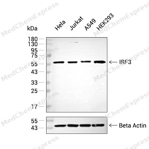 Western blot analysis of extracts from Hela (lane 2(20μg) , Jurkat (lane 3(20μg), A549 (lane 4(20μg) and HEK293(lane 5(20μg) using IRF3(HY-P80504) Rabbit mAb. Proteins were transferred to a PVDF membrane and blocked with 5% non-fat milk in TBST for 2 hour at room temperature. The primary antibody (1/1000) and Loading control antibody (Beta Actin, HY-P80438, 1/10000) was used in 5% non-fat milk in TBST at 4°C overnight. Goat Anti-Mouse IgG-HRP Secondary Antibody (1/10000) was used for 1 hour at room temperature.
 -
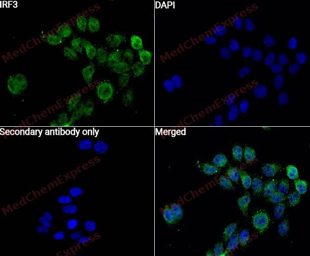 Immunocytochemistry analysis of Hela cells labeling IRF3 with IRF3 Antibody (HY-P80504)at 1/50 dilution. Cells were fixed in 4% paraformaldehyde for 15 minutes at room temperature, permeabilized with 0.1% Triton X-100 for 10 minutes at room temperature, then blocked with QuickBlock™ Blocking Buffer for Immunol Staining for 10 min at room temperature. Cells were then incubated with IRF3 Antibody (HY-P80504) at 1/50 dilution in QuickBlock™ Blocking Buffer for Immunol Staining at 4 ℃. Alexa Fluor® 488-conjugated AffiniPure Goat Anti-Rabbit IgG H&L(HY-P8002, Green) was used as the secondary antibody at 1/1,000 dilution. PBS instead of the primary antibody was used as the secondary antibody only control. The Nuclear counterstain was DAPI (Blue).
 -
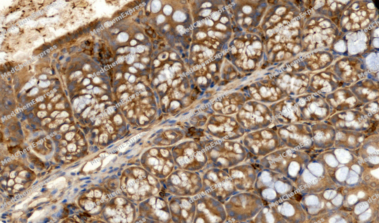 Immunohistochemical analysis of paraffin-embedded Mouse colon tissue using IRF3 Antibody. The section was pre-treated using heat mediated antigen retrieval with sodium citrate buffer (pH 6.0) for 8 minutes. The tissues were blocked in QuickBlock for 20 minutes at room temperature, washed with ddH2O and PBS, and then probed with the primary antibody (HY-P80504, 1/500) in 4℃ overnight. The detection was performed using an HRP conjugated compact polymer system. DAB was used as the chromogen. Tissues were counterstained with hematoxylin and mounted with DPX.
 -
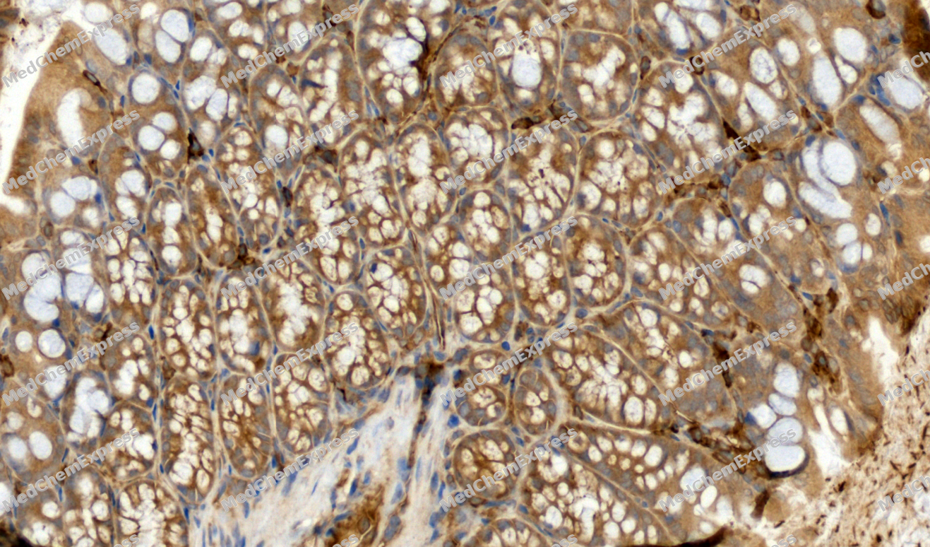
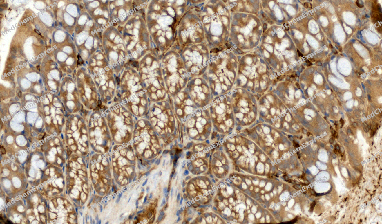 Immunohistochemical analysis of paraffin-embedded Mouse colon tissue using IRF3 Antibody. The section was pre-treated using heat mediated antigen retrieval with sodium citrate buffer (pH 6.0) for 8 minutes. The tissues were blocked in QuickBlock for 20 minutes at room temperature, washed with ddH2O and PBS, and then probed with the primary antibody (HY-P80504, 1/500) in 4℃ overnight. The detection was performed using an HRP conjugated compact polymer system. DAB was used as the chromogen. Tissues were counterstained with hematoxylin and mounted with DPX.
 -
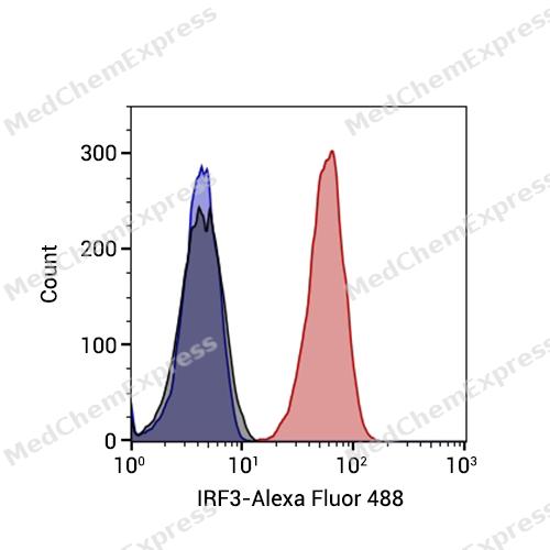 Flow cytometric analysis of 1X10^6 Jurkat cells labeling IRF3 Antibody (HY-P80504, red). Cells were fixed with 4% paraformaldehyde and permeabilised with 90% methanol. Then stained with the primary antibody at 1/1000 dilution for an hour at 4℃. Alexa Fluor® 488-conjugated AffiniPure Goat Anti-Rabbit IgG H&L (HY-P8002) was used as the secondary antibody at 1/1,000 dilution for 30 minutes at 4℃. Rabbit IgG Isotype Control (HY-P80879, blue) was used as the isotype control, cells without incubation with primary antibody were used as the unlabeled control (black).


Background
-
Function
IRF3 is key transcriptional regulator of type I interferon (IFN)-dependent immune responses which plays a critical role in the innate immune response against DNA and RNA viruses. Regulates the transcription of type I IFN genes (IFN-alpha and IFN-beta) and IFN-stimulated genes (ISG) by binding to an interferon-stimulated response element (ISRE) in their promoters. Acts as a more potent activator of the IFN-beta (IFNB) gene than the IFN-alpha (IFNA) gene and plays a critical role in both the early and late phases of the IFNA/B gene induction. Found in an inactive form in the cytoplasm of uninfected cells and following viral infection, double-stranded RNA (dsRNA), or toll-like receptor (TLR) signaling, is phosphorylated by IKBKE and TBK1 kinases. This induces a conformational change, leading to its dimerization and nuclear localization and association with CREB binding protein (CREBBP) to form dsRNA-activated factor 1 (DRAF1), a complex which activates the transcription of the type I IFN and ISG genes. Can activate distinct gene expression programs in macrophages and can induce significant apoptosis in primary macrophages. In response to Sendai virus infection, is recruited by TOMM70:HSP90AA1 to mitochondrion and forms an apoptosis complex TOMM70:HSP90AA1:IRF3:BAX inducing apoptosis. Key transcription factor regulating the IFN response during SARS-CoV-2 infection[1][2][3][4][5][6][7][8][9][10][11][12][13][14][15].
-
Subcellular Localization
Cytoplasm; Nucleus; Mitochondrion
-
Expression
Tissue_specificity:Expressed constitutively in a variety of tissues. -
Subunit
Monomer (PubMed:16846591, PubMed:16979567, PubMed:20049431, PubMed:36603579). Homodimer; phosphorylation-induced (PubMed:22394562, PubMed:25636800, PubMed:26347139, PubMed:36603579).
-
SwissProt ID
-
Synonyms
Interferon regulatory factor 3, IRF-3, IRF3
-
Research Field
Immunology
Documentation
-
Data Sheet (263 KB)
-
SDS (251 KB)
- English - EN (251 KB)
- Français - FR (251 KB)
- Deutsch - DE (251 KB)
- Norwegian - NO (251 KB)
- Español - ES (251 KB)
- Swedish - SV (251 KB)
- Italian - IT (251 KB)
- Korean - KR (251 KB)
- Portuguese - PT (251 KB)
-
User Guide for Antibodies (1077 KB)
References
[1]. Tanaka Y, et al. STING specifies IRF3 phosphorylation by TBK1 in the cytosolic DNA signaling pathway. Sci Signal. 2012 Mar 6;5(214):ra20. [Content Brief]
[2]. Wang S, et al. Herpes simplex virus 1 serine/threonine kinase US3 hyperphosphorylates IRF3 and inhibits beta interferon production. J Virol. 2013 Dec;87(23):12814-27. [Content Brief]
[3]. Liu S, et al. Phosphorylation of innate immune adaptor proteins MAVS, STING, and TRIF induces IRF3 activation. Science. 2015 Mar 13;347(6227):aaa2630. [Content Brief]
[4]. Zhao B, et al. Structural basis for concerted recruitment and activation of IRF-3 by innate immune adaptor proteins. Proc Natl Acad Sci U S A. 2016 Jun 14;113(24):E3403-12. [Content Brief]
[5]. Li D, et al. DDX56 inhibits type I interferon by disrupting assembly of IRF3-IPO5 to inhibit IRF3 nucleus import. J Cell Sci. 2019 Jul 24;133(5):. [Content Brief]
[6]. Yang S, et al. Metabolic enzyme UAP1 mediates IRF3 pyrophosphorylation to facilitate innate immune response. Mol Cell. 2023 Jan 19;83(2):298-313.e8. [Content Brief]
[7]. Au WC, et al. Identification of a member of the interferon regulatory factor family that binds to the interferon-stimulated response element and activates expression of interferon-induced genes. Proc Natl Acad Sci U S A. 1995 Dec 5;92(25):11657-61. [Content Brief]
[8]. Yoneyama M, et al. Control of IRF-3 activation by phosphorylation. J Interferon Cytokine Res. 2002 Jan;22(1):73-6. [Content Brief]
[9]. Solis M, et al. Distinct functions of IRF-3 and IRF-7 in IFN-alpha gene regulation and control of anti-tumor activity in primary macrophages. Biochem Pharmacol. 2006 Nov 30;72(11):1469-76. [Content Brief]
[10]. Honda K, et al. Type I interferon [corrected] gene induction by the interferon regulatory factor family of transcription factors. Immunity. 2006 Sep;25(3):349-60. [Content Brief]
[11]. Savitsky D, et al. Regulation of immunity and oncogenesis by the IRF transcription factor family. Cancer Immunol Immunother. 2010 Apr;59(4):489-510. [Content Brief]
[12]. Zhang Q, et al. Inborn errors of type I IFN immunity in patients with life-threatening COVID-19. Science. 2020 Oct 23;370(6515):. [Content Brief]
[13]. Qin BY, et al. Crystal structure of IRF-3 in complex with CBP. Structure. 2005 Sep;13(9):1269-77. [Content Brief]
[14]. Yin X, et al. MDA5 Governs the Innate Immune Response to SARS-CoV-2 in Lung Epithelial Cells. Cell Rep. 2021 Jan 12;34(2):108628. [Content Brief]
[15]. Wei B, et al. Tom70 mediates Sendai virus-induced apoptosis on mitochondria. J Virol. 2015 Apr;89(7):3804-18. [Content Brief]

 Powered by Bioz
