



Phospho-GSK3 beta(Ser 9) Antibody (YA192)
(Synonyms: Glycogen synthase kinase-3 beta, GSK-3 beta, Serine/threonine-protein kinase GSK3B, GSK3B)
Based on 1 Customer Validation
Phospho-GSK3 beta(Ser 9) Antibody (YA192) is a Rabbit-derived and non-conjugated IgG monoclonal antibody, targeting to Phospho-GSK3 beta(Ser 9).
-
Host:
Rabbit
-
Isotype:
IgG
-
Application:
WB, ICC/IF, IHC-P
-
Reactivity :
Human
-
Formulation:
Supplied in 1*TBS (pH7.4), 0.05% BSA and 40% Glycerol. Preservative: 0.05% Sodium Azide.
-
Conjugation:
Non-conjugated


To place orders, for customer services and technical support, please contact: MedChemExpress USA
Tel: 609-228-6898 E-mail: [email protected] [email protected]





Applications
| Application |
WB

WB: Western Blot
|
ICC/IF
ICC/IF: Immunocytochemistry/
Immunofluorescence |
IHC-P
IHC-P: Immunohistochemistry-Paraffin
|
|---|---|---|---|
| Dilution Ratio | 1:500-1:2000 | 1:50-1:200 | 1:50-1:200 |


Product Details
Phospho-GSK3 beta(Ser 9) Antibody (YA192) is a Rabbit-derived and non-conjugated IgG monoclonal antibody, targeting to Phospho-GSK3 beta(Ser 9).
-
Host Rabbit

-
Clonality Recombinant,Monoclonal

-
Species ReactivityHuman
-
Observed Molecular WeightObserved band size: 47 kDa
Note: Due to possible protein modifications or aggregation, the molecular weight should be confirmed by actual measurement, and the predicted value is for reference only.

-
Calculated Molecular Weight Predicted band size: 47 kDa

Synthetic phosphopeptide corresponding to residues surrounding Ser9 of Human GSK3 beta.AA range:1-50.
Endogenous
Protein A affinity purified.
Non-conjugated
Phosphorylated
IgG


Product Properties
-
Appearance
Solution
-
Formulation
Supplied in 1*TBS (pH7.4), 0.05% BSA and 40% Glycerol. Preservative: 0.05% Sodium Azide.
-
Concentration
Batch-dependent, Please check the COA for the concentration of each lot. Check Lot Concentration
-
Storage & Stability
Stored at -20°C for 1 year. Avoid repeated freeze / thaw cycles.
-
Shipping
Shipping with blue ice.


Verification Images
-
 Western blot analysis was performed on protein extracts (25 μg) from HeLa (lane 2), HeLa (lane 3, Calyculin A (HY-18983), 10 nM, 15 mins), HeLa (lane 4, Calyculin A (HY-18983), 25 nM, 15 mins), HeLa (lane 5, Calyculin A (HY-18983), 50 nM, 15 mins), K562 (lane 6), and HT-1080 (lane 7) using Phospho-GSK3 beta(Ser 9) antibody. Proteins were transferred onto a 0.22 μm PVDF membrane using a wet transfer system at a voltage of 350 mA for 90 mins. The membrane was then blocked with 5% BSA in TBST (HY-K1025) for 1 h at room temperature. The primary antibody (1:1000) and loading control antibody GAPDH Antibody (HRP) (HY-P80954A) (1:5000) were diluted in 5% BSA in TBST and incubated with the membrane overnight at 4°C. After washing, the membrane of primary antibody was incubated with HRP-conjugated goat anti-rabbit/mouse IgG secondary antibody (HY-P8001/HY-P8004) (1:5000) diluted in 5% nonfat milk in TBST for 1 h at room temperature. Protein bands were visualized using an Ultra High Sensitivity ECL detection kit (HY-K1005).
 -
 Immunohistochemical analysis of paraffin-embedded human salivary gland tissue using Phospho-GSK3 beta (Ser 9) Antibody (HY-P80462, 1/200). The section was pretreated using heat mediated antigen retrieval with sodium citrate buffer (pH 6.0) for 8 minutes. The tissues were blocked with quick block buffer for 0.5 hours at room temperature, washed with PBS and PBST, and then incubated with the primary antibody overnight at 4℃. The detection was performed using an HRP conjugated compact polymer system. DAB was used as the chromogen. Tissues were counterstained with hematoxylin and mounted with DPX.
 -
 Immunohistochemical analysis of paraffin-embedded human small intestine tissue using Phospho-GSK3 beta (Ser 9) Antibody (HY-P80462, 1/200). The section was pretreated using heat mediated antigen retrieval with sodium citrate buffer (pH 6.0) for 8 minutes. The tissues were blocked with quick block buffer for 0.5 hours at room temperature, washed with PBS and PBST, and then incubated with the primary antibody overnight at 4℃. The detection was performed using an HRP conjugated compact polymer system. DAB was used as the chromogen. Tissues were counterstained with hematoxylin and mounted with DPX.
 -
 Immunohistochemical analysis of paraffin-embedded human placenta tissue using Phospho-GSK3 beta (Ser 9) Antibody (HY-P80462, 1/200). The section was pretreated using heat mediated antigen retrieval with sodium citrate buffer (pH 6.0) for 8 minutes. The tissues were blocked with quick block buffer for 0.5 hours at room temperature, washed with PBS and PBST, and then incubated with the primary antibody overnight at 4℃. The detection was performed using an HRP conjugated compact polymer system. DAB was used as the chromogen. Tissues were counterstained with hematoxylin and mounted with DPX.
 -
 Immunohistochemical analysis of paraffin-embedded human epididymis tissue using Phospho-GSK3 beta (Ser 9) Antibody (HY-P80462, 1/200). The section was pretreated using heat mediated antigen retrieval with sodium citrate buffer (pH 6.0) for 8 minutes. The tissues were blocked with quick block buffer for 0.5 hours at room temperature, washed with PBS and PBST, and then incubated with the primary antibody overnight at 4℃. The detection was performed using an HRP conjugated compact polymer system. DAB was used as the chromogen. Tissues were counterstained with hematoxylin and mounted with DPX.
 -
 Immunohistochemical analysis of paraffin-embedded human prostate tissue using Phospho-GSK3 beta (Ser 9) Antibody (HY-P80462, 1/200). The section was pretreated using heat mediated antigen retrieval with sodium citrate buffer (pH 6.0) for 8 minutes. The tissues were blocked with quick block buffer for 0.5 hours at room temperature, washed with PBS and PBST, and then incubated with the primary antibody overnight at 4℃. The detection was performed using an HRP conjugated compact polymer system. DAB was used as the chromogen. Tissues were counterstained with hematoxylin and mounted with DPX.
 -
 Immunohistochemical analysis of paraffin-embedded human fallopian tube tissue using Phospho-GSK3 beta (Ser 9) Antibody (HY-P80462, 1/200). The section was pretreated using heat mediated antigen retrieval with sodium citrate buffer (pH 6.0) for 8 minutes. The tissues were blocked with quick block buffer for 0.5 hours at room temperature, washed with PBS and PBST, and then incubated with the primary antibody overnight at 4℃. The detection was performed using an HRP conjugated compact polymer system. DAB was used as the chromogen. Tissues were counterstained with hematoxylin and mounted with DPX.
 -
 Immunohistochemical analysis of paraffin-embedded human stomach tissue using Phospho-GSK3 beta (Ser 9) Antibody (HY-P80462, 1/200). The section was pretreated using heat mediated antigen retrieval with sodium citrate buffer (pH 6.0) for 8 minutes. The tissues were blocked with quick block buffer for 0.5 hours at room temperature, washed with PBS and PBST, and then incubated with the primary antibody overnight at 4℃. The detection was performed using an HRP conjugated compact polymer system. DAB was used as the chromogen. Tissues were counterstained with hematoxylin and mounted with DPX.
 -
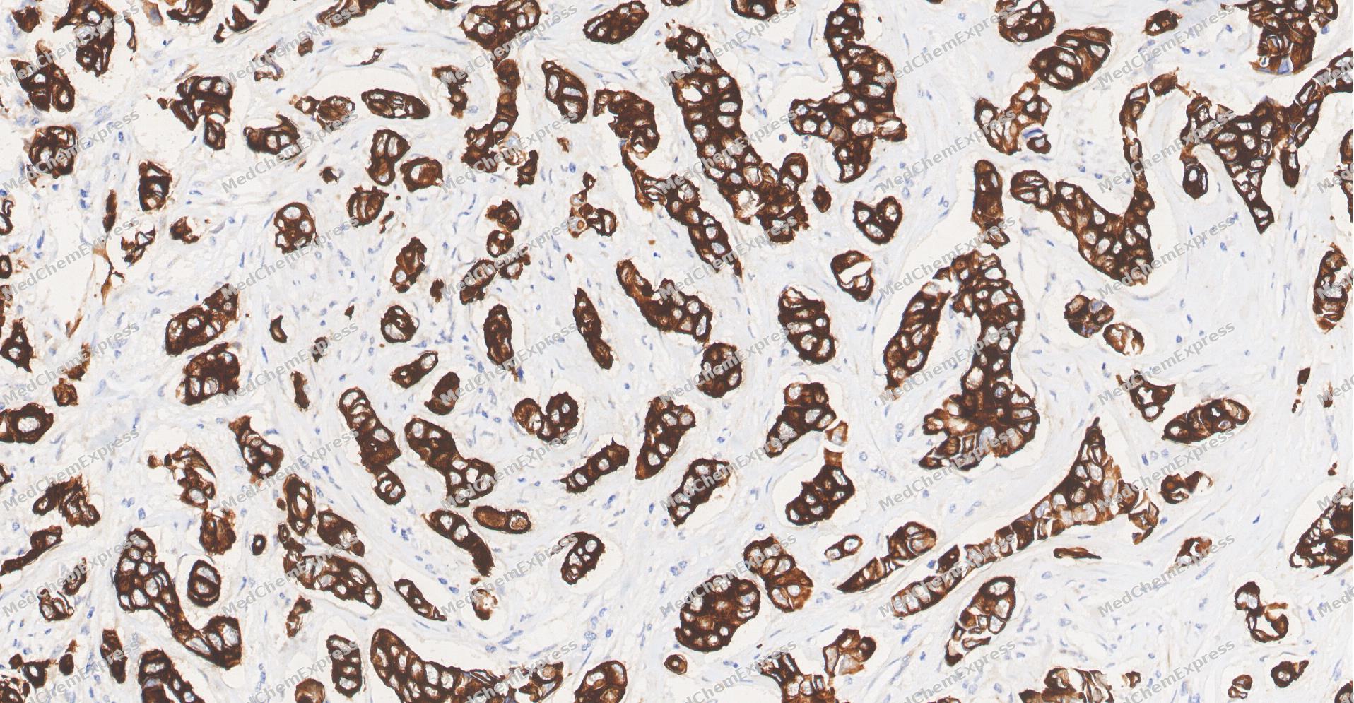 Immunohistochemical analysis of paraffin-embedded human Breast Cancer tissue using Phospho-GSK3 beta(Ser 9) antibody was performed. The section was pretreated using high-temperature and high-pressure mediated EDTA antigen retrieval buffer (pH 9.0), for 5 minutes. The tissues were incubated with primary antibody (HY-P80462, 1:100 dilution) at room temperature for 60 minutes. Detection was performed using an HRP conjugated compact polymer system. DAB was used as the chromogen. The tissues were counterstained with hematoxylin and mounted with neutral balsam mounting medium.
 -
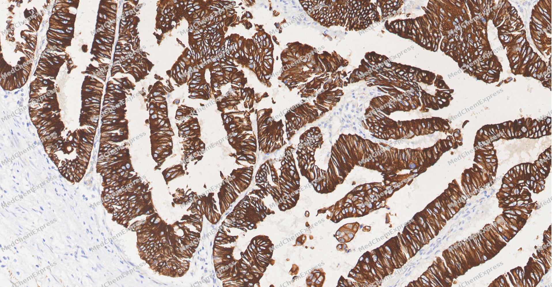 Immunohistochemical analysis of paraffin-embedded human Colon cancer tissue using Phospho-GSK3 beta(Ser 9) antibody was performed. The section was pretreated using high-temperature and high-pressure mediated EDTA antigen retrieval buffer (pH 9.0), for 5 minutes. The tissues were incubated with primary antibody (HY-P80462, 1:100 dilution) at room temperature for 60 minutes. Detection was performed using an HRP conjugated compact polymer system. DAB was used as the chromogen. The tissues were counterstained with hematoxylin and mounted with neutral balsam mounting medium.
 -
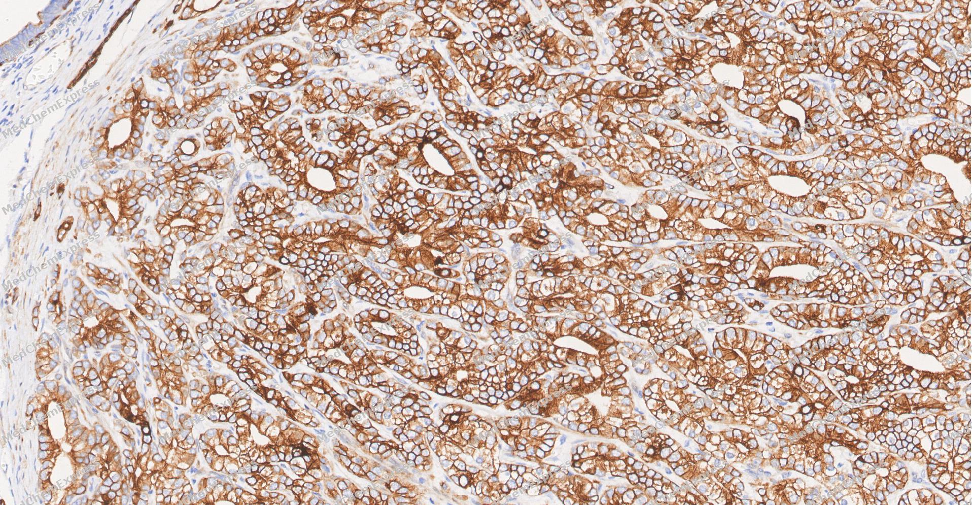 Immunohistochemical analysis of paraffin-embedded human Prostate Cancer tissue using Phospho-GSK3 beta(Ser 9) antibody was performed. The section was pretreated using high-temperature and high-pressure mediated EDTA antigen retrieval buffer (pH 9.0), for 5 minutes. The tissues were incubated with primary antibody (HY-P80462, 1:100 dilution) at room temperature for 60 minutes. Detection was performed using an HRP conjugated compact polymer system. DAB was used as the chromogen. The tissues were counterstained with hematoxylin and mounted with neutral balsam mounting medium.
 -
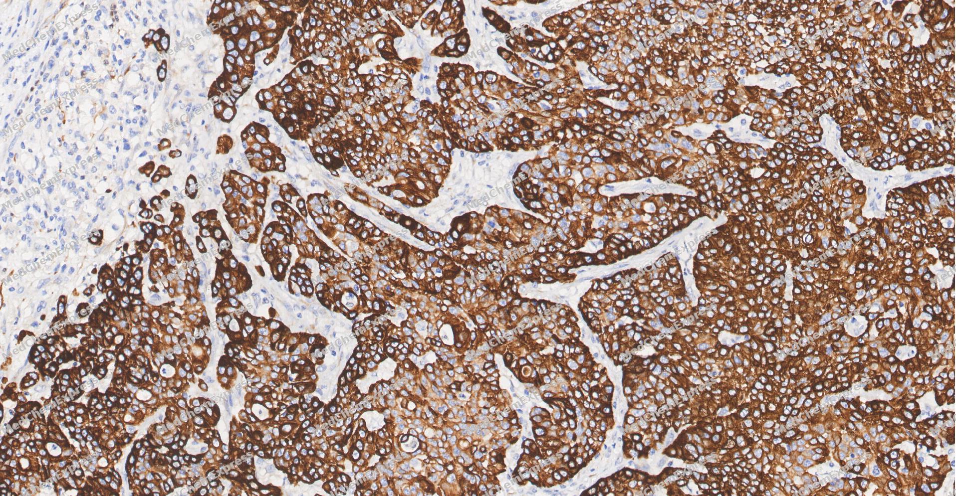 Immunohistochemical analysis of paraffin-embedded human Gastric Cancer tissue using Phospho-GSK3 beta(Ser 9) antibody was performed. The section was pretreated using high-temperature and high-pressure mediated EDTA antigen retrieval buffer (pH 9.0), for 5 minutes. The tissues were incubated with primary antibody (HY-P80462, 1:100 dilution) at room temperature for 60 minutes. Detection was performed using an HRP conjugated compact polymer system. DAB was used as the chromogen. The tissues were counterstained with hematoxylin and mounted with neutral balsam mounting medium.
 -
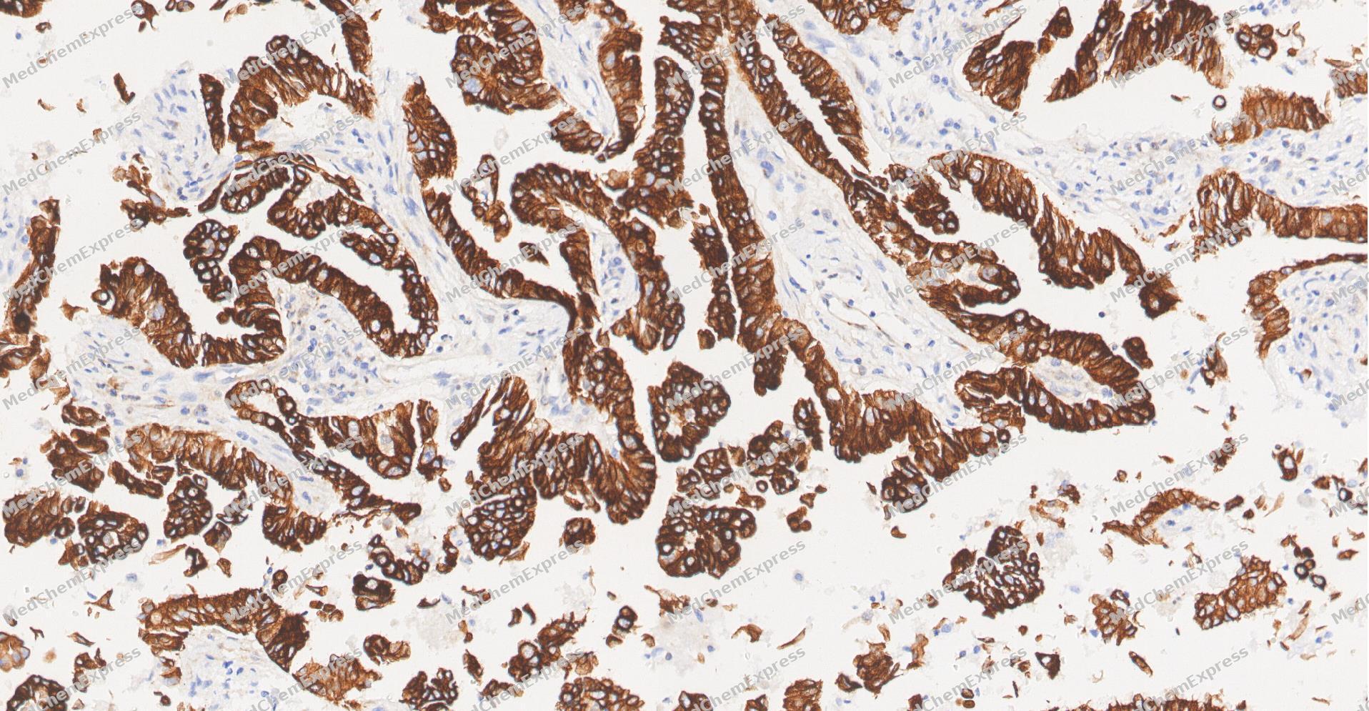 Immunohistochemical analysis of paraffin-embedded human Lung Adenocarcinoma tissue using Phospho-GSK3 beta(Ser 9) antibody was performed. The section was pretreated using high-temperature and high-pressure mediated EDTA antigen retrieval buffer (pH 9.0), for 5 minutes. The tissues were incubated with primary antibody (HY-P80462, 1:100 dilution) at room temperature for 60 minutes. Detection was performed using an HRP conjugated compact polymer system. DAB was used as the chromogen. The tissues were counterstained with hematoxylin and mounted with neutral balsam mounting medium.
 -
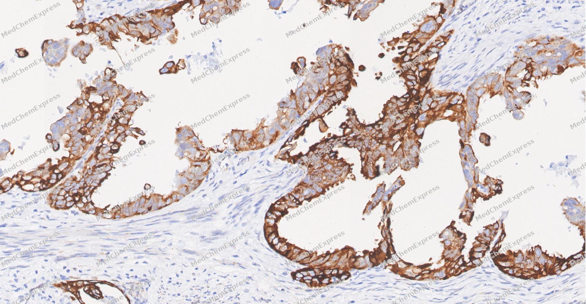 Immunohistochemical analysis of paraffin-embedded human Endometrial Carcinoma tissue using Phospho-GSK3 beta(Ser 9) antibody was performed. The section was pretreated using high-temperature and high-pressure mediated EDTA antigen retrieval buffer (pH 9.0), for 5 minutes. The tissues were incubated with primary antibody (HY-P80462, 1:100 dilution) at room temperature for 60 minutes. Detection was performed using an HRP conjugated compact polymer system. DAB was used as the chromogen. The tissues were counterstained with hematoxylin and mounted with neutral balsam mounting medium.


Background
-
Function
GSK3 beta is a Constitutively active protein kinase that acts as a negative regulator in the hormonal control of glucose homeostasis, Wnt signaling and regulation of transcription factors and microtubules, by phosphorylating and inactivating glycogen synthase (GYS1 or GYS2), EIF2B, CTNNB1/beta-catenin, APC, AXIN1, DPYSL2/CRMP2, JUN, NFATC1/NFATC, MAPT/TAU and MACF1. Requires primed phosphorylation of the majority of its substrates. In skeletal muscle, contributes to insulin regulation of glycogen synthesis by phosphorylating and inhibiting GYS1 activity and hence glycogen synthesis. May also mediate the development of insulin resistance by regulating activation of transcription factors. Regulates protein synthesis by controlling the activity of initiation factor 2B (EIF2BE/EIF2B5) in the same manner as glycogen synthase. In Wnt signaling, GSK3B forms a multimeric complex with APC, AXIN1 and CTNNB1/beta-catenin and phosphorylates the N-terminus of CTNNB1 leading to its degradation mediated by ubiquitin/proteasomes. Phosphorylates JUN at sites proximal to its DNA-binding domain, thereby reducing its affinity for DNA. Phosphorylates NFATC1/NFATC on conserved serine residues promoting NFATC1/NFATC nuclear export, shutting off NFATC1/NFATC gene regulation, and thereby opposing the action of calcineurin. Phosphorylates MAPT/TAU on 'Thr-548', decreasing significantly MAPT/TAU ability to bind and stabilize microtubules. MAPT/TAU is the principal component of neurofibrillary tangles in Alzheimer disease. Plays an important role in ERBB2-dependent stabilization of microtubules at the cell cortex. Phosphorylates MACF1, inhibiting its binding to microtubules which is critical for its role in bulge stem cell migration and skin wound repair. Probably regulates NF-kappa-B (NFKB1) at the transcriptional level and is required for the NF-kappa-B-mediated anti-apoptotic response to TNF-alpha (TNF/TNFA). Negatively regulates replication in pancreatic beta-cells, resulting in apoptosis, loss of beta-cells and diabetes. Through phosphorylation of the anti-apoptotic protein MCL1, may control cell apoptosis in response to growth factors deprivation. Phosphorylates MUC1 in breast cancer cells, decreasing the interaction of MUC1 with CTNNB1/beta-catenin. Is necessary for the establishment of neuronal polarity and axon outgrowth. Phosphorylates MARK2, leading to inhibition of its activity. Phosphorylates SIK1 at 'Thr-182', leading to sustainment of its activity. Phosphorylates ZC3HAV1 which enhances its antiviral activity. Phosphorylates SNAI1, leading to its ubiquitination and proteasomal degradation. Phosphorylates SFPQ at 'Thr-687' upon T-cell activation. Phosphorylates NR1D1 st 'Ser-55' and 'Ser-59' and stabilizes it by protecting it from proteasomal degradation. Regulates the circadian clock via phosphorylation of the major clock components including BMAL1, CLOCK and PER2. Phosphorylates FBXL2 at 'Thr-404' and primes it for ubiquitination by the SCF(FBXO3) complex and proteasomal degradation. Phosphorylates CLOCK AT 'Ser-427' and targets it for proteasomal degradation. Phosphorylates BMAL1 at 'Ser-17' and 'Ser-21' and primes it for ubiquitination and proteasomal degradation. Phosphorylates OGT at 'Ser-3' or 'Ser-4' which positively regulates its activity. Phosphorylates MYCN in neuroblastoma cells which may promote its degradation. Regulates the circadian rhythmicity of hippocampal long-term potentiation and BMAL1 and PER2 expression. Acts as a regulator of autophagy by mediating phosphorylation of KAT5/TIP60 under starvation conditions, activating KAT5/TIP60 acetyltransferase activity and promoting acetylation of key autophagy regulators, such as ULK1 and RUBCNL/Pacer. Negatively regulates extrinsic apoptotic signaling pathway via death domain receptors. Promotes the formation of an anti-apoptotic complex, made of DDX3X, BRIC2 and GSK3B, at death receptors, including TNFRSF10B. The anti-apoptotic function is most effective with weak apoptotic signals and can be overcome by stronger stimulation. Phosphorylates E2F1, promoting the interaction between E2F1 and USP11, stabilizing E2F1 and promoting its activity. Phosphorylates mTORC2 complex component RICTOR at 'Ser-1235' in response to endoplasmic stress, inhibiting mTORC2. Phosphorylates mTORC2 complex component RICTOR at 'Thr-1695' which facilitates FBXW7-mediated ubiquitination and subsequent degradation of RICTOR. Phosphorylates FXR1, promoting FXR1 ubiquitination by the SCF(FBXO4) complex and FXR1 degradation by the proteasome. Phosphorylates interleukin-22 receptor subunit IL22RA1, preventing its proteasomal degradation[1][2][3][4][5][6][7][8][9][10][11][12][13][14][15][16][17][18][19][20][21][22][23][24][25][26].
-
Subcellular Localization
Cytoplasm; Nucleus; Cell membrane
-
Expression
Tissue_specificity:It is expressed in the testes, thymus, prostate, and ovaries, and expressed weakly in the lungs, brain, and kidneys. It co-localizes with EIF2AK2/PKR and TAU in the brain tissue of Alzheimer's disease (AD) . -
Isoforms & Post-Translational Modification
P49841 has 2 isomers: P49841-1: 46744 Da (predicted); P49841-2: 48034 Da (predicted).
Phosphorylated by AKT1 and ILK1. Upon insulin-mediated signaling, the activated PKB/AKT1 protein kinase phosphorylates and deactivates GSK3B, resulting in the dephosphorylation and activation of GYS1. Activated by phosphorylation at Tyr-216 (PubMed:25169422). Inactivated by phosphorylation at Ser-9 (Probable). Phosphorylated in a circadian manner in the hippocampus (By similarity);Mono-ADP-ribosylation by PARP10 negatively regulates kinase activity;Palmitoylated. Palmitoylation by ZDHHC4 prevents AKT1-mediated phosphorylation -
Subunit
Monomer. Interacts with ARRB2, DISC1 and ZBED3 (By similarity). Interacts with CABYR, MMP2, MUC1, NIN and PRUNE1. Interacts with AXIN1; the interaction mediates hyperphosphorylation of CTNNB1 leading to its ubiquitination and destruction. Interacts with and phosphorylates SNAI1. Interacts with DNM1L (via a C-terminal domain). Found in a complex composed of MACF1, APC, AXIN1, CTNNB1 and GSK3B (By similarity). Interacts with SGK3. Interacts with DAB2IP (via C2 domain); the interaction stimulates GSK3B kinase activation. Interacts (via C2 domain) with PPP2CA. Interacts with the CLOCK-BMAL1 heterodimer (PubMed:19946213). Interacts with the BMAL1 (PubMed:28903391). Interacts with CTNND2 (PubMed:19706605). Interacts with NCYM (PubMed:24391509). The complex composed, at least, of APC, CTNNB1 and GSK3B interacts with JPT1; the interaction requires the inactive form of GSK3B (phosphorylated at 'Ser-9') (PubMed:25169422). Forms a complex composed of PRKAR2A or PRKAR2B, GSK3B and GSKIP through GSKIP interaction; facilitates PKA-induced phosphorylation and regulates GSK3B activity (PubMed:20007971, PubMed:25920809, PubMed:27484798). Interacts with GSKIP (PubMed:16981698). Interacts with GID8 (PubMed:28829046). Interacts with PIWIL2 (By similarity). Interacts with LMBR1L (PubMed:31073040). Interacts with DDX3X (PubMed:18846110). Interacts with BIRC2 (PubMed:18846110). Interacts with TNFRSF10B; TNFRSF10B stimulation inhibits GSK3B kinase activity (PubMed:18846110). Interacts with RICTOR; the interaction results in phosphorylation of RICTOR at 'Thr-1695' by GSK3B which facilitates FBXW7-mediated ubiquitination and subsequent degradation of RICTOR (PubMed:25897075). Found in a complex with SLC39A6, SLC39A10 and with GSK3B that controls NCAM1 phosphorylation (By similarity). Interacts with PKP3 (via ARM repeats); the interaction may be involved in PKP3 protein degradation (PubMed:34058472)
-
SwissProt ID
-
Synonyms
Glycogen synthase kinase-3 beta, GSK-3 beta, Serine/threonine-protein kinase GSK3B, GSK3B
-
Research Field
Neuroscience


Documentation
-
Data Sheet (268 KB)
-
SDS (252 KB)
- English - EN (252 KB)
- Français - FR (252 KB)
- Deutsch - DE (252 KB)
- Norwegian - NO (252 KB)
- Español - ES (252 KB)
- Swedish - SV (252 KB)
- Italian - IT (252 KB)
- Korean - KR (252 KB)
- Portuguese - PT (252 KB)
-
User Guide for Antibodies (1077 KB)
References
[1]. Frame S, et al. A common phosphate binding site explains the unique substrate specificity of GSK3 and its inactivation by phosphorylation. Mol Cell. 2001 Jun;7(6):1321-7. [Content Brief]
[2]. Dajani R, et al. Structural basis for recruitment of glycogen synthase kinase 3beta to the axin-APC scaffold complex. EMBO J. 2003 Feb 3;22(3):494-501. [Content Brief]
[3]. Cho JH, et al. Primed phosphorylation of tau at Thr231 by glycogen synthase kinase 3beta (GSK3beta) plays a critical role in regulating tau's ability to bind and stabilize microtubules. J Neurochem. 2004 Jan;88(2):349-58. [Content Brief]
[4]. Yin L, et al. Nuclear receptor Rev-erbalpha is a critical lithium-sensitive component of the circadian clock. Science. 2006 Feb 17;311(5763):1002-5. [Content Brief]
[5]. Boyle WJ, et al. Activation of protein kinase C decreases phosphorylation of c-Jun at sites that negatively regulate its DNA-binding activity. Cell. 1991 Feb 8;64(3):573-84. [Content Brief]
[6]. Zaoui K, et al. ErbB2 receptor controls microtubule capture by recruiting ACF7 to the plasma membrane of migrating cells. Proc Natl Acad Sci U S A. 2010 Oct 26;107(43):18517-22. [Content Brief]
[7]. Beals CR, et al. Nuclear export of NF-ATc enhanced by glycogen synthase kinase-3. Science. 1997 Mar 28;275(5308):1930-4. [Content Brief]
[8]. Welsh GI, et al. Glycogen synthase kinase-3 is rapidly inactivated in response to insulin and phosphorylates eukaryotic initiation factor eIF-2B. Biochem J. 1993 Sep 15;294 ( Pt 3)(Pt 3):625-9. [Content Brief]
[9]. Li Y, et al. Interaction of glycogen synthase kinase 3beta with the DF3/MUC1 carcinoma-associated antigen and beta-catenin. Mol Cell Biol. 1998 Dec;18(12):7216-24. [Content Brief]
[10]. Castaño Z, et al. The neuron-specific isoform of glycogen synthase kinase-3beta is required for axon growth. J Neurochem. 2010 Apr;113(1):117-30. [Content Brief]
[11]. Hashimoto YK, et al. Importance of autophosphorylation at Ser186 in the A-loop of salt inducible kinase 1 for its sustained kinase activity. J Cell Biochem. 2008 Aug 1;104(5):1724-39. [Content Brief]
[12]. Sun L, et al. Glycogen synthase kinase 3β (GSK3β) modulates antiviral activity of zinc-finger antiviral protein (ZAP). J Biol Chem. 2012 Jun 29;287(27):22882-8. [Content Brief]
[13]. Zhou BP, et al. Dual regulation of Snail by GSK-3beta-mediated phosphorylation in control of epithelial-mesenchymal transition. Nat Cell Biol. 2004 Oct;6(10):931-40. [Content Brief]
[14]. Yook JI, et al. Wnt-dependent regulation of the E-cadherin repressor snail. J Biol Chem. 2005 Mar 25;280(12):11740-8. [Content Brief]
[15]. Jin Y, et al. FBXO11 promotes ubiquitination of the Snail family of transcription factors in cancer progression and epidermal development. Cancer Lett. 2015 Jun 28;362(1):70-82. [Content Brief]
[16]. Liu Y, et al. SPSB3 targets SNAIL for degradation in GSK-3β phosphorylation-dependent manner and regulates metastasis. Oncogene. 2018 Feb 8;37(6):768-776. [Content Brief]
[17]. Heyd F, et al. Phosphorylation-dependent regulation of PSF by GSK3 controls CD45 alternative splicing. Mol Cell. 2010 Oct 8;40(1):126-37. [Content Brief]
[18]. Spengler ML, et al. A serine cluster mediates BMAL1-dependent CLOCK phosphorylation and degradation. Cell Cycle. 2009 Dec 15;8(24):4138-46. [Content Brief]
[19]. Lu Y, et al. Cancer/testis antigen PIWIL2 suppresses circadian rhythms by regulating the stability and activity of BMAL1 and CLOCK. Oncotarget. 2017 Aug 15;8(33):54913-54924. [Content Brief]
[20]. Suenaga Y, et al. NCYM, a Cis-antisense gene of MYCN, encodes a de novo evolved protein that inhibits GSK3β resulting in the stabilization of MYCN in human neuroblastomas. PLoS Genet. 2014 Jan;10(1):e1003996. [Content Brief]
[21]. Cheng X, et al. Pacer Is a Mediator of mTORC1 and GSK3-TIP60 Signaling in Regulation of Autophagosome Maturation and Lipid Metabolism. Mol Cell. 2019 Feb 21;73(4):788-802.e7. [Content Brief]
[22]. Sun M, et al. Identification of an antiapoptotic protein complex at death receptors. Cell Death Differ. 2008 Dec;15(12):1887-900. [Content Brief]
[23]. García-Alvarez G, et al. Glycogen synthase kinase-3beta binds to E2F1 and regulates its transcriptional activity. Biochim Biophys Acta. 2007 Mar;1773(3):375-82. [Content Brief]
[24]. Wang D, et al. Phosphorylated E2F1 is stabilized by nuclear USP11 to drive Peg10 gene expression and activate lung epithelial cells. J Mol Cell Biol. 2018 Feb 1;10(1):60-73. [Content Brief]
[25]. Chen CH, et al. ER stress inhibits mTORC2 and Akt signaling through GSK-3β-mediated phosphorylation of rictor. Sci Signal. 2011 Feb 22;4(161):ra10. [Content Brief]
[26]. Koo J, et al. Rictor Undergoes Glycogen Synthase Kinase 3 (GSK3)-dependent, FBXW7-mediated Ubiquitination and Proteasomal Degradation. J Biol Chem. 2015 May 29;290(22):14120-9. [Content Brief]

 Powered by Bioz
