



Phospho-JAK2 (Tyr1007/1008) Antibody (YA6449)
(Synonyms: JAK2; Tyrosine-protein kinase JAK2; Janus kinase 2; JAK-2)Based on 1 publication(s) in Google Scholar
Phospho-JAK2 (Tyr1007/1008) Antibody (YA6449) is a Rabbit-derived and non-conjugated IgG monoclonal antibody, targeting to Phospho-JAK2 (Tyr1007/1008).
-
Host:
Rabbit
-
Isotype:
IgG
-
Application:
WB, IHC-P, ICC/IF, IP, FC
-
Reactivity :
Human, Mouse, Rat
-
Formulation:
Supplied in Rabbit IgG in 10mM phosphate buffered saline , pH 7.4, 150mM sodium chloride, 0.05% BSA, 0.02% sodium azide and 50% glycerol.
-
Conjugation:
Non-conjugated


To place orders, for customer services and technical support, please contact: MedChemExpress USA
Tel: 609-228-6898 E-mail: [email protected] [email protected]




Publications Citing Use of MedChemExpress (MCE) Phospho-JAK2 (Tyr1007/1008) Antibody (YA6449)
More

Applications
| Application |
WB

WB: Western Blot
|
IHC-P
IHC-P: Immunohistochemistry-Paraffin
|
ICC/IF
ICC/IF: Immunocytochemistry/
Immunofluorescence |
IP
IP: Immunoprecipitation
|
FC
FC: Flow Cytometry
|
|---|---|---|---|---|---|
| Dilution Ratio | 1:500-1:1000 | 1:50-1:100 | 1:50-1:200 | 1:20 | 1:50-1:100 |


Product Details
Phospho-JAK2 (Tyr1007/1008) Antibody (YA6449) is a Rabbit-derived and non-conjugated IgG monoclonal antibody, targeting to Phospho-JAK2 (Tyr1007/1008).
-
Host Rabbit

-
Clonality Monoclonal,Recombinant

-
Species ReactivityHuman, Mouse, Rat
-
Calculated Molecular Weight Predicted band size: 131 kDa

Entrez Gene: 3716 Human ; 3717 Human ; 16451 Mouse ; 16452 Mouse ; 24514 Rat
SwissProt: O60674 Human ; P23458 Human ; P52332 Mouse ; Q62120 Mouse ; Q62689 Rat
OMIM: 600880 Human
A synthesized peptide derived from human Phospho-JAK2 (Y1007 + Y1008) KE-pY-pY-KV.
affinity purified
Non-conjugated
Unmodified
IgG


Product Properties
-
Appearance
Solution
-
Formulation
Supplied in Rabbit IgG in 10mM phosphate buffered saline , pH 7.4, 150mM sodium chloride, 0.05% BSA, 0.02% sodium azide and 50% glycerol.
-
Concentration
Batch-dependent, Please check the COA for the concentration of each lot. Check Lot Concentration
-
Storage & Stability
Stored at -20°C for 1 year. Avoid repeated freeze / thaw cycles.
-
Shipping
Shipping with blue ice.


Publications (1)
-
Journal Impact Factor
-
Most Recent
-
J Nanobiotechnology
Engineered Panax notoginseng polysaccharide micelles inhibit macrophage polarization and delay the progression of rheumatoid arthritis via JAK2-STAT3 signaling pathway. [Abstract]2025 Jul 14;23(1):509. PMID: 40660236


Verification Images
-
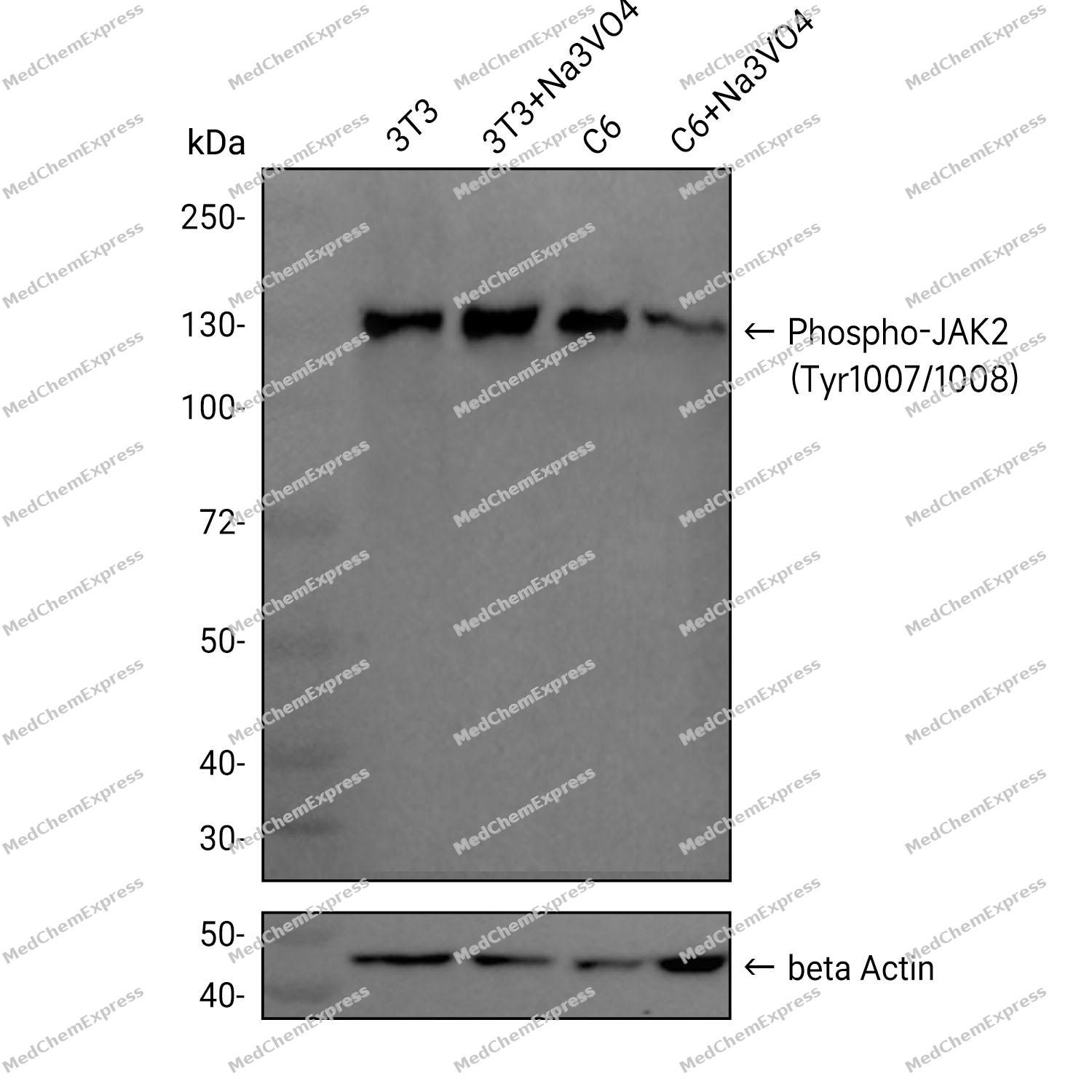 Western blot analysis was performed on extracts from 3T3 (lane 1, 15 μg), 3T3+Na3VO4 (lane 2, 15 μg), C6 (lane 3, 15 μg), and C6+Na3VO4 (lane 4, 15 μg) using Phospho-JAK2 (Tyr1007/1008) Rabbit mAb.Proteins were transferred to a PVDF membrane and blocked with 5% non-fat milk in TBST at 4°C overnight.The primary antibody (1:1000 dilution) and the loading control antibody (beta Actin, HY-P80438, 1:20000 dilution) were incubated in 5% non-fat milk in TBST for 1 hour at 37°C.Goat Anti-Rabbit IgG-HRP Secondary Antibody (1:20000 dilution) was then applied for 40 minutes at 37°C.
 -
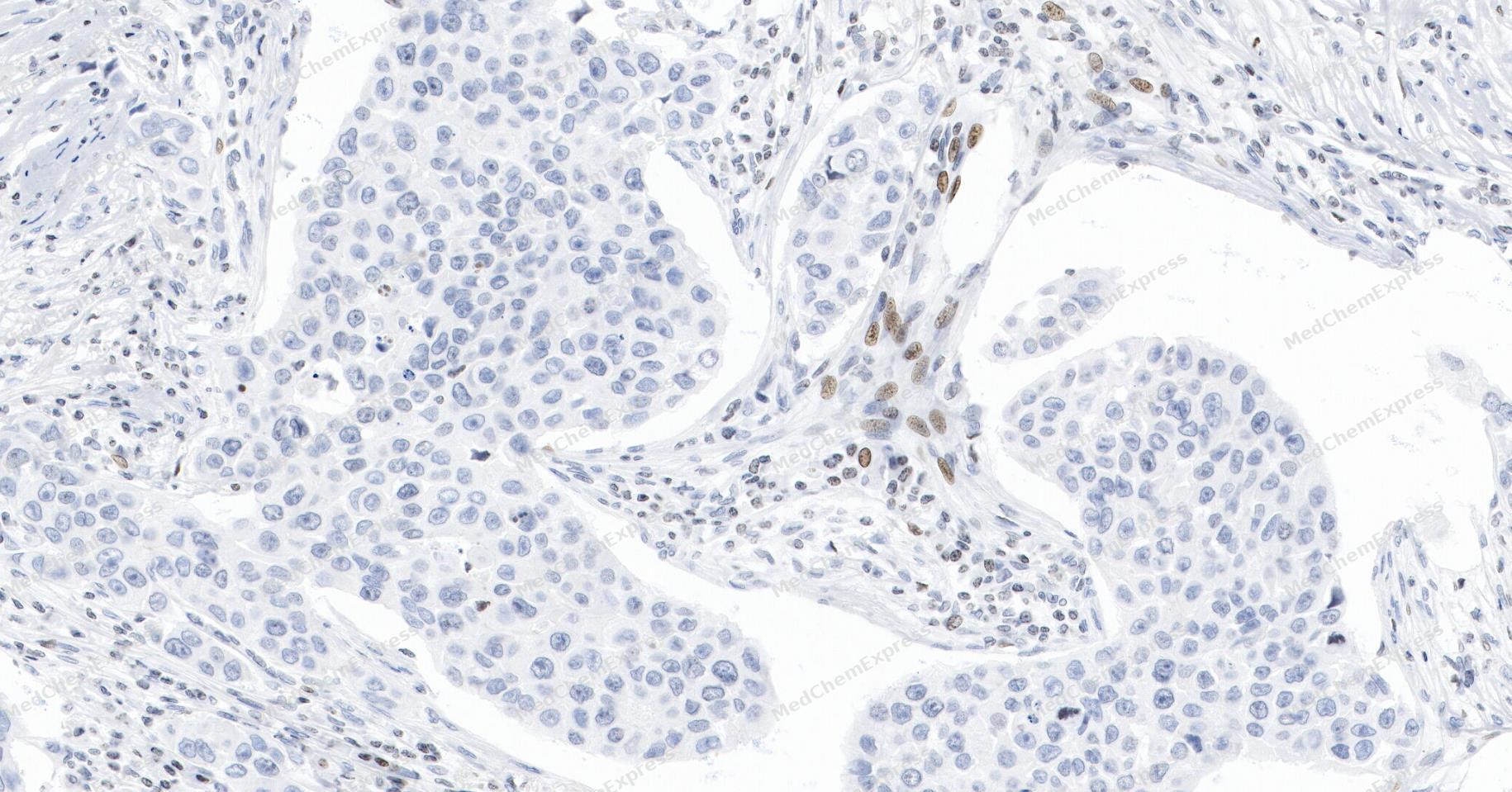 Immunohistochemical analysis of paraffin-embedded human breast cancer tissue using Phospho-JAK2 (Tyr1007/1008) Antibody (YA6449). The section was pre-treated using heat mediated antigen retrieval with Tris/EDTA buffer (pH 9.0) for 20 minutes. The tissues were probed with the primary antibody (HY-P80829A, 1/75) overnight at 4℃. The detection was performed using an HRP conjugated compact polymer system. DAB was used as the chromogen. Tissues were counterstained with hematoxylin and mounted with neutral balsam.
 -
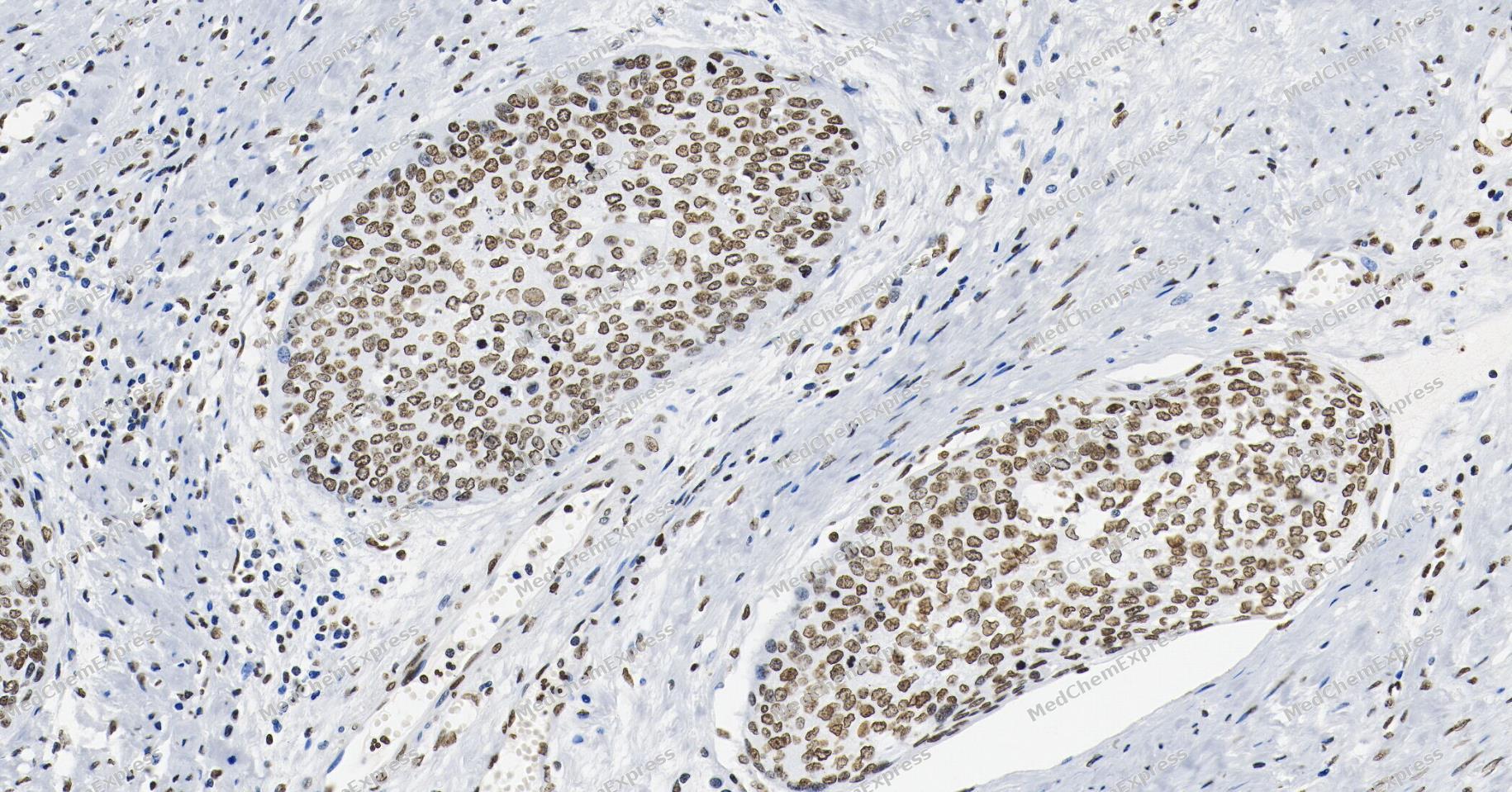 Immunohistochemical analysis of paraffin-embedded human cervical cancer tissue using Phospho-JAK2 (Tyr1007/1008) Antibody (YA6449). The section was pre-treated using heat mediated antigen retrieval with Tris/EDTA buffer (pH 9.0) for 20 minutes. The tissues were probed with the primary antibody (HY-P80829A, 1/75) overnight at 4℃. The detection was performed using an HRP conjugated compact polymer system. DAB was used as the chromogen. Tissues were counterstained with hematoxylin and mounted with neutral balsam.
 -
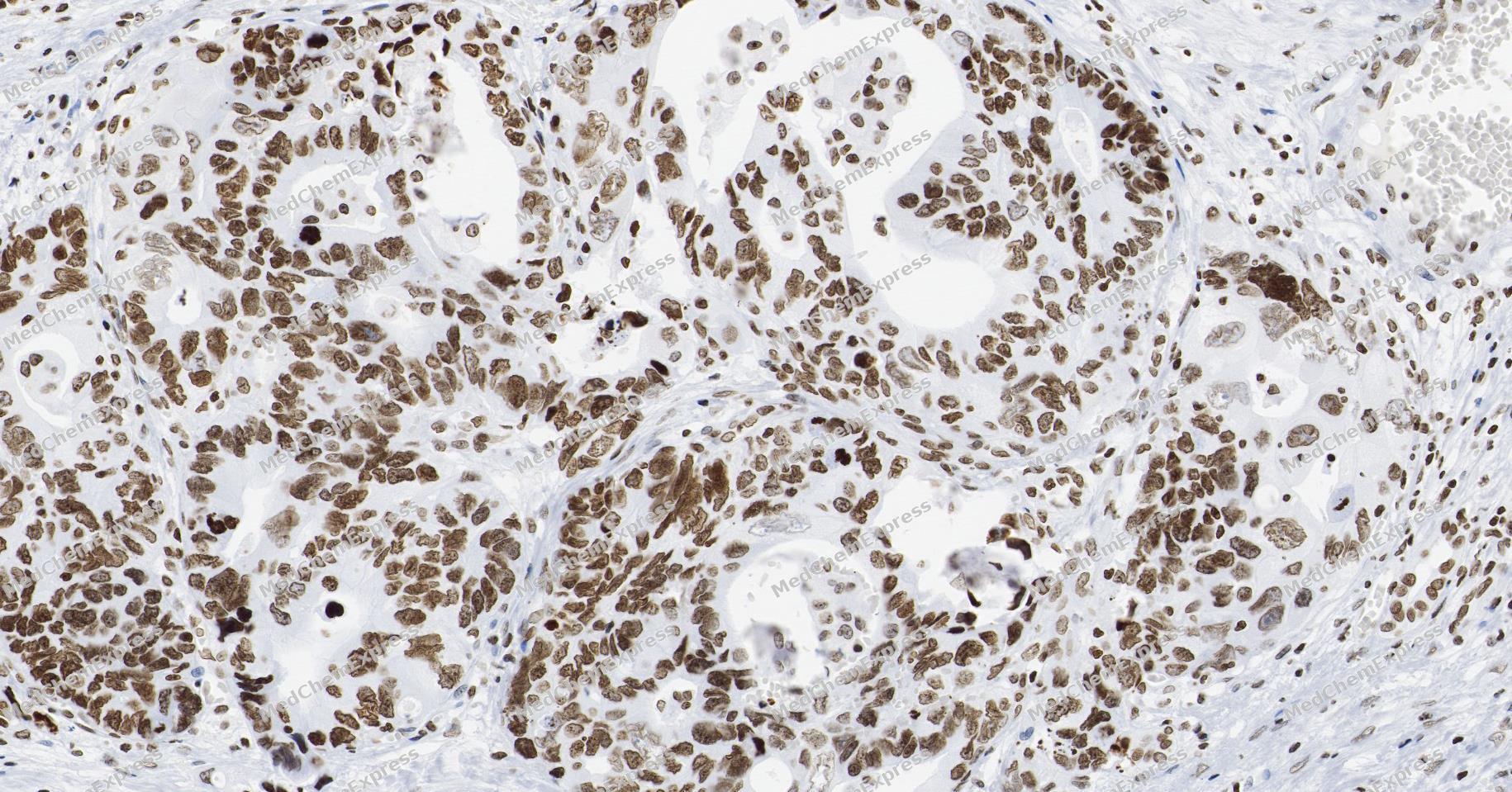 Immunohistochemical analysis of paraffin-embedded human liver cancer tissue using Phospho-JAK2 (Tyr1007/1008) Antibody (YA6449). The section was pre-treated using heat mediated antigen retrieval with Tris/EDTA buffer (pH 9.0) for 20 minutes. The tissues were probed with the primary antibody (HY-P80829A, 1/75) overnight at 4℃. The detection was performed using an HRP conjugated compact polymer system. DAB was used as the chromogen. Tissues were counterstained with hematoxylin and mounted with neutral balsam.
 -
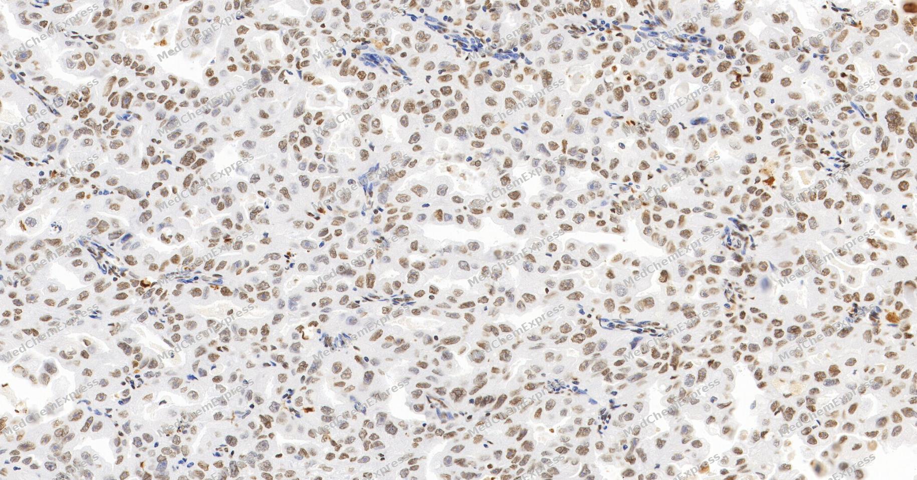 Immunohistochemical analysis of paraffin-embedded human ovarian cancer tissue using Phospho-JAK2 (Tyr1007/1008) Antibody (YA6449). The section was pre-treated using heat mediated antigen retrieval with Tris/EDTA buffer (pH 9.0) for 20 minutes. The tissues were probed with the primary antibody (HY-P80829A, 1/75) overnight at 4℃. The detection was performed using an HRP conjugated compact polymer system. DAB was used as the chromogen. Tissues were counterstained with hematoxylin and mounted with neutral balsam.
 -
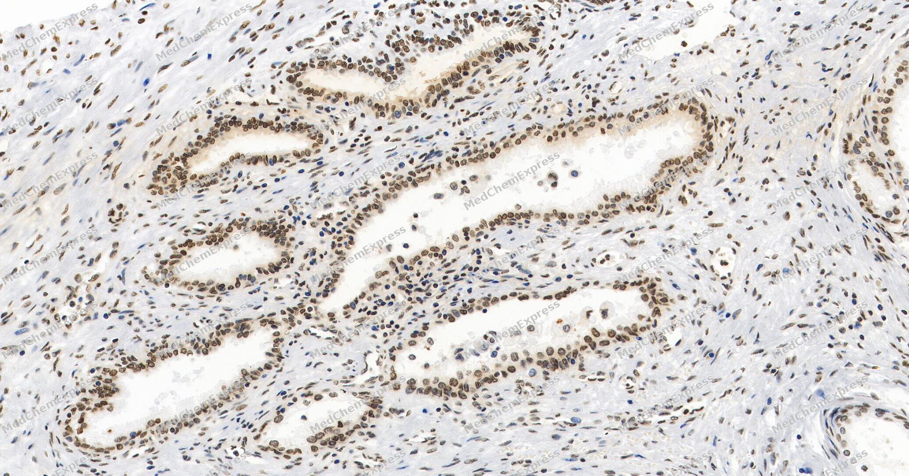 Immunohistochemical analysis of paraffin-embedded human prostate cancer tissue using Phospho-JAK2 (Tyr1007/1008) Antibody (YA6449). The section was pre-treated using heat mediated antigen retrieval with Tris/EDTA buffer (pH 9.0) for 20 minutes. The tissues were probed with the primary antibody (HY-P80829A, 1/75) overnight at 4℃. The detection was performed using an HRP conjugated compact polymer system. DAB was used as the chromogen. Tissues were counterstained with hematoxylin and mounted with neutral balsam.
 -
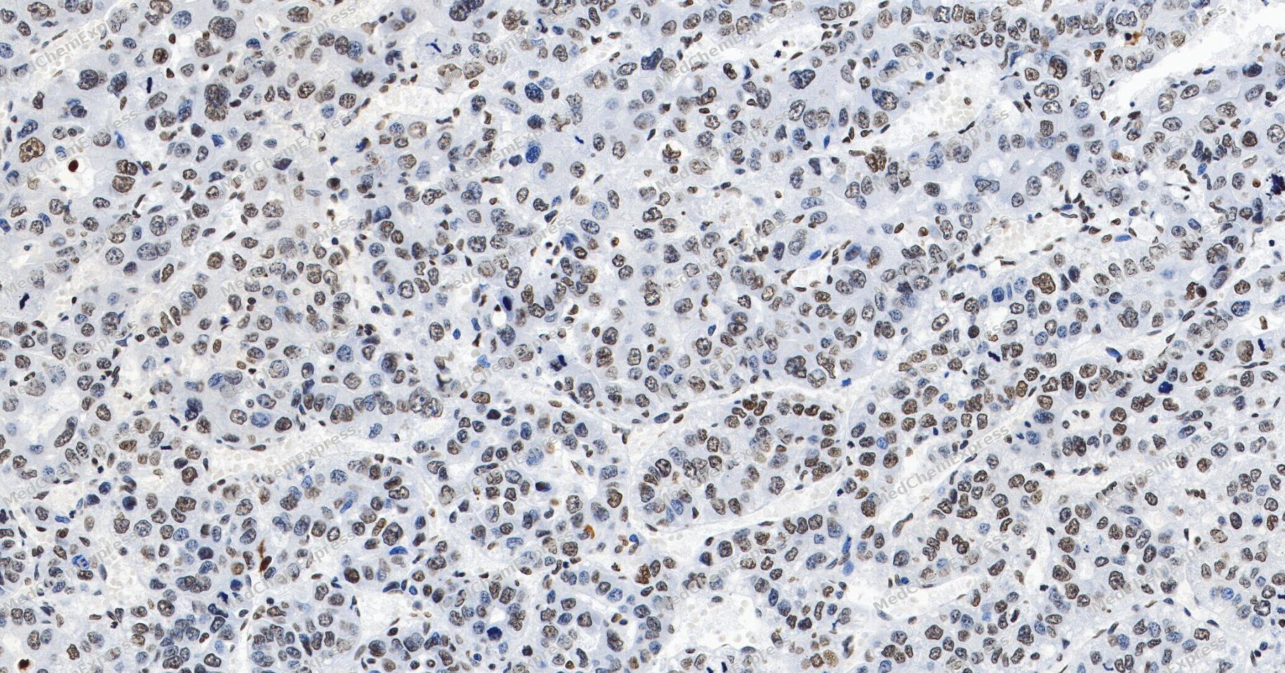 Immunohistochemical analysis of paraffin-embedded human hepatocellular carcinoma tissue using Phospho-JAK2 (Tyr1007/1008) Antibody (YA6449). The section was pre-treated using heat mediated antigen retrieval with Tris/EDTA buffer (pH 9.0) for 20 minutes. The tissues were probed with the primary antibody (HY-P80829A, 1/75) overnight at 4℃. The detection was performed using an HRP conjugated compact polymer system. DAB was used as the chromogen. Tissues were counterstained with hematoxylin and mounted with neutral balsam.
 -
 Immunohistochemical analysis of paraffin-embedded human Lung Adenocarcinoma tissue using Phospho-JAK2 (Tyr1007/1008) antibody (YA6449) was performed. The section was pretreated using high-temperature and high-pressure mediated EDTA antigen retrieval buffer (pH 9.0), for 5 minutes. The tissues were incubated with primary antibody (HY-P80829A, 1:100 dilution) at room temperature for 60 minutes. Detection was performed using an HRP conjugated compact polymer system. DAB was used as the chromogen. The tissues were counterstained with hematoxylin and mounted with neutral balsam mounting medium.
 -
 Immunohistochemical analysis of paraffin-embedded human Ovarian Cancer tissue using Phospho-JAK2 antibody was performed. The section was pretreated using high-temperature and high-pressure mediated EDTA antigen retrieval buffer (pH 9.0), for 5 minutes. The tissues were incubated with primary antibody (HY-P80829A, 1:100 dilution) at room temperature for 60 minutes. Detection was performed using an HRP conjugated compact polymer system. DAB was used as the chromogen. The tissues were counterstained with hematoxylin and mounted with neutral balsam mounting medium.
 -
 Immunohistochemical analysis of paraffin-embedded human Testis tissue using Phospho-JAK2 antibody was performed. The section was pretreated using high-temperature and high-pressure mediated EDTA antigen retrieval buffer (pH 9.0), for 5 minutes. The tissues were incubated with primary antibody (HY-P80829A, 1:100 dilution) at room temperature for 60 minutes. Detection was performed using an HRP conjugated compact polymer system. DAB was used as the chromogen. The tissues were counterstained with hematoxylin and mounted with neutral balsam mounting medium.
 -
 Immunohistochemical analysis of paraffin-embedded human Kidney cancer tissue using Phospho-JAK2 antibody was performed. The section was pretreated using high-temperature and high-pressure mediated EDTA antigen retrieval buffer (pH 9.0), for 5 minutes. The tissues were incubated with primary antibody (HY-P80829A, 1:100 dilution) at room temperature for 60 minutes. Detection was performed using an HRP conjugated compact polymer system. DAB was used as the chromogen. The tissues were counterstained with hematoxylin and mounted with neutral balsam mounting medium.
 -
 Immunohistochemical analysis of paraffin-embedded human Esophageal Carcinoma tissue using Phospho-JAK2 antibody was performed. The section was pretreated using high-temperature and high-pressure mediated EDTA antigen retrieval buffer (pH 9.0), for 5 minutes. The tissues were incubated with primary antibody (HY-P80829A, 1:100 dilution) at room temperature for 60 minutes. Detection was performed using an HRP conjugated compact polymer system. DAB was used as the chromogen. The tissues were counterstained with hematoxylin and mounted with neutral balsam mounting medium.
 -
 Immunohistochemical analysis of paraffin-embedded human Prostate Cancer tissue using Phospho-JAK2 antibody was performed. The section was pretreated using high-temperature and high-pressure mediated EDTA antigen retrieval buffer (pH 9.0), for 5 minutes. The tissues were incubated with primary antibody (HY-P80829A, 1:100 dilution) at room temperature for 60 minutes. Detection was performed using an HRP conjugated compact polymer system. DAB was used as the chromogen. The tissues were counterstained with hematoxylin and mounted with neutral balsam mounting medium.
 -
 Immunocytochemistry analysis of HeLa cells labeling Phospho-JAK2 (Tyr1007/1008) with Phospho-JAK2 (Tyr1007/1008) Antibody (HY-P80829A) at 1/50 dilution. Cells were fixed in 4% paraformaldehyde for 15 minutes at room temperature, permeabilized with 0.1% Triton X-100 in PBS for 15 minutes at room temperature, then blocked with quick block buffer for 10 minutes at room temperature. Cells were then incubated with Phospho-JAK2 (Tyr1007/1008) Antibody (HY-P80829A) at 1/50 dilution in quick block buffer overnight at 4 ℃. AF488-conjugated Goat Anti-Rabbit IgG H&L(HY-P8002, Green) was used as the secondary antibody at 1/1,000 dilution. PBS instead of the primary antibody was used as the secondary antibody only control. The Nuclear counterstain was DAPI (Blue).
 -
 Immunocytochemistry analysis of HeLa cells labeling Phospho-JAK2 (Tyr1007/1008) with Phospho-JAK2 (Tyr1007/1008) Antibody (HY-P80829A) at 1/100 dilution. Cells were fixed in 4% paraformaldehyde for 15 minutes at room temperature, permeabilized with 0.1% Triton X-100 in PBS for 15 minutes at room temperature, then blocked with quick block buffer for 10 minutes at room temperature. Cells were then incubated with Phospho-JAK2 (Tyr1007/1008) Antibody (HY-P80829A) at 1/100 dilution in quick block buffer overnight at 4 ℃. AF488-conjugated Goat Anti-Rabbit IgG H&L(HY-P8002, Green) was used as the secondary antibody at 1/1,000 dilution. PBS instead of the primary antibody was used as the secondary antibody only control. The Nuclear counterstain was DAPI (Blue).
 -
 Immunocytochemistry analysis of Hela cells labeling Phospho-JAK2 (Tyr1007/1008) with Phospho-JAK2 (Tyr1007/1008) Antibody (HY-P80829A) at 1/100 dilution . Cells were fixed in 4% paraformaldehyde for 15 minutes at room temperature, permeabilized with 0.1% Triton X-100 in PBS for 15 minutes at room temperature, then blocked with quick block buffer for 10 minutes at room temperature. Cells were then incubated with Phospho-JAK2 (Tyr1007/1008) Antibody (HY-P80829A) at 1/100 dilution in quick block buffer overnight at 4 ℃. AF488-conjugated Goat Anti-Rabbit IgG H&L(HY-P8002, Green) was used as the secondary antibody at 1/1,000 dilution. PBS instead of the primary antibody was used as the secondary antibody only control. The Nuclear counterstain was DAPI (Blue).
 -
 Immunocytochemistry analysis of Hela cells labeling Phospho-JAK2 (Tyr1007/1008) with Phospho-JAK2 (Tyr1007/1008) Antibody (HY-P80829A) at 1/200 dilution . Cells were fixed in 4% paraformaldehyde for 15 minutes at room temperature, permeabilized with 0.1% Triton X-100 in PBS for 15 minutes at room temperature, then blocked with quick block buffer for 10 minutes at room temperature. Cells were then incubated with Phospho-JAK2 (Tyr1007/1008) Antibody (HY-P80829A) at 1/200 dilution in quick block buffer overnight at 4 ℃. AF488-conjugated Goat Anti-Rabbit IgG H&L(HY-P8002, Green) was used as the secondary antibody at 1/1,000 dilution. PBS instead of the primary antibody was used as the secondary antibody only control. The Nuclear counterstain was DAPI (Blue).
 -
 Tyramide signaling amplification based immunofluorescence was performed on paraffin-embedded human ovarian carcinoma tissue using Phospho-JAK2 (Tyr1007/1008) antibody. Antigen retrieval was performed in EDTA buffer pH 9.0 (95 °C, 20 min) followed by cooling to RT. Then incubated with primary antibody (HY-P80829A, 1:200 dilution) at room temperature for 60 minutes and HRP conjugated secondary antibody for 10 minutes. Fluorescence was then developed with Vari Fluor 532 TSA (200×)(HY-D1832). The tissues were counterstained with DAPI and mounted with Anti-fade mounting medium.
 -
 Tyramide signaling amplification based immunofluorescence was performed on paraffin-embedded human ovarian carcinoma tissue using Phospho-JAK2 (Tyr1007/1008) antibody. Antigen retrieval was performed in EDTA buffer pH 9.0 (95 °C, 20 min) followed by cooling to RT. Then incubated with primary antibody (HY-P80829A, 1:200 dilution) at room temperature for 60 minutes and HRP conjugated secondary antibody for 10 minutes. Fluorescence was then developed with Vari Fluor 532 TSA (200×)(HY-D1832). The tissues were counterstained with DAPI and mounted with Anti-fade mounting medium.
 -
 Tyramide signaling amplification based immunofluorescence was performed on paraffin-embedded human ovarian carcinoma tissue using Phospho-JAK2 (Tyr1007/1008) antibody. Antigen retrieval was performed in EDTA buffer pH 9.0 (95 °C, 20 min) followed by cooling to RT. Then incubated with primary antibody (HY-P80829A, 1:200 dilution) at room temperature for 60 minutes and HRP conjugated secondary antibody for 10 minutes. Fluorescence was then developed with Vari Fluor 532 TSA (200×)(HY-D1832). The tissues were counterstained with DAPI and mounted with Anti-fade mounting medium.
 -
 Tyramide signaling amplification based immunofluorescence was performed on paraffin-embedded human ovarian carcinoma tissue using Phospho-JAK2 (Tyr1007/1008) antibody. Antigen retrieval was performed in EDTA buffer pH 9.0 (95 °C, 20 min) followed by cooling to RT. Then incubated with primary antibody (HY-P80829A, 1:200 dilution) at room temperature for 60 minutes and HRP conjugated secondary antibody for 10 minutes. Fluorescence was then developed with Vari Fluor 532 TSA (200×)(HY-D1832). The tissues were counterstained with DAPI and mounted with Anti-fade mounting medium.
 -
 Tyramide signaling amplification based immunofluorescence was performed on paraffin-embedded human ovarian carcinoma tissue using Phospho-JAK2 (Tyr1007/1008) antibody. Antigen retrieval was performed in EDTA buffer pH 9.0 (95 °C, 20 min) followed by cooling to RT. Then incubated with primary antibody (HY-P80829A, 1:200 dilution) at room temperature for 60 minutes and HRP conjugated secondary antibody for 10 minutes. Fluorescence was then developed with Vari Fluor 532 TSA (200×)(HY-D1832). The tissues were counterstained with DAPI and mounted with Anti-fade mounting medium.
 -
 Tyramide signaling amplification based immunofluorescence was performed on paraffin-embedded human ovarian carcinoma tissue using Phospho-JAK2 (Tyr1007/1008) antibody. Antigen retrieval was performed in EDTA buffer pH 9.0 (95 °C, 20 min) followed by cooling to RT. Then incubated with primary antibody (HY-P80829A, 1:200 dilution) at room temperature for 60 minutes and HRP conjugated secondary antibody for 10 minutes. Fluorescence was then developed with Vari Fluor 532 TSA (200×)(HY-D1832). The tissues were counterstained with DAPI and mounted with Anti-fade mounting medium.


Background
-
Function
JAK2 is a non-receptor tyrosine kinase in the JAK1-JAK2-JAK3-TYK2 family and acts as a cytoplasmic signaling component of cytokine receptors[1]. Mechanistically, cytokine-mediated activation of JAK2 promotes receptor phosphorylation, recruits STAT proteins, and drives STAT phosphorylation-dependent transcriptional signaling[1][2]. This pathway supports cell growth, survival, development, differentiation, hematopoiesis, immune regulation, and homeostasis[2][3]. In disease models, gain-of-function JAK2 V617F in the pseudokinase domain causes constitutive activity and is strongly linked to myeloproliferative neoplasms and leukemias[1][4]. Compared with related isoforms, JAK3 is essential for common γ-chain cytokine receptor signaling in T- and B-cell development, whereas complete mouse deletion of JAK1 or JAK2 is not compatible with life[5][6]. For experimental applications, ruxolitinib is a selective oral JAK1/JAK2 inhibitor, and JAK2-focused compounds support studies of mutant signaling, cytokine responses, splenomegaly, angiogenesis, and inhibitor resistance[7][8][9].
-
Subcellular Localization
Endomembrane system; Peripheral membrane protein; Cytoplasm; Nucleus
-
Expression
Tissue_specificity:Commonly expressed in most organizations -
Subunit
Interacts with EPOR, LYN, SIRPA, SH2B1 and TEC (By similarity). Interacts with IL23R (PubMed:12023369). Interacts with SKB1 (PubMed:10531356). Interacts with STAM2 (PubMed:10899310). Interacts with IFNGR2 (via intracellular domain) (PubMed:7615558, PubMed:7673114). Interacts with LEPR (Isoform B) (By similarity). Interacts with HSP90AB1; promotes functional activation in a heat shock-dependent manner (PubMed:20353823). Interacts with STRA6 (PubMed:21368206). Interacts with RHEX; this interaction occurs in a erythropoietin (EPO)-dependent manner (PubMed:25092874). Interacts with ASB2; the interaction targets JAK2 for Notch-induced proteasomal degradation (PubMed:21119685). Interacts with MPL/TPOR (PubMed:15899890)
-
SwissProt ID
-
Synonyms
JAK2; Tyrosine-protein kinase JAK2; Janus kinase 2; JAK-2
Documentation
-
Data Sheet (263 KB)
-
SDS (251 KB)
- English - EN (251 KB)
- Français - FR (251 KB)
- Deutsch - DE (251 KB)
- Norwegian - NO (251 KB)
- Español - ES (251 KB)
- Swedish - SV (251 KB)
- Italian - IT (251 KB)
- Korean - KR (251 KB)
- Portuguese - PT (251 KB)
-
User Guide for Antibodies (1077 KB)
References
[1]. Hubbard SR. Mechanistic Insights into Regulation of JAK2 Tyrosine Kinase. Front Endocrinol (Lausanne). 2018 Jan 5;8:361. doi: 10.3389/fendo.2017.00361. PMID: 29379470; PMCID: PMC5770812. et al. Mechanistic Insights into Regulation of JAK2 Tyrosine Kinase. Front Endocrinol (Lausanne). 2018 Jan 5;8:361. [Content Brief]
[2]. Ihle JN, et al. Jaks and Stats in signaling by the cytokine receptor superfamily. Trends Genet. 1995 Feb;11(2):69-74. [Content Brief]
[3]. Rawlings JS, et al. The JAK/STAT signaling pathway. J Cell Sci. 2004 Mar 15;117(Pt 8):1281-3. [Content Brief]
[4]. Gäbler K, et al. JAK2 mutants (e.g., JAK2V617F) and their importance as drug targets in myeloproliferative neoplasms. JAKSTAT. 2013 Jul 1;2(3):e25025. [Content Brief]
[5]. Suzuki K, et al. Janus kinase 3 (Jak3) is essential for common cytokine receptor gamma chain (gamma(c))-dependent signaling: comparative analysis of gamma(c), Jak3, and gamma(c) and Jak3 double-deficient mice. Int Immunol. 2000 Feb;12(2):123-32. [Content Brief]
[6]. Ghoreschi K, et al. Janus kinases in immune cell signaling. Immunol Rev. 2009 Mar;228(1):273-87. [Content Brief]
[7]. Naqvi K, et al. A potential role of ruxolitinib in leukemia. Expert Opin Investig Drugs. 2011 Aug;20(8):1159-66. [Content Brief]
[8]. Hart S, et al. SB1518, a novel macrocyclic pyrimidine-based JAK2 inhibitor for the treatment of myeloid and lymphoid malignancies. Leukemia. 2011 Nov;25(11):1751-9. [Content Brief]
[9]. Cheng Z, et al. Angiogenesis in JAK2 V617F positive myeloproliferative neoplasms and ruxolitinib decrease VEGF, HIF-1 enesis in JAK2 V617F positive cells. Leuk Lymphoma. 2018 Jan;59(1):196-203. [Content Brief]

 Powered by Bioz
