



S100B Antibody
(Synonyms: NEF; Protein S100 B; Protein S100-B; S 100 calcium binding protein beta chain; S 100 protein beta chain; S-100 protein beta chain; S-100 protein subunit beta; S100; S100 calcium binding protein beta (neural); S100 calcium-binding protein B; S100 protein b)Based on 1 Customer Validation
S100B Antibody is a Rabbit-derived and non-conjugated IgG polyclonal antibody, targeting to S100B.
-
Host:
Rabbit
-
Isotype:
IgG
-
Application:
WB, IHC-P, ICC/IF, IP
-
Reactivity :
Human, Mouse, Rat, Goat
-
Formulation:
Supplied in 50mM Tris-Glycine(pH 7.4), 0.15M NaCl, 40%Glycerol, 0.01% sodium azide and 0.05% BSA.
-
Conjugation:
Non-conjugated


To place orders, for customer services and technical support, please contact: MedChemExpress USA
Tel: 609-228-6898 E-mail: [email protected] [email protected]





Applications
| Application |
WB

WB: Western Blot
|
IHC-P
IHC-P: Immunohistochemistry-Paraffin
|
ICC/IF
ICC/IF: Immunocytochemistry/
Immunofluorescence |
IP
IP: Immunoprecipitation
|
|---|---|---|---|---|
| Dilution Ratio | 1:500-1:1000 | 1:50-1:100 | 1:50-1:200 | 1:20 |


Product Details
S100B Antibody is a Rabbit-derived and non-conjugated IgG polyclonal antibody, targeting to S100B.
-
Host Rabbit

-
Clonality Polyclonal

-
Species ReactivityHuman, Mouse, Rat, Goat
-
Observed Molecular WeightObserved band size: 11 kDa
Note: Due to possible protein modifications or aggregation, the molecular weight should be confirmed by actual measurement, and the predicted value is for reference only.

-
Calculated Molecular Weight Predicted band size: 11 kDa

Entrez Gene: 6285 Human ; 20203 Mouse ; 25742 Rat
SwissProt: P04271 Human ; P50114 Mouse ; P04631 Rat
OMIM: 176990 Human
Synthetic peptide corresponding to Human S100B.The exact sequence is proprietary to MCE.
Endogenous
affinity purified
Non-conjugated
Unmodified
IgG


Product Properties
-
Appearance
Solution
-
Formulation
Supplied in 50mM Tris-Glycine(pH 7.4), 0.15M NaCl, 40%Glycerol, 0.01% sodium azide and 0.05% BSA.
-
Concentration
Batch-dependent, Please check the COA for the concentration of each lot. Check Lot Concentration
-
Storage & Stability
Stored at -20°C for 1 year. Avoid repeated freeze / thaw cycles.
-
Shipping
Shipping with blue ice.


Verification Images
-
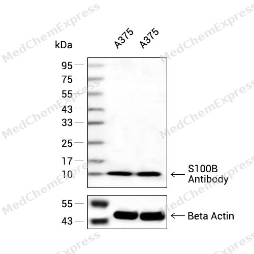 Western blot analysis of extracts from A375(lane 2(20ug) and A375(lane 3(40ug) using S100B Antibody (HY-P80891) Rabbit mAb. Proteins were transferred to a PVDF membrane and blocked with 5% non-fat milk in TBST for 2 hour at room temperature. The primary antibody (1/1000) and Loading control antibody (Beta Actin, HY-P80438, 1/10000) was used in 5% non-fat milk in TBST at 4°C overnight. Goat Anti-Mouse/Rabbit IgG-HRP Secondary Antibody (1/10000) was used for 1 hour at room temperature.
 -
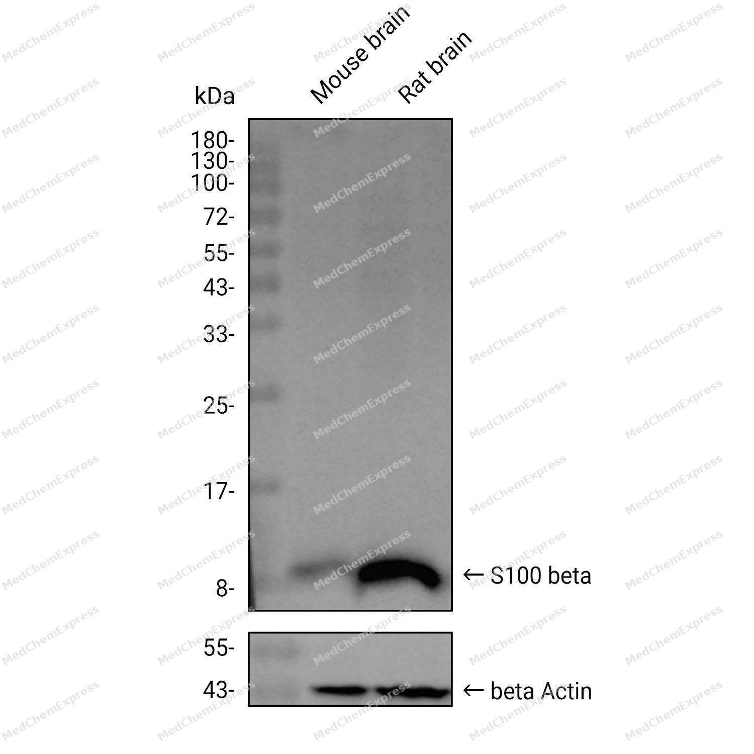 Western blot analysis was performed on extracts from Mouse brain (lane 1, 15 μg), and Rat brain (lane 2, 15 μg) using S100 beta Rabbit pAb.Proteins were transferred to a PVDF membrane and blocked with 5% non-fat milk in TBST at 4°C overnight.The primary antibody (1:2000 dilution) and the loading control antibody (beta-Actin, HY-P80438, 1:20000 dilution) were incubated in 5% non-fat milk in TBST for 1 hour at 37°C.Goat Anti-Rabbit IgG-HRP Secondary Antibody (1:20000 dilution) was then applied for 40 minutes at 37°C.
 -
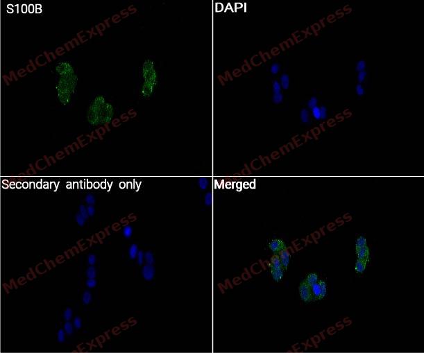 Immunocytochemistry analysis of T-47D cells labeling S100B with S100B Antibody (HY-P80891) at 1/50 dilution. Cells were fixed in 4% paraformaldehyde for 15 minutes at room temperature, then blocked with QuickBlock™ Blocking Buffer for Immunol Staining for 10 min at room temperature. Cells were then incubated with S100B Antibody (HY-P80891) at 1/50 dilution in QuickBlock™ Blocking Buffer for Immunol Staining at 4 ℃. Alexa Fluor® 488-conjugated AffiniPure Goat Anti-Rabbit IgG H&L(HY-P8002, Green) was used as the secondary antibody at 1/1,000 dilution. PBS instead of the primary antibody was used as the secondary antibody only control. The Nuclear counterstain was DAPI (Blue).
 -
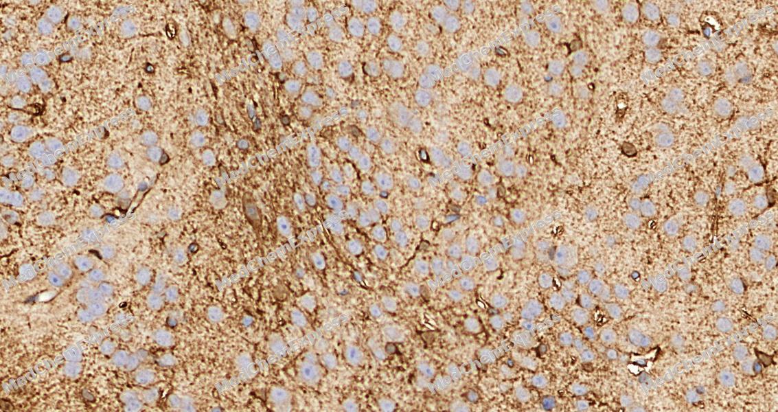 Immunohistochemical analysis of paraffin-embedded mouse brain tissue using S100B Antibody. The section was pre-treated using heat mediated antigen retrieval with Tris-EDTA buffer (pH 9.0) for 8 minutes. The tissues were blocked in QuickBlock for 20 minutes at room temperature, washed with ddH2O and PBS, and then probed with the primary antibody at 1/100 dilution in 4℃ overnight. The detection was performed using an HRP conjugated compact polymer system. DAB was used as the chromogen. Tissues were counterstained with hematoxylin and mounted with DPX.
 -
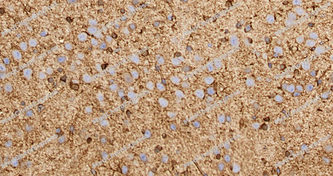 Immunohistochemical analysis of paraffin-embedded mouse brain tissue using S100B Antibody. The section was pre-treated using heat mediated antigen retrieval with Tris-EDTA buffer (pH 9.0) for 8 minutes. The tissues were blocked in QuickBlock for 20 minutes at room temperature, washed with ddH2O and PBS, and then probed with the primary antibody at 1/100 dilution in 4℃ overnight. The detection was performed using an HRP conjugated compact polymer system. DAB was used as the chromogen. Tissues were counterstained with hematoxylin and mounted with DPX.


Background
-
Function
S100B is a member of the S100 EF-hand calcium-binding protein family and is highly enriched in astrocytes, where it functions as an intracellular calcium sensor and extracellular signaling molecule that regulates cellular differentiation, growth, and neural homeostasis[1][2]. Mechanistically, extracellular S100B interacts with the receptor for advanced glycation end products (RAGE), activating signaling pathways that influence neuronal and glial responses, neuroinflammation, and cell survival processes[3][4]. Through these activities, S100B participates in the regulation of neurite extension, astrocyte maturation, oligodendrocyte development, and neural network function during central nervous system development and maintenance[2][4]. In disease contexts, altered S100B expression or release is associated with traumatic brain injury, neurodegenerative disorders, Alzheimer's disease, and other neurological conditions, and circulating S100B has therefore become a widely investigated biomarker of central nervous system injury and blood-brain barrier dysfunction[1][5]. Compared with related S100 family members, S100B is distinguished by its predominant enrichment in astrocytes and its extensive characterization as both a pathogenic mediator and a clinical biomarker in neurological disorders[1][2]. For experimental applications, modulation of S100B-dependent signaling has been explored through strategies targeting S100B interactions with RAGE or other binding partners, and S100B antagonists have been used to investigate the contribution of this protein to disease-associated cellular responses[3].
-
Subcellular Localization
Cytoplasm; Nucleus; Secreted
-
Expression
Tissue_specificity:Although S100 is a major component of water-soluble brain proteins, it is also present in a variety of other tissues. -
Subunit
Dimer of either two alpha chains, or two beta chains, or one alpha and one beta chain (PubMed:12480931, PubMed:20950652). The S100B dimer binds two molecules of STK38 (By similarity). Interacts with CACYBP in a calcium-dependent manner (By similarity). Interacts with ATAD3A; this interaction probably occurs in the cytosol prior to ATAD3A mitochondrial targeting (PubMed:20351179). Interacts with S100A6 (PubMed:9925766). The S100B dimer interacts with two molecules of CAPZA1 (PubMed:12480931). Interacts with AGER (PubMed:20943659). Interacts with PPP5C (via TPR repeats); the interaction is calcium-dependent and modulates PPP5C activity (PubMed:22399290). Interacts with TPPP; this interaction inhibits TPPP dimerization (PubMed:33831707). Interacts with isoform CLSTN3beta of CLSTN3; interaction promotes secretion (By similarity)
-
SwissProt ID
-
Synonyms
NEF; Protein S100 B; Protein S100-B; S 100 calcium binding protein beta chain; S 100 protein beta chain; S-100 protein beta chain; S-100 protein subunit beta; S100; S100 calcium binding protein beta (neural); S100 calcium-binding protein B; S100 protein b
-
Research Field
Cell Biology


Documentation
-
Data Sheet (262 KB)
-
SDS (252 KB)
- English - EN (252 KB)
- Français - FR (252 KB)
- Deutsch - DE (252 KB)
- Norwegian - NO (252 KB)
- Español - ES (252 KB)
- Swedish - SV (252 KB)
- Italian - IT (252 KB)
- Korean - KR (252 KB)
- Portuguese - PT (252 KB)
-
User Guide for Antibodies (1077 KB)
References
[1]. Michetti F, et al. The S100B Protein: A Multifaceted Pathogenic Factor More Than a Biomarker. Int J Mol Sci. 2023 May 31;24(11):9605. [Content Brief]
[2]. Hernández-Ortega K, et al. S100B actions on glial and neuronal cells in the developing brain: an overview. Front Neurosci. 2024 Jul 4;18:1425525. [Content Brief]
[3]. Dowarha D, et al. S100B as an Antagonist To Interfere with the Interface Area Flanked by S100A11 and RAGE V Domain. ACS Omega. 2018 Aug 22;3(8):9689-9698. [Content Brief]
[4]. Leclerc E, et al. The S100B/RAGE Axis in Alzheimer's Disease. Cardiovasc Psychiatry Neurol. 2010;2010:539581. [Content Brief]
[5]. Thelin EP, et al. A review of the clinical utility of serum S100B protein levels in the assessment of traumatic brain injury. Acta Neurochir (Wien). 2017 Feb;159(2):209-225. [Content Brief]

 Powered by Bioz
