



KCL-286

Based on 1 Customer Validation
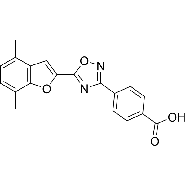
KCL-286 (C286) is an orally active and brain-penetrant retinoic acid receptor (RAR) β2 agonist (EC50 = 1.9 nM). KCL-286 targets RARβ2 with good selectivity over RAR α (EC50 = 26 nM) and RAR γ (EC50 = 11 nM). KCL-286 activates RARβ2 in the injured neurons. KCL-286 induces axonal regeneration of both spinal and sensory nerves through the inhibitory environment of the CNS, modulates neuroinflammation and extracellular matrix molecules. KCL-286 can modulate the expression of CSPGs by neuronal secretion of decorin which promotes myelination and aids axonal growth. KCL-286 can be studied in research for area such as spinal cord injury and traumatic nerve injury.
For research use only. We do not sell to patients.
- Purity : 98.16%
- CAS No.: 1952276-71-9
- Formula: C19H14N2O4
- Molecular Weight:334.33
-
Storage:Powder -20°C, 3 years , 4°C, 2 years ; In solvent -80°C, 6 months , -20°C, 1 month

To place orders, for customer services and technical support, please contact: MedChemExpress USA
Tel: 609-228-6898 E-mail: [email protected] [email protected]


-
 Biological Activity
-
Chemical Information
-
Solvent & Solubility
- Protocols
- Purity & Documentation
- References
-
 Help & FAQs


-
HY-108527
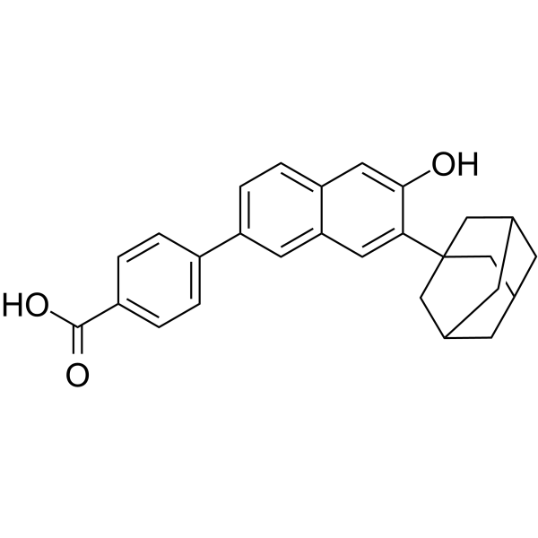
CD1530RARγ Agonist -
HY-107436
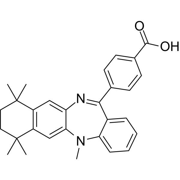
LE135RARβ/RARα Antagonist -
HY-107397
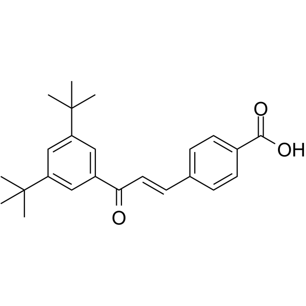
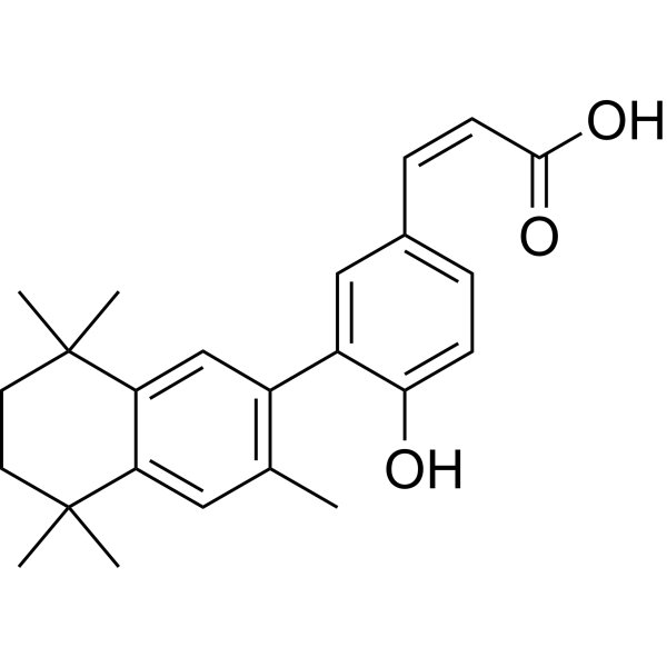
Ch55RAR-α/RAR-β Agonist -
HY-107399
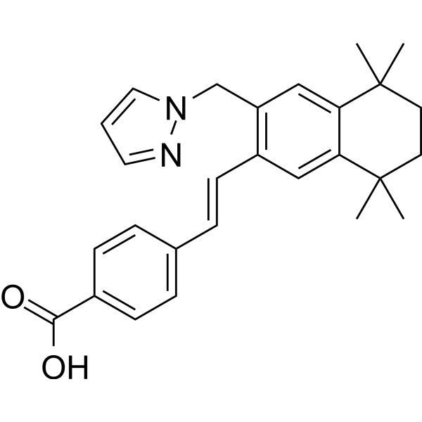
CD3254RXR Agonist -
HY-15682
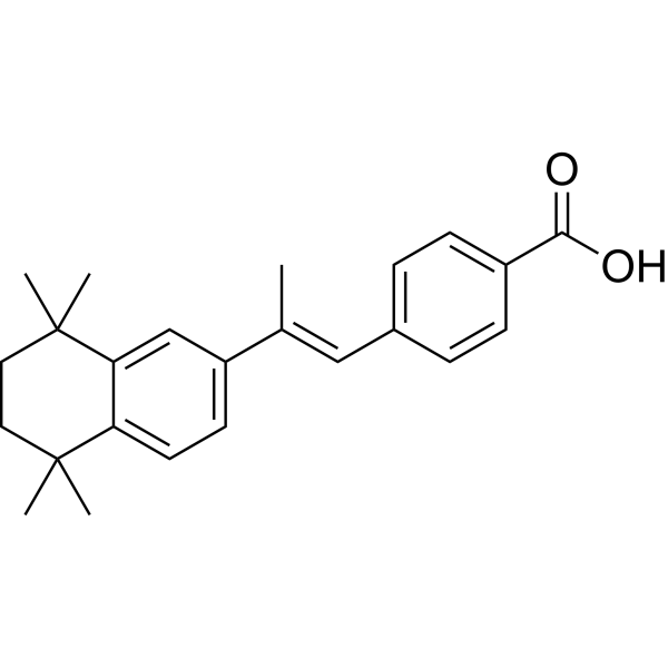
TTNPBApoptosis Inducer -
HY-108521
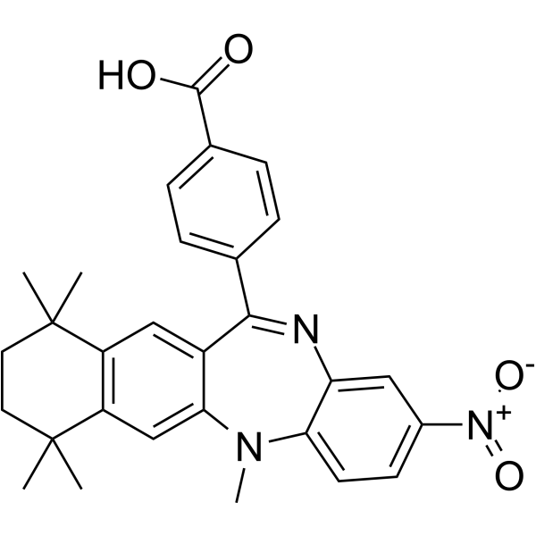
HX531RXR Antagonist -
HY-14799
PalovaroteneRARγ Agonist -
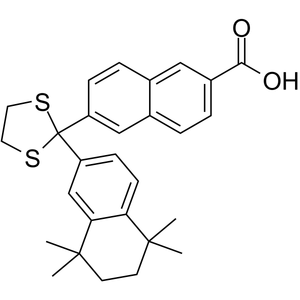
HY-108530
MM11253RARγ Antagonist -
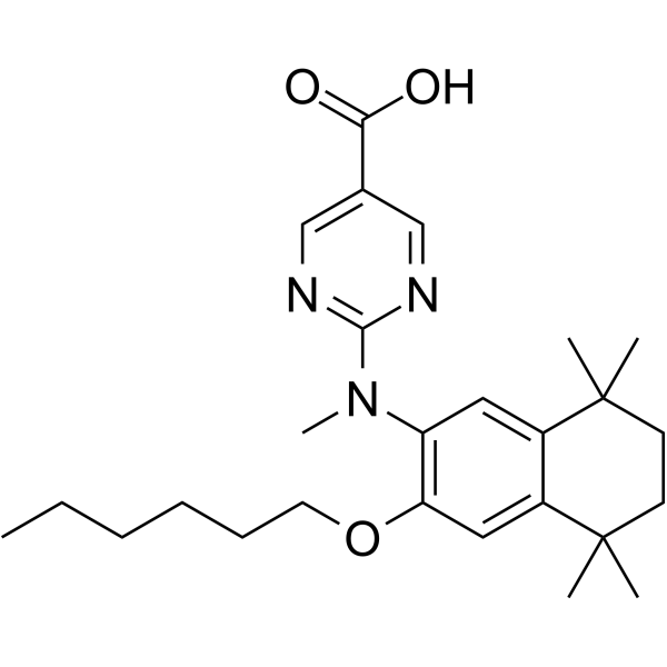
HY-108522
PA452RXR Antagonist -
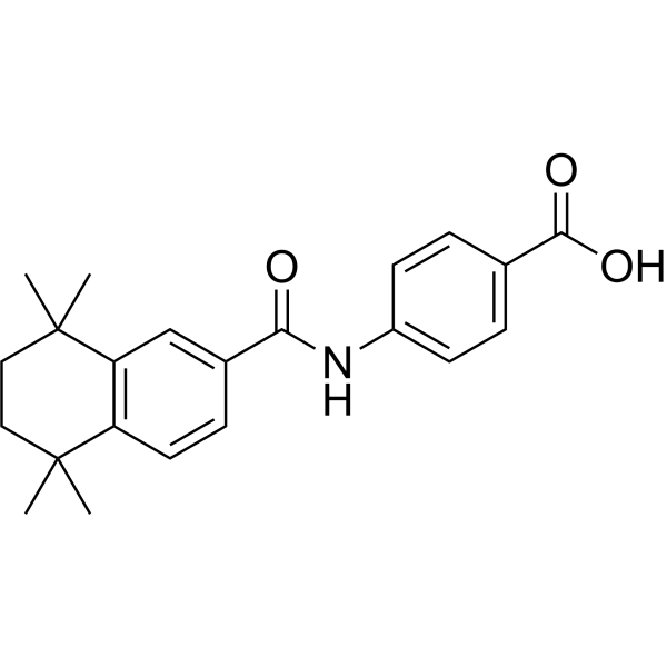
HY-10475
AM580RARα Agonist -
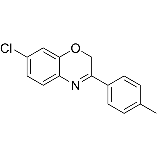
HY-101106
AR7RARA/RARα Antagonist -
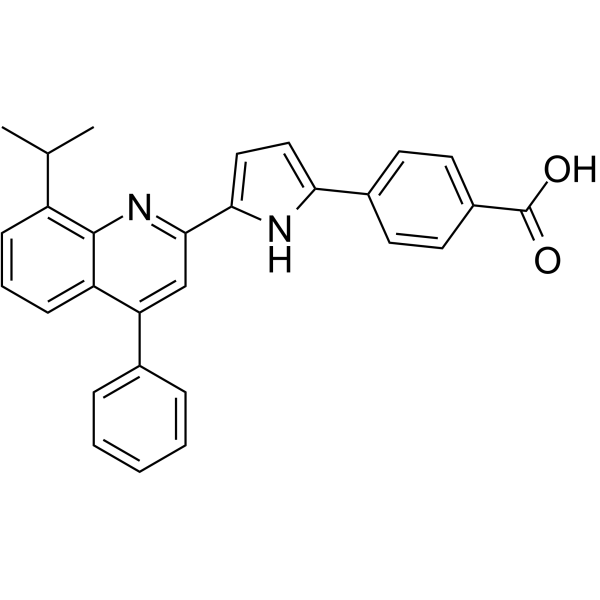
HY-108531
ER 50891RARα Antagonist


Biological Activity
Description
IC50 & Target
RAR α (EC50 = 26 nM);RAR β (EC50 = 1.9 nM);RAR γ (EC50 = 11 nM)[1]
Cellular Effect
|
Cell Line

|
Type | Value | Description | References |
|---|---|---|---|---|
| COS-7 | EC50 |
1.9 nM
Compound: 10
|
Transactivation of GAL4-fused mouse RARbeta-LBD expressed in COS-7 cells after 24 hrs by bright-Glo reagent based assay
Transactivation of GAL4-fused mouse RARbeta-LBD expressed in COS-7 cells after 24 hrs by bright-Glo reagent based assay
|
[PMID: 30792038] |
| COS-7 | EC50 |
13 nM
Compound: 10
|
Transactivation of GAL4-fused mouse RARgamma-LBD expressed in COS-7 cells after 24 hrs by bright-Glo reagent based assay
Transactivation of GAL4-fused mouse RARgamma-LBD expressed in COS-7 cells after 24 hrs by bright-Glo reagent based assay
|
[PMID: 30792038] |
| COS-7 | EC50 |
26 nM
Compound: 10
|
Transactivation of GAL4-fused mouse RARalpha-LBD expressed in COS-7 cells after 24 hrs by bright-Glo reagent based assay
Transactivation of GAL4-fused mouse RARalpha-LBD expressed in COS-7 cells after 24 hrs by bright-Glo reagent based assay
|
[PMID: 30792038] |
| HEK293 | EC50 |
2.05 nM
Compound: 10
|
Transactivation of GAL4 DBD-fused human RARbeta-LBD expressed in HEK293 cells by beta-lactamase reporter gene based assay
Transactivation of GAL4 DBD-fused human RARbeta-LBD expressed in HEK293 cells by beta-lactamase reporter gene based assay
|
[PMID: 30792038] |
In Vitro
KCL-286 (0.1 μM, 72 h) increases the neurite density and the number of synapses to a significant extent which activates RARβ2[2].
MedChemExpress (MCE) has not independently confirmed the accuracy of these methods. They are for reference only. Further protocols information, click here.
In Vivo
KCL-286 (3 mg/kg, p.o., every other days for 4 weeks) improves the hind limb locomotor function in moderate thoracic SC contusion rat model[2].
KCL-286 (3 mg/kg, p.o., every other days for 4 weeks) achieves full locomotor and sensory recovery after 3 weeks in C5-T1 dorsal root avulsion rat model[2].
MedChemExpress (MCE) has not independently confirmed the accuracy of these methods. They are for reference only.
-
Animal Model:Non-injured or avulsed male Sprague-Dawley rats[2]
-
Dosage:3 mg/kg, every other days for 4 weeks
-
Administration:Oral gavage (p.o.)
-
Result:Resulted in 82 genes presenting a return to their basal level and 99 genes being strongly upregulated in injured model.
Resulted in 58 genes returning to their basal level after being significantly downregulated under injured conditions.
Significantly modified genes associated with ECM, paths within this signaling complex were both upregulated (those pertaining to the modulation of the cholinergic system, ion transport, and ion channels) or downregulated by the drug after injury.
Highly upregulated the ligand-gated ion channel.
Regulated the G-protein coupled receptor signaling cascades and G-protein coupled receptor protein pathways and chemokine signalling.
Upregulated RARβ in the cell bodies and axons of all neuron subtypes.
-
Animal Model:Moderate thoracic SC contusion male Sprague-Dawley rat model[2]
-
Dosage:3 mg/kg, every other days for 4 weeks
-
Administration:Oral gavage (p.o.)
-
Result:Significantly reduced the tissue loss that progressively occurs at sub-acute and chronic stages following the injury.
Significantly reduced gliosis, shown by reduction in OX42 expression and neuronal loss assessed by neuronal nuclear protein (NeuN) levels.
Significantly lowered the expression of reactive astrocytes Glial fibrillary acidic protein and microglia Ionized Calcium Binding Adaptor Molecule 1.
Significantly upregulated decorin expression and lowered CSPGs in contused rats.


Chemical Information
-
CAS No. 1952276-71-9
-
Appearance Solid
-
Molecular Weight 334.33
-
Formula C19H14N2O4
-
Color White to off-white
-
SMILES
CC1=CC=C(C)C2=C1OC(C3=NC(C4=CC=C(C(O)=O)C=C4)=NO3)=C2
-
Synonyms
C286
-
Shipping
Room temperature in continental US; may vary elsewhere.
-
Storage
Powder -20°C 3 years 4°C 2 years In solvent -80°C 6 months -20°C 1 month


Solvent & Solubility
In Vitro:
DMSO : 6.25 mg/mL (18.69 mM; ultrasonic and warming and heat to 60°C; Hygroscopic DMSO has a significant impact on the solubility of product, please use newly opened DMSO)
Please refer to the solubility information to select the appropriate solvent. Once prepared, please aliquot and store the solution to prevent product inactivation from repeated freeze-thaw cycles.
Storage method and period of stock solution: -80°C, 6 months; -20°C, 1 month. When stored at -80°C, please use it within 6 months. When stored at -20°C, please use it within 1 month.
Please refer to the solubility information to select the appropriate solvent. Once prepared, please aliquot and store the solution to prevent product inactivation from repeated freeze-thaw cycles.
Storage method and period of stock solution: -80°C, 6 months; -20°C, 1 month. When stored at -80°C, please use it within 6 months. When stored at -20°C, please use it within 1 month.
Concentration (start) × Volume (start) = Concentration (final) × Volume (final)
In Vivo:
Select the appropriate dissolution method based on your experimental animal and administration route.
- For the following dissolution methods, please ensure to first prepare a clear stock solution using an In Vitro approach and then sequentially add co-solvents:
- To ensure reliable experimental results, the clarified stock solution can be appropriately stored based on storage conditions. As for the working solution for In Vivo experiments, it is recommended to prepare freshly and use it on the same day.
- The percentages shown for the solvents indicate their volumetric ratio in the final prepared solution. If precipitation or phase separation occurs during preparation, heat and/or sonication can be used to aid dissolution.
In Vivo Dissolution Calculator
Please enter the basic information of animal experiments:
-
-
-
-
Recommended: Prepare an additional quantity of animals to account for potential losses during experiments.
Please enter your animal formula composition:
-
%DMSO +
Recommended: Keep the proportion of DMSO in working solution below 2% if your animal is weak.
-
%+
-
+%Tween-80 + +
-
%Saline +
The co-solvents required include: DMSO, . All of co-solvents are available by MedChemExpress (MCE). , Tween 80. All of co-solvents are available by MedChemExpress (MCE).
Working solution concentration: 0.22 mg/mL
Method for preparing stock solution: mg drug dissolved in μL DMSO. Stock solution concentration: mg/mL.
1. Take μL DMSO stock solution;
2. Add μL .
μL , mix evenly;
3. Then add μL Tween 80, mix evenly;
4. Then add μL
Please ensure that the stock solution in the first step is dissolved to a clear state, and add co-solvents in sequence. You can use ultrasonic heating (ultrasonic cleaner, recommended frequency 20-40 kHz), vortexing, etc. to assist dissolution.

Protocols
-
Fibrosis/Collagen Morphometry
Fibrosis and collagen morphometry is based on the quantitative visualization of fibrillar collagen deposition in tissue sections using histochemical stains such as Sirius Red (Picrosirius Red) or Masson's trichrome, followed by image-based or polarization-enhanced analysis to estimate collagen proportional area as a surrogate of extracellular matrix accumulation during fibrotic remodeling. Sirius Red combined with polarized light microscopy enhances detection of collagen fibers due to birefringence properties, enabling more specific visualization of collagen type I and III fibrils compared to conventional bright-field histology, while whole-section or region-restricted digital morphometry reduces field-selection bias in fibrosis assessment. Alternative quantitative approaches include second harmonic generation (SHG) and two-photon excited fluorescence microscopy, which enable label-free detection of fibrillar collagen and have been validated against histological staining and biochemica
-
Collagen: Sirius Red Staining
Sirius Red or picrosirius red staining is a histochemical method for visualizing collagen-rich extracellular matrix in tissue sections, and collagen fibers are detected as red-stained structures under bright-field microscopy with enhanced birefringence under polarized light. Picrosirius red is useful for assessing total collagen organization, distribution, and fibrosis burden, but polarized color should not be interpreted as a definitive collagen type I versus type III readout because color is affected by fiber orientation, thickness, and packing.
-
RNA extraction experimental
By lysing cells, releasing RNA, and removing impurities such as proteins and DNA, high-purity RNA products are finally obtained. The commonly used traditional method is the guanidine isothiocyanate/phenol/chloroform method (Trizol), which is suitable for a variety of animal materials including animal tissues, microorganisms, cultured cells, etc., and most plant materials.
-
ECM-Embedded Organoid (Matrigel/Dome) Culture
ECM-embedded organoid dome culture embeds epithelial stem cells, crypts, organoid fragments, or tumor-derived epithelial cells in a basement-membrane-like hydrogel such as Matrigel, allowing 3D growth, self-organization, lumen formation, budding or cystic morphogenesis, and lineage maintenance under defined niche-factor-containing medium. The primary readouts are organoid establishment efficiency, growth, morphology, passaging capacity, lineage-marker expression, and, when fluorescently labeled lines are used, microscopy- or flow-cytometry-based quantification of population behavior in 3D culture.
-
Matrigel/ECM Transwell Invasion Assay
The Matrigel/ECM Transwell invasion assay measures the ability of cells to move toward a chemoattractant while crossing an extracellular-matrix barrier placed on a porous membrane; therefore, the readout reflects both chemotactic motility and matrix invasion rather than migration alone. Matrigel is a basement-membrane-rich matrix derived from Engelbreth-Holm-Swarm mouse sarcoma and has been used as a reconstituted basement membrane barrier in chemoinvasion assays. The assay readout is generated by quantifying cells that reach the underside of the insert membrane or lower compartment after incubation, commonly by staining and counting invaded cells or by fluorescence-based quantification.
-
Research Protocol for Neurological Diseases
PINK1/Parkin-mediated mitophagy pathway is a mitochondrial quality-control signaling axis in which mitochondrial depolarization stabilizes PINK1 on damaged mitochondria, activates Parkin recruitment and E3 ubiquitin ligase activity, promotes ubiquitination of outer mitochondrial membrane proteins, recruits selective autophagy adaptors, and drives lysosomal degradation of damaged mitochondria. In neurological disease research, this pathway is experimentally important because neurons, especially dopaminergic neurons, are highly dependent on mitochondrial integrity, and defective mitochondrial turnover can lead to mitochondrial dysfunction, oxidative stress, impaired neuronal survival, α-synuclein accumulation, and neuroinflammatory damage-associated signals. The genetic disease link is strongest in Parkinson’s disease because mutations in PRKN/parkin cause autosomal recessive juvenile parkinsonism, mutations in PINK1 cause hereditary early-onset Parkinson’s disease, and Drosophila studie
-
Matrigel Transwell/Boyden Chamber Invasion Assay
Matrigel Transwell/Boyden chamber invasion assay measures the ability of cells to degrade or traverse an extracellular matrix-coated porous membrane and move from an upper chamber toward a chemoattractant in a lower chamber. Invasion is distinguished from migration by coating the membrane with Matrigel or basement membrane matrix; uncoated inserts measure migration, while coated inserts require cells to cross an ECM barrier before reaching the underside of the membrane.


Purity & Documentation
-
Data Sheet (279 KB)
-
SDS (393 KB)
- English - EN (393 KB)
- Français - FR (393 KB)
- Deutsch - DE (393 KB)
- Norwegian - NO (393 KB)
- Español - ES (393 KB)
- Swedish - SV (393 KB)
- Italian - IT (393 KB)
- Korean - KR (393 KB)
- Portuguese - PT (393 KB)
-
Handling Instructions (2659 KB)
References
[1]. Goncalves MB, et al. Phase 1 safety, tolerability, pharmacokinetics and pharmacodynamic results of KCL-286, a novel retinoic acid receptor-β agonist for treatment of spinal cord injury, in male healthy participants. Br J Clin Pharmacol. 2023 Jul 15.. [Content Brief]
[2]. Goncalves, M. B., (2024). C286, an orally available retinoic acid receptor β agonist drug, regulates multiple pathways to achieve spinal cord injury repair. Frontiers in molecular neuroscience, 17, 1411384. [Content Brief]
[3]. Borthwick, A. D., et al., (2020). Recent advances in the design of RAR α and RAR β agonists as orally bioavailable drugs. A review. Bioorganic & medicinal chemistry, 28(20), 115664. [Content Brief]


Complete Stock Solution Preparation Table
Please refer to the solubility information to select the appropriate solvent. Once prepared, please aliquot and store the solution to prevent product inactivation from repeated freeze-thaw cycles.
Storage method and period of stock solution: -80°C, 6 months; -20°C, 1 month. When stored at -80°C, please use it within 6 months. When stored at -20°C, please use it within 1 month.
| Optional Solvent | Concentration Solvent Mass | 1 mg | 5 mg | 10 mg | 25 mg |
|---|---|---|---|---|---|
| DMSO | 1 mM | 2.9911 mL | 14.9553 mL | 29.9106 mL | 74.7764 mL |
| 5 mM | 0.5982 mL | 2.9911 mL | 5.9821 mL | 14.9553 mL | |
| 10 mM | 0.2991 mL | 1.4955 mL | 2.9911 mL | 7.4776 mL | |
| 15 mM | 0.1994 mL | 0.9970 mL | 1.9940 mL | 4.9851 mL |


KCL-286 Related Classifications
HY-111573 Related Classifications
 Powered by Bioz