



APG-2449

Based on 1 Customer Validation
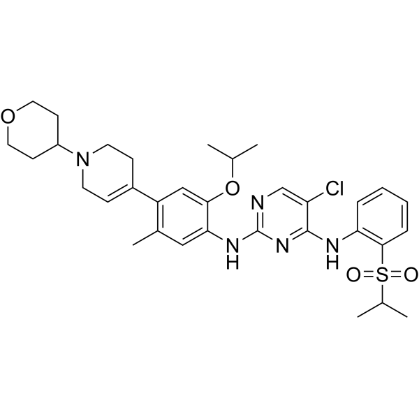
APG-2449 is an orally active inhibitor for BCL-2 and multikinase (ALK/FAK/ROS1) with potent antitumor activities. APG-2449 reduces cell viability and enhances apoptosis in acute myeloid leukemia cells in vitro. APG-2449 decreases activation of FAK and its downstream effectors. APG-2449 can be studied in research for mesothelioma tumor, non-small cell lung cancer, ovarian cancer, hematologic and solid malignancies.
For research use only. We do not sell to patients.
- Purity : 99.08%
- CAS No.: 2196186-84-0
- Formula: C33H42ClN5O4S
- Molecular Weight:640.24
-
Storage:Powder -20°C, 3 years , 4°C, 2 years ; In solvent -80°C, 6 months , -20°C, 1 month

To place orders, for customer services and technical support, please contact: MedChemExpress USA
Tel: 609-228-6898 E-mail: [email protected] [email protected]


-
 Biological Activity
-
Chemical Information
-
Solvent & Solubility
- Protocols
- Purity & Documentation
- References
-
 Help & FAQs




Biological Activity
Description
In Vitro
APG-2449 inhibits ALK, ROS1, and FAK with Kd values if 1.6, 0.81, and 5.4 nM, respectively[1].
APG-2449 exhibits moderate antiproliferative activity in cells without ALK or ROS1 rearrangements but with detectable expression of FAK protein (e.g. IC50 = 3.55 μM in NCI-H1975 and 2.71 μM in PA-1)[1].
APG-2449 (0-10 μM, 6-24 h) exerts anti-cancer effect mediated by suppressing ALK- or ROS1-driven signaling pathway in HCC78 cells carrying SLC34A2-ROS1 fusion[1].
APG-2449 (0-10 μM, 4 h) downregulates p-FAK and downstream signaling factors in PA-1 ovarian cancer cells[1].
APG-2449 (1-5 μM, 72 h) dose-dependently decreases numbers of CD44+ and ALDH1+ SKOV-3 ovarian cancer stem cells[1].
APG-2449 (0.5-1 μM, 14 d) significantly decreases the colony formation in KYSE-150 and KYSE-520 cells[4].
APG-2449 (1-2 μM, 24 h) inhibits the migration ability of esophageal squamous cell carcinoma (ESCC) cells[4].
APG-2449 (0-2 μM, 24-72 h) significantly suppresses cell viability when combined with Ibrutinib (HY-10997) compared with single-agent in KYSE-150 and KYSE-520 cells[4].
MedChemExpress (MCE) has not independently confirmed the accuracy of these methods. They are for reference only.
-
Cell Line:HCC78 cells carrying SLC34A2-ROS1 fusion
-
Concentration:2.5, 5 and 10 μM
-
Incubation Time:6 or 24 h
-
Result:Induced downregulation of p-ROS1, p-AKT, p-ERK1/2, and p-STAT3 after 24 h.
In Vivo
APG-2449 (25-100 mg/kg, p.o., once daily) exerts potent antitumor activity in severe combined immunodeficient (SCID) mice bearing KARPAS-299 CDX tumors with NPM-ALK fusion[1].
APG-2449 (50-150 mg/kg, p.o., 15-22 d) exerts potent and durable antitumor activity against secondary ALK- and ROS1-resistant mutations in tumor-bearing mice model[1].
APG-2449 (50-150 mg/kg, p.o., 22 d) shows dose-dependent antitumor activity in SCID mice bearing PA-1 CDX tumors[1].
APG-2449 (20-60 mg/kg, p.o., 22 d) demonstrates potent antitumor activity in combination with Osimertinib (HY-15772) and Trametinib (HY-10999) in PC-9/OR xenograft models[1].
APG-2449 (50-100 mg/kg, p.o., once daily for 2 weeks) significantly suppresses tumor growth in both KYSE-150 and KYSE-520 tumor cells xenograft models at 100 mg/kg[4].
MedChemExpress (MCE) has not independently confirmed the accuracy of these methods. They are for reference only.
-
Animal Model:Nude mice bearing NSCLC H3122 CDX tumors with EML4-ALK fusion[1]
-
Dosage:25, 50 and 100 mg/kg
-
Administration:Oral gavage (p.o.), once daily
-
Result:Had anti-tumor activity in a dose-dependent manner.
Did not lead to severe body weight loss.
-
Animal Model:Severe combined immunodeficient (SCID) mice bearing KARPAS-299 CDX tumors with NPM-ALK fusion[1]
-
Dosage:25, 50 and 100 mg/kg
-
Administration:Oral gavage (p.o.)
-
Result:Exerted dose-dependent suppression of tumor phosphorylated ALK, Ak strain transforming, extracellular signal-regulated kinase and signal transducer and activator of transcription 24 hours after single dose administration.
Resulted in dose-proportional increases in APG-2449 plasma and tumor exposures.
- Detection of Apoptosis
- TUNEL staining for apoptotic DNA fragmentation
- Cell Cytotoxicity Assay
- Mammalian live/dead viability and cytotoxicity staining
- Annexin V plus membrane-impermeant dye apoptosis staining
- Apoptosis Solutions
- CCK-8/WST-8 Cell Proliferation Assay
- MTT Cell Proliferation Assay
- Cell Viability Determination by MTT Colorimetric Assay
Clinical Trial
| NCT Number | Sponsor | Condition | Start Date |
Phase

|
|---|---|---|---|---|
| NCT01329991 | 2011-05 | PHASE1 |


Chemical Information
-
CAS No. 2196186-84-0
-
Appearance Solid
-
Molecular Weight 640.24
-
Formula C33H42ClN5O4S
-
Color Off-white to light yellow
-
SMILES
O=S(C1=C(NC2=NC(NC3=CC(C)=C(C4=CCN(C5CCOCC5)CC4)C=C3OC(C)C)=NC=C2Cl)C=CC=C1)(C(C)C)=O
-
Shipping
Room temperature in continental US; may vary elsewhere.
-
Storage
Powder -20°C 3 years 4°C 2 years In solvent -80°C 6 months -20°C 1 month


Solvent & Solubility
In Vitro:
DMSO : 25 mg/mL (39.05 mM; Need ultrasonic; Hygroscopic DMSO has a significant impact on the solubility of product, please use newly opened DMSO)
Please refer to the solubility information to select the appropriate solvent. Once prepared, please aliquot and store the solution to prevent product inactivation from repeated freeze-thaw cycles.
Storage method and period of stock solution: -80°C, 6 months; -20°C, 1 month. When stored at -80°C, please use it within 6 months. When stored at -20°C, please use it within 1 month.
Please refer to the solubility information to select the appropriate solvent. Once prepared, please aliquot and store the solution to prevent product inactivation from repeated freeze-thaw cycles.
Storage method and period of stock solution: -80°C, 6 months; -20°C, 1 month. When stored at -80°C, please use it within 6 months. When stored at -20°C, please use it within 1 month.
Concentration (start) × Volume (start) = Concentration (final) × Volume (final)
In Vivo:
Select the appropriate dissolution method based on your experimental animal and administration route.
- For the following dissolution methods, please ensure to first prepare a clear stock solution using an In Vitro approach and then sequentially add co-solvents:
- To ensure reliable experimental results, the clarified stock solution can be appropriately stored based on storage conditions. As for the working solution for In Vivo experiments, it is recommended to prepare freshly and use it on the same day.
- The percentages shown for the solvents indicate their volumetric ratio in the final prepared solution. If precipitation or phase separation occurs during preparation, heat and/or sonication can be used to aid dissolution.
Add each solvent one by one: 10% DMSO 40% PEG300 5% Tween-80 45% Saline
Solubility: ≥ 2.5 mg/mL (3.90 mM); Clear solution
This protocol yields a clear solution of ≥ 2.5 mg/mL (saturation unknown).
Taking 1 mL working solution as an example, add 100 μL DMSO stock solution (25.0 mg/mL) to 400 μL PEG300, and mix evenly; then add 50 μL Tween-80 and mix evenly; then add 450 μL Saline to adjust the volume to 1 mL.
Preparation of Saline: Dissolve 0.9 g sodium chloride in ddH₂O and dilute to 100 mL to obtain a clear Saline solution.
In Vivo Dissolution Calculator
Please enter the basic information of animal experiments:
-
-
-
-
Recommended: Prepare an additional quantity of animals to account for potential losses during experiments.
Please enter your animal formula composition:
-
%DMSO +
Recommended: Keep the proportion of DMSO in working solution below 2% if your animal is weak.
-
%+
-
+%Tween-80 + +
-
%Saline +
The co-solvents required include: DMSO, . All of co-solvents are available by MedChemExpress (MCE). , Tween 80. All of co-solvents are available by MedChemExpress (MCE).
Working solution concentration: 0.22 mg/mL
Method for preparing stock solution: mg drug dissolved in μL DMSO. Stock solution concentration: mg/mL.
1. Take μL DMSO stock solution;
2. Add μL .
μL , mix evenly;
3. Then add μL Tween 80, mix evenly;
4. Then add μL
Please ensure that the stock solution in the first step is dissolved to a clear state, and add co-solvents in sequence. You can use ultrasonic heating (ultrasonic cleaner, recommended frequency 20-40 kHz), vortexing, etc. to assist dissolution.


Purity & Documentation
-
Data Sheet (279 KB)
-
SDS (251 KB)
- English - EN (251 KB)
- Français - FR (251 KB)
- Deutsch - DE (251 KB)
- Norwegian - NO (251 KB)
- Español - ES (251 KB)
- Swedish - SV (251 KB)
- Italian - IT (251 KB)
- Korean - KR (251 KB)
- Portuguese - PT (251 KB)
-
Handling Instructions (2659 KB)
References
[1]. Fang DD, et al. Discovery of a novel ALK/ROS1/FAK inhibitor, APG-2449, in preclinical non-small cell lung cancer and ovarian cancer models. BMC Cancer. 2022 Jul 11;22(1):752. [Content Brief]
[4]. Luo, Q. Y., et al., (2021). A multi-kinase inhibitor APG-2449 enhances the antitumor effect of ibrutinib in esophageal squamous cell carcinoma via EGFR/FAK pathway inhibition. Biochemical pharmacology, 183, 114318. [Content Brief]


Complete Stock Solution Preparation Table
Please refer to the solubility information to select the appropriate solvent. Once prepared, please aliquot and store the solution to prevent product inactivation from repeated freeze-thaw cycles.
Storage method and period of stock solution: -80°C, 6 months; -20°C, 1 month. When stored at -80°C, please use it within 6 months. When stored at -20°C, please use it within 1 month.
| Optional Solvent | Concentration Solvent Mass | 1 mg | 5 mg | 10 mg | 25 mg |
|---|---|---|---|---|---|
| DMSO | 1 mM | 1.5619 mL | 7.8096 mL | 15.6191 mL | 39.0479 mL |
| 5 mM | 0.3124 mL | 1.5619 mL | 3.1238 mL | 7.8096 mL | |
| 10 mM | 0.1562 mL | 0.7810 mL | 1.5619 mL | 3.9048 mL | |
| 15 mM | 0.1041 mL | 0.5206 mL | 1.0413 mL | 2.6032 mL | |
| 20 mM | 0.0781 mL | 0.3905 mL | 0.7810 mL | 1.9524 mL | |
| 25 mM | 0.0625 mL | 0.3124 mL | 0.6248 mL | 1.5619 mL | |
| 30 mM | 0.0521 mL | 0.2603 mL | 0.5206 mL | 1.3016 mL |


APG-2449 Related Classifications
HY-155163 Related Classifications
 Powered by Bioz