



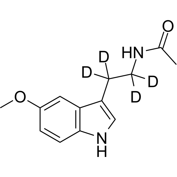
Melatonin-d4

Based on 1 Customer Validation
Melatonin-d4 is deuterium labeled Melatonin. Melatonin is a hormone made by the pineal gland that can activates melatonin receptor. Antioxidative and anti-inflammatory properties. Melatonin is a selective ATF-6 inhibitor and induces human hepatoma cell apoptosis through COX-2 downregulation.
Nur für Forschungszwecke. Wir verkaufen nicht an Patienten.
- Reinheit : 99.61%
- CAS. Nr.: 66521-38-8
- Formel: C13H12D4N2O2
- Molecular Weight:236.30
-
Speicherung:Powder -20°C, 3 years , 4°C, 2 years ; In solvent -80°C, 6 months , -20°C, 1 month

To place orders, for customer services and technical support, please contact: MedChemExpress USA
Tel: 609-228-6898 E-mail: [email protected] [email protected]


-
 Biologische Aktivität
-
Chemical Information
-
Lösungsmittel & Löslichkeit
- Protokoll
- Reinheit & Dokumentation
- Verweise
-
 Help & FAQs


-
HY-15142
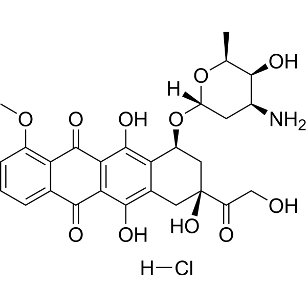
Doxorubicin hydrochlorideTopoisomerase Inhibitor -
HY-A0014
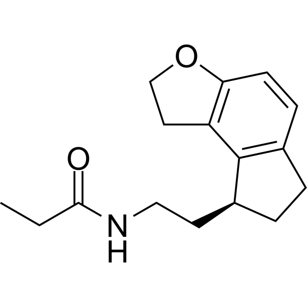
RamelteonMT1/2 Inhibitor -

HY-B0075R
Melatonin (standard)Melatonin Receptor Activator -
HY-14608S7
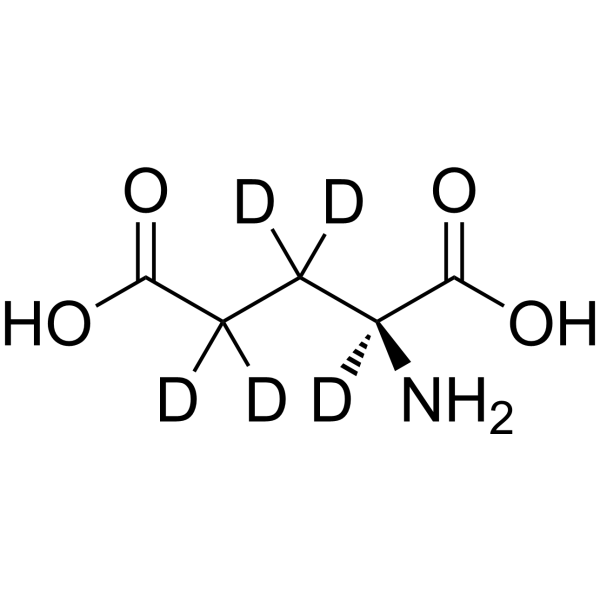
L-Glutamic acid-d5Stable Isotope -
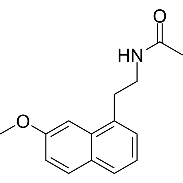
HY-17038
AgomelatineMT1/2 Agonist -
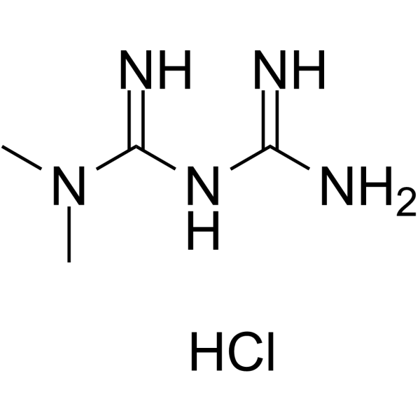
HY-17471A
Metformin hydrochlorideAntihyperglycemic Agent -
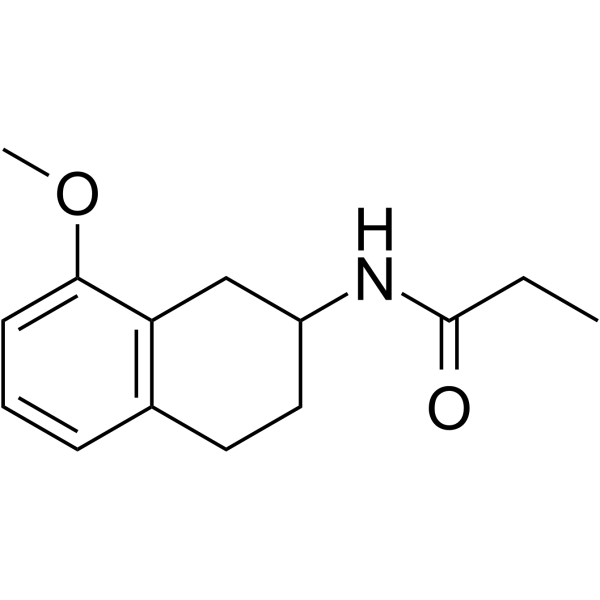
HY-101358
8-M-PDOTMT2 Receptor Agonist -

HY-A0014R
Ramelteon (Standard)MT1/2 Inhibitor -
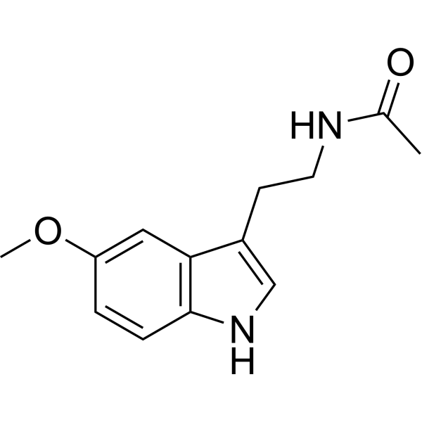
HY-B0075
MelatoninMelatonin Receptor Activator -

HY-17038R
Agomelatine (Standard)MT1/2 Agonist -
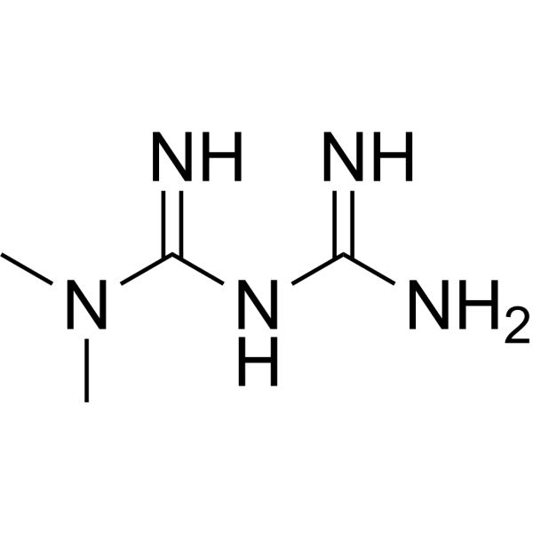
HY-B0627
MetforminAntihyperglycemic Agent -
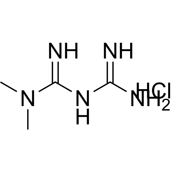
HY-17471AR
Metformin hydrochloride (Standard)Analytical Standard


Biologische Aktivität
Beschreibung
In Vitro
Stable heavy isotopes of hydrogen, carbon, and other elements have been incorporated into drug molecules, largely as tracers for quantitation during the drug development process. Deuteration has gained attention because of its potential to affect the pharmacokinetic and metabolic profiles of drugs[1].
MedChemExpress (MCE) has not independently confirmed the accuracy of these methods. They are for reference only.
Anwendung
1. This compound can be used as a tracer
2. This compound can be used as an internal standard for quantitative analysis by NMR, GC-MS, or LC-MS.


Chemical Information
-
CAS. Nr. 66521-38-8
-
Unlabeled CAS 73-31-4
-
Appearance Solid
-
Molecular Weight 236.30
-
Formel C13H12D4N2O2
-
Color White to off-white
-
SMILES
CC(NC([2H])([2H])C([2H])([2H])C1=CNC2=C1C=C(OC)C=C2)=O
-
Synonyms
N-Acetyl-5-methoxytryptamine-d4
-
Versand
Room temperature in continental US; may vary elsewhere.
-
Speicherung
Powder -20°C 3 years 4°C 2 years In solvent -80°C 6 months -20°C 1 month


Lösungsmittel & Löslichkeit
In Vitro:
DMSO : 100 mg/mL (423.19 mM; Need ultrasonic; Hygroscopic DMSO has a significant impact on the solubility of product, please use newly opened DMSO)
Please refer to the solubility information to select the appropriate solvent. Once prepared, please aliquot and store the solution to prevent product inactivation from repeated freeze-thaw cycles.
Storage method and period of stock solution: -80°C, 6 months; -20°C, 1 month. When stored at -80°C, please use it within 6 months. When stored at -20°C, please use it within 1 month.
Please refer to the solubility information to select the appropriate solvent. Once prepared, please aliquot and store the solution to prevent product inactivation from repeated freeze-thaw cycles.
Storage method and period of stock solution: -80°C, 6 months; -20°C, 1 month. When stored at -80°C, please use it within 6 months. When stored at -20°C, please use it within 1 month.
Konzentration (Stammlösung) × Volumen (Stammlösung) = Konzentration (Ziellösung) × Volumen (Ziellösung)
In Vivo:
Select the appropriate dissolution method based on your experimental animal and administration route.
- For the following dissolution methods, please ensure to first prepare a clear stock solution using an In Vitro approach and then sequentially add co-solvents:
- To ensure reliable experimental results, the clarified stock solution can be appropriately stored based on storage conditions. As for the working solution for In Vivo experiments, it is recommended to prepare freshly and use it on the same day.
- The percentages shown for the solvents indicate their volumetric ratio in the final prepared solution. If precipitation or phase separation occurs during preparation, heat and/or sonication can be used to aid dissolution.
Add each solvent one by one: 10% DMSO 40% PEG300 5% Tween-80 45% Saline
Solubility: ≥ 2.5 mg/mL (10.58 mM); Clear solution
This protocol yields a clear solution of ≥ 2.5 mg/mL (saturation unknown).
Taking 1 mL working solution as an example, add 100 μL DMSO stock solution (25.0 mg/mL) to 400 μL PEG300, and mix evenly; then add 50 μL Tween-80 and mix evenly; then add 450 μL Saline to adjust the volume to 1 mL.
Preparation of Saline: Dissolve 0.9 g sodium chloride in ddH₂O and dilute to 100 mL to obtain a clear Saline solution.
Add each solvent one by one: 10% DMSO 90% (20% SBE-β-CD in Saline)
Solubility: ≥ 2.5 mg/mL (10.58 mM); Clear solution
This protocol yields a clear solution of ≥ 2.5 mg/mL (saturation unknown).
Taking 1 mL working solution as an example, add 100 μL DMSO stock solution (25.0 mg/mL) to 900 μL 20% SBE-β-CD in Saline, and mix evenly.
Preparation of 20% SBE-β-CD in Saline (4°C, storage for one week): 2 g SBE-β-CD powder is dissolved in 10 mL Saline, completely dissolve until clear.
In Vivo Dissolution Calculator
Please enter the basic information of animal experiments:
-
-
-
-
Recommended: Prepare an additional quantity of animals to account for potential losses during experiments.
Please enter your animal formula composition:
-
%DMSO +
Recommended: Keep the proportion of DMSO in working solution below 2% if your animal is weak.
-
%+
-
+%Tween-80 + +
-
%Saline +
The co-solvents required include: DMSO, . All of co-solvents are available by MedChemExpress (MCE). , Tween 80. All of co-solvents are available by MedChemExpress (MCE).
Working solution concentration: 0.22 mg/mL
Method for preparing stock solution: mg drug dissolved in μL DMSO. Stock solution concentration: mg/mL.
1. Take μL DMSO stock solution;
2. Add μL .
μL , mix evenly;
3. Then add μL Tween 80, mix evenly;
4. Then add μL
Please ensure that the stock solution in the first step is dissolved to a clear state, and add co-solvents in sequence. You can use ultrasonic heating (ultrasonic cleaner, recommended frequency 20-40 kHz), vortexing, etc. to assist dissolution.


Reinheit & Dokumentation
-
Data Sheet (277 KB)
-
SDS (419 KB)
- English - EN (419 KB)
- Français - FR (419 KB)
- Deutsch - DE (419 KB)
- Norwegian - NO (419 KB)
- Español - ES (419 KB)
- Swedish - SV (419 KB)
- Italian - IT (419 KB)
- Korean - KR (419 KB)
- Portuguese - PT (419 KB)
-
Handling Instructions (2659 KB)
Verweise
[1]. Kilic U, et al. Particular phosphorylation of PI3K/Akt on Thr308 via PDK-1 and PTEN mediates melatonin's neuroprotective activity after focal cerebral ischemia in mice. Redox Biol. 2017 Apr 5;12:657-665 [Content Brief]
[2]. Hu C, et al. Neuroprotective effect of melatonin on soluble Aβ1-42-induced cortical neurodegeneration via Reelin-Dab1 signaling pathway. Neurol Res. 2017 Apr 7:1-1 [Content Brief]
[3]. Rahim I, et al. Melatonin administration to wild-type mice and non-treated NLRP3 mutant mice share similar inhibition of the inflammatory response during sepsis. J Pineal Res. 2017 Mar 31 [Content Brief]
[4]. Bu LJ, et al. Melatonin, a novel selective ATF-6 inhibitor, induces human hepatoma cell apoptosis through COX-2 downregulation. World J Gastroenterol. 2017 Feb 14;23(6):986-998. [Content Brief]


Complete Stock Solution Preparation Table
Please refer to the solubility information to select the appropriate solvent. Once prepared, please aliquot and store the solution to prevent product inactivation from repeated freeze-thaw cycles.
Storage method and period of stock solution: -80°C, 6 months; -20°C, 1 month. When stored at -80°C, please use it within 6 months. When stored at -20°C, please use it within 1 month.
| Optional Solvent | Concentration Solvent Mass | 1 mg | 5 mg | 10 mg | 25 mg |
|---|---|---|---|---|---|
| DMSO | 1 mM | 4.2319 mL | 21.1595 mL | 42.3191 mL | 105.7977 mL |
| 5 mM | 0.8464 mL | 4.2319 mL | 8.4638 mL | 21.1595 mL | |
| 10 mM | 0.4232 mL | 2.1160 mL | 4.2319 mL | 10.5798 mL | |
| 15 mM | 0.2821 mL | 1.4106 mL | 2.8213 mL | 7.0532 mL | |
| 20 mM | 0.2116 mL | 1.0580 mL | 2.1160 mL | 5.2899 mL | |
| 25 mM | 0.1693 mL | 0.8464 mL | 1.6928 mL | 4.2319 mL | |
| 30 mM | 0.1411 mL | 0.7053 mL | 1.4106 mL | 3.5266 mL | |
| 40 mM | 0.1058 mL | 0.5290 mL | 1.0580 mL | 2.6449 mL | |
| 50 mM | 0.0846 mL | 0.4232 mL | 0.8464 mL | 2.1160 mL | |
| 60 mM | 0.0705 mL | 0.3527 mL | 0.7053 mL | 1.7633 mL | |
| 80 mM | 0.0529 mL | 0.2645 mL | 0.5290 mL | 1.3225 mL | |
| 100 mM | 0.0423 mL | 0.2116 mL | 0.4232 mL | 1.0580 mL |


Melatonin-d4 Related Classifications
HY-B0075S Related Classifications
 Powered by Bioz