



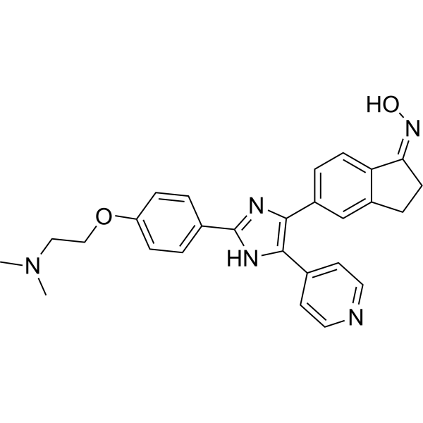
SB-590885
Based on 9 publication(s) in Google Scholar
SB-590885 is a BRAF/c-Raf kinase inhibitor that selectively targets B-Raf, and it amplifies the ERK/MAPK signaling pathway in RAS-activated cells. SB-590885 effectively inhibits the malignant proliferation, transformation and tumorigenicity of oncogenic B-Raf cells; it also induces the proliferation of erythroid progenitor cells, delays their differentiation and promotes hemoglobin synthesis, thereby improving ineffective erythropoiesis and reducing apoptosis. SB-590885 exerts a synergistic effect with TGF-β inhibitors and glucocorticoids, significantly promoting the formation of erythroid colonies in cells from patients with Diamond-Blackfan anemia (DBA). SB-590885 is mainly used in relevant studies on DBA, cisplatin-induced myelosuppression-related anemia, and pan-cancers such as melanoma and colorectal cancer.
Nur für Forschungszwecke. Wir verkaufen nicht an Patienten.
- Reinheit : 99.77%
- CAS. Nr.: 405554-55-4
- Formel: C27H27N5O2
- Molecular Weight:453.54
-
Speicherung:Powder -20°C, 3 years , 4°C, 2 years ; In solvent -80°C, 2 years , -20°C, 1 year

To place orders, for customer services and technical support, please contact: MedChemExpress USA
Tel: 609-228-6898 E-mail: [email protected] [email protected]


-
 Biologische Aktivität
-
Chemical Information
-
Lösungsmittel & Löslichkeit
- Protokoll
- Reinheit & Dokumentation
- Verweise
-
 Help & FAQs


-
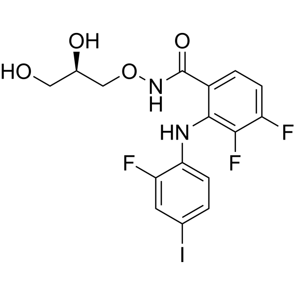
HY-10254
MirdametinibMEK Inhibitor -

HY-10182
LaduviglusibGSK-3 Inhibitor -
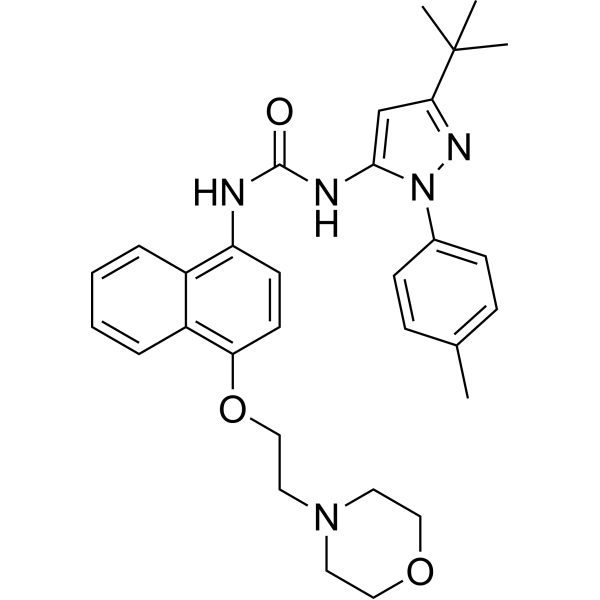
HY-10320
Doramapimodp38 MAPK Inhibitor -
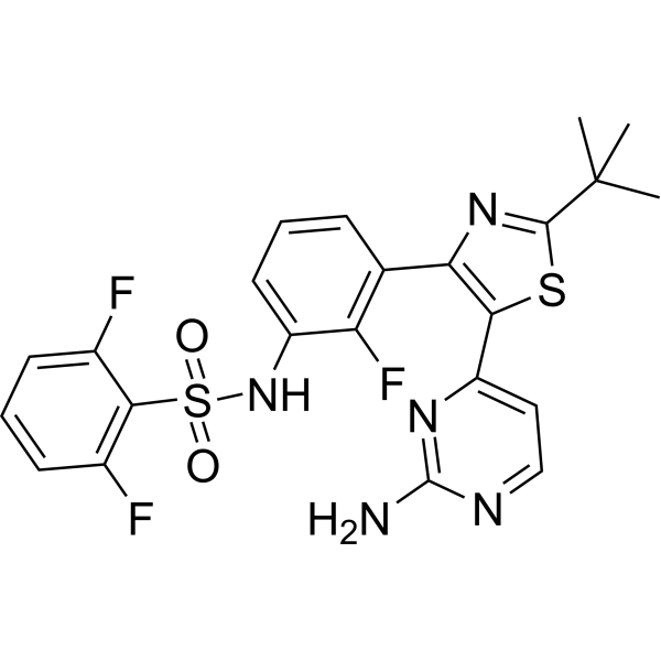
HY-14660
DabrafenibBRAF Inhibitor -
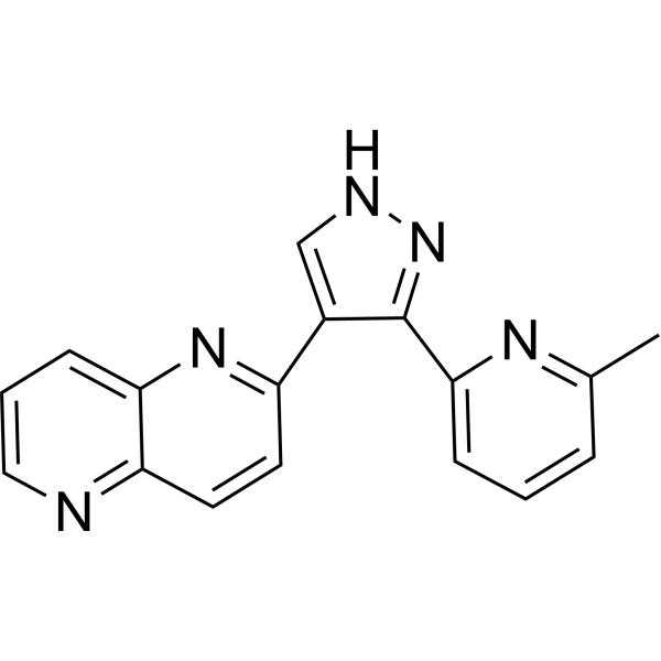
HY-13012
RepSoxTGFβR-1/ALK5 Inhibitor -
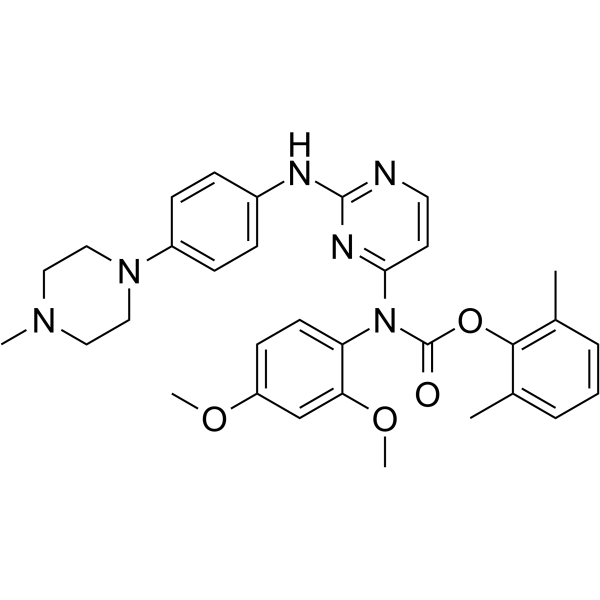
HY-12299
WH-4-023Src Inhibitor -
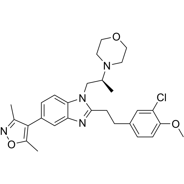
HY-15826
SGC-CBP30CBP/p300 Inhibitor -
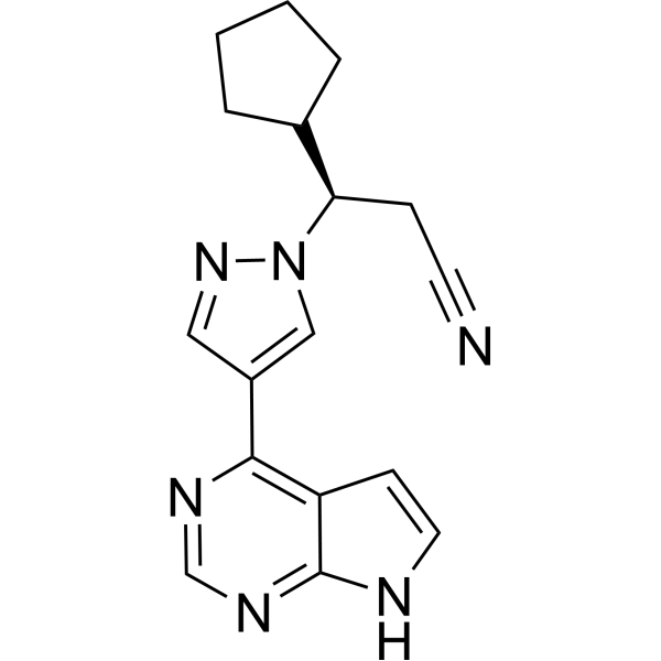
HY-50856
RuxolitinibJAK1/2 Inhibitor -
HY-12848
SAGSmoothened Agonist -
HY-12292
IM-12GSK-3β Inhibitor -

HY-10071
Y-27632ROCK Inhibitor -
HY-10585
Valproic acidHDAC Inhibitor
Publications Citing Use of MedChemExpress (MCE) SB-590885
More- Signal Transduct Target Ther. 2024 Dec 2;9(1):338. [Abstract]
- Cell Stem Cell. 2025 Oct 2;32(10):1509-1527.e9. [Abstract]
- Oncogene. 2024 Jun;43(27):2078-2091. [Abstract]
- Antioxidants (Basel). 2026 May 12;15(5):612. [Abstract]
- Mol Syst Biol. 2015 Mar 26;11(3):797. [Abstract]
- Cancers (Basel). 2020 Jun 10;12(6):1516. [Abstract]
- bioRxiv. 2026 Jan 17.
- bioRxiv. 2025 May 3:2025.04.29.651188. [Abstract]
- ACS Comb Sci. 2019 Dec 9;21(12):805-816. [Abstract]
 Customer Validation & Images

-
Cell Proliferation/Viability Assay
-
Microbiological Assay
-
WB
-
WB
-
IP


Biologische Aktivität
Beschreibung
IC50 & Target
[1]|
B-Raf 0.16 nM (Ki) |
C-Raf 1.72 nM (Ki) |
Cellular Effect
|
Cell Line

|
Type | Value | Description | References |
|---|---|---|---|---|
| A-375 | IC50 |
370 nM
Compound: 2; SB-590885
|
Cytotoxicity against human A375 cells harboring BRAF V600E mutant after 48 hrs by CellTiter-Glo assay
Cytotoxicity against human A375 cells harboring BRAF V600E mutant after 48 hrs by CellTiter-Glo assay
|
[PMID: 29461827] |
| A549 | EC50 |
0.028 μM
Compound: 6, SB-590885
|
Inhibition of B-Raf V600E mutant-mediated Erk phosphorylation in human A549 cells after 60 mins by Western blot analysis
Inhibition of B-Raf V600E mutant-mediated Erk phosphorylation in human A549 cells after 60 mins by Western blot analysis
|
[PMID: 22222036] |
| COLO 205 | EC50 |
0.028 μM
Compound: 6, SB-590885
|
Inhibition of B-Raf V600E mutant-mediated Erk phosphorylation in human Colo205 cells after 60 mins by Western blot analysis
Inhibition of B-Raf V600E mutant-mediated Erk phosphorylation in human Colo205 cells after 60 mins by Western blot analysis
|
[PMID: 22222036] |
| HCT-116 | EC50 |
1.1 μM
Compound: 6, SB-590885
|
Growth inhibition of human HCT116 cells expressing wild-type B-Raf and K-Ras2 G13D mutant after 72 hrs by WST-1 assay
Growth inhibition of human HCT116 cells expressing wild-type B-Raf and K-Ras2 G13D mutant after 72 hrs by WST-1 assay
|
[PMID: 22222036] |
| HFF | EC50 |
1.1 μM
Compound: 6, SB-590885
|
Inhibition of B-Raf-mediated Erk phosphorylation in human HFF cells after 60 mins by Western blot analysis
Inhibition of B-Raf-mediated Erk phosphorylation in human HFF cells after 60 mins by Western blot analysis
|
[PMID: 22222036] |
| HMEC | EC50 |
1.1 μM
Compound: 6, SB-590885
|
Inhibition of B-Raf-mediated Erk phosphorylation in human HMEC cells after 60 mins by Western blot analysis
Inhibition of B-Raf-mediated Erk phosphorylation in human HMEC cells after 60 mins by Western blot analysis
|
[PMID: 22222036] |
| HT-29 | EC50 |
0.028 μM
Compound: 6, SB-590885
|
Inhibition of B-Raf V600E mutant-mediated Erk phosphorylation in human HT-29 cells after 60 mins by Western blot analysis
Inhibition of B-Raf V600E mutant-mediated Erk phosphorylation in human HT-29 cells after 60 mins by Western blot analysis
|
[PMID: 22222036] |
| PrEC | EC50 |
1.1 μM
Compound: 6, SB-590885
|
Inhibition of B-Raf-mediated Erk phosphorylation in human PREC cells after 60 mins by Western blot analysis
Inhibition of B-Raf-mediated Erk phosphorylation in human PREC cells after 60 mins by Western blot analysis
|
[PMID: 22222036] |
| SK-MEL-2 | EC50 |
1.1 μM
Compound: 6, SB-590885
|
Growth inhibition of human SK-MEL-2 cells expressing wild-type B-Raf and N-Ras2 Q61R mutant after 72 hrs by WST-1 assay
Growth inhibition of human SK-MEL-2 cells expressing wild-type B-Raf and N-Ras2 Q61R mutant after 72 hrs by WST-1 assay
|
[PMID: 22222036] |
In Vitro
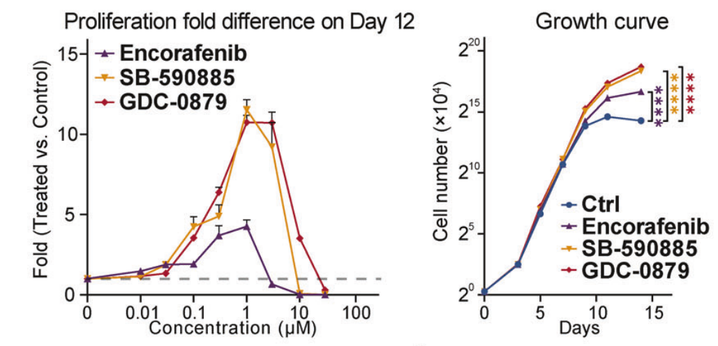
SB-590885(1 μM; 12 d) increases the cell number by more than 10-fold compared with the control group in erythroid differentiation culture of UCB-CD34+ hematopoietic stem/progenitor cells, while transiently delaying the erythroid differentiation process[1].
SB-590885(1 μM; 14 d) significantly increases the area of BFU-E erythroid colonies and the total number of erythroid cells, without affecting the proportion of colonies of other lineages. It also alleviates ineffective erythropoiesis caused by cytokine deprivation, reduces apoptosis, and promotes cell proliferation[1].
MedChemExpress (MCE) has not independently confirmed the accuracy of these methods. They are for reference only.


Chemical Information
-
CAS. Nr. 405554-55-4
-
Appearance Solid
-
Molecular Weight 453.54
-
Formel C27H27N5O2
-
Color White to yellow
-
SMILES
O/N=C1CCC2=CC(C3=C(NC(C4=CC=C(C=C4)OCCN(C)C)=N3)C5=CC=NC=C5)=CC=C/12
-
Versand
Room temperature in continental US; may vary elsewhere.
-
Speicherung
Powder -20°C 3 years 4°C 2 years In solvent -80°C 2 years -20°C 1 year


Publications (9)
-
Journal Impact Factor
-
Most Recent
-
Signal Transduct Target Ther
BRAF inhibitors enhance erythropoiesis and treat anemia through paradoxical activation of MAPK signaling. [Abstract]2024 Dec 2;9(1):338. PMID: 39617757
SB-590885 purchased from MedChemExpress. Usage Cited in: Signal Transduct Target Ther. 2024 Dec 2;9(1):338. [Abstract]
BRAF inhibitors promoted the self-renewal of primary erythroid progenitors in vitro. The drug dose-response assay for UCB-CD34+-derived erythroid culture was conducted, with total cell numbers counted on Day 12. The graph illustrates the fold difference in proliferation between the GDC-treated and control (DMSO) groups on Day 12. The dashed line indicates the fold change for the control group. All experiments used control (DMSO), SB-590885 (HY-10966) at 1 μM, GDC-0879 (HY-50864) at 2 μM, and Encorafenib (HY-15605) at 0.5 μM.
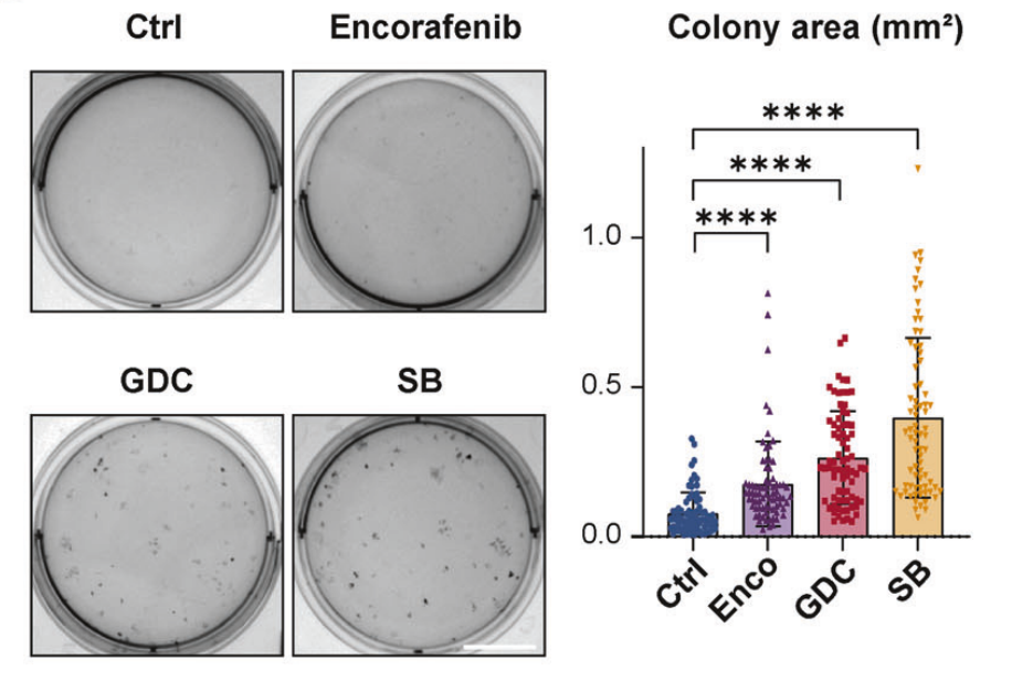
SB-590885 purchased from MedChemExpress. Usage Cited in: Signal Transduct Target Ther. 2024 Dec 2;9(1):338. [Abstract]
Statistical analysis of the area of 75 individual erythroid colonies in panel on Day 14.All experiments used control (DMSO), SB-590885 (HY-10966) at 1 μM, GDC-0879 (HY-50864) at 2 μM, and Encorafenib (HY-15605) at 0.5 μM.
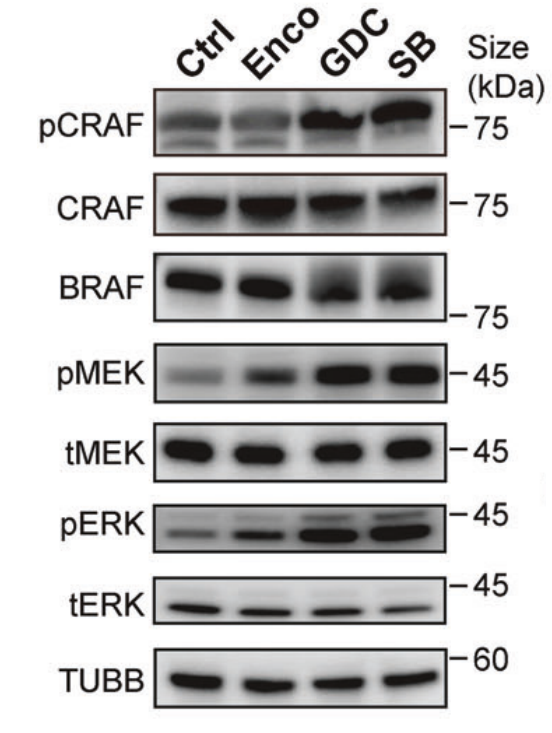
SB-590885 purchased from MedChemExpress. Usage Cited in: Signal Transduct Target Ther. 2024 Dec 2;9(1):338. [Abstract]
Immunoblotting of MAPK signaling cascade proteins in UCB-CD34+-derived erythroblasts cultured under normal conditions and treated on Day 9 with Encorafenib (HY-15605; 0.5 μM), GDC-0879 (HY-50864; 2 μM), or SB-590885 (HY-10966; 0.5 μM) for 30 min.
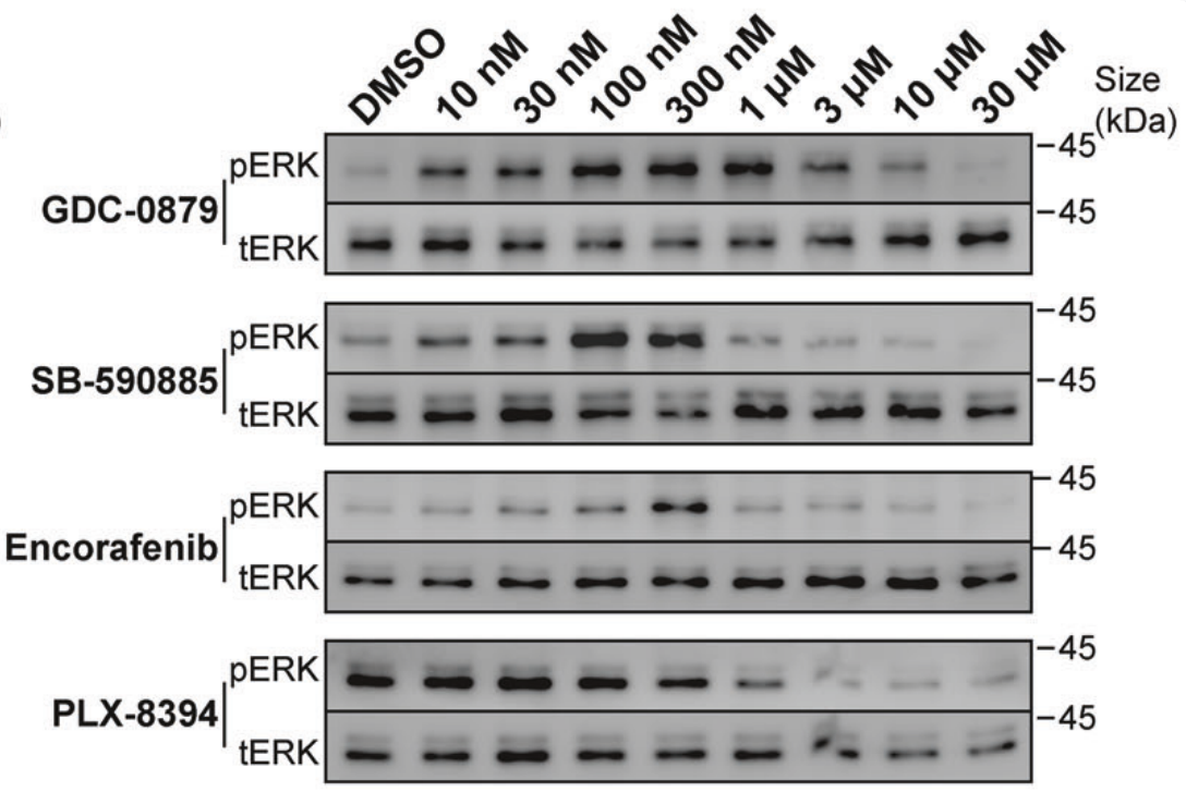
SB-590885 purchased from MedChemExpress. Usage Cited in: Signal Transduct Target Ther. 2024 Dec 2;9(1):338. [Abstract]
Levels of phosphorylated and total ERK proteins in UCB-CD34+-derived erythroblasts on Day 9, cultured under normal conditions and treated with different BRAF inhibitors (SB-590885 (HY-10966), GDC-0879 (HY-50864), Encorafenib (HY-15605)) for 30 min.
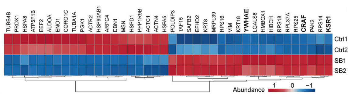
SB-590885 purchased from MedChemExpress. Usage Cited in: Signal Transduct Target Ther. 2024 Dec 2;9(1):338. [Abstract]
Heatmap of the top 20 proteins that were most significantly upregulated and downregulated respectively by 3 × Flag-BRAF interaction proteins in the control group (DMSO) and the 30-minute 1 μM SB (SB-590885)-treated group, identified through flag-affinity immunoprecipitation-mass spectrometry (IP-MS) of 3 × Flag-BRAF in BRAF-overexpressing K562 cells. The components of the RAF protein dimer complex are bolded. cutoff, p < 0.05. g Volcano plot of 3 × Flag-BRAF interacting proteins in IP-MS of SB-treated and control groups in K562 cells. cutoff: p-adj < 0.05, foldchange > 2. The components of the RAF protein dimer complex are bolded.
-
Cell Stem Cell
An inducible model of human post-implantation development derived from primed and naive stem cells. [Abstract]2025 Oct 2;32(10):1509-1527.e9. PMID: 40885193 -
Oncogene
RAF1 facilitates KIT signaling and serves as a potential treatment target for gastrointestinal stromal tumor. [Abstract]2024 Jun;43(27):2078-2091. PMID: 38760447 -
Antioxidants (Basel)
A Novel SIRT1 Activator Hydroxygenkwanin Alleviates Osteoporosis by Inhibiting Ferroptosis and Lactylation in Skeletal Stem/Progenitor Cells. [Abstract]2026 May 12;15(5):612. PMID: 42193234 -
Mol Syst Biol
Systematic analysis of BRAF(V600E) melanomas reveals a role for JNK/c-Jun pathway in adaptive resistance to drug-induced apoptosis. [Abstract]2015 Mar 26;11(3):797. PMID: 25814555 -
Cancers (Basel)
Dual Targeting of BRAF and mTOR Signaling in Melanoma Cells with Pyridinyl Imidazole Compounds. [Abstract]2020 Jun 10;12(6):1516. PMID: 32531927 -
-
bioRxiv
A structure-based modelling approach identifies effective drug combinations for RAS-mutant acute myeloid leukemia. [Abstract]2025 May 3:2025.04.29.651188. PMID: 40654850 -
ACS Comb Sci
Benzimidazolyl-pyrazolo[3,4- b]pyridinones, Selective Inhibitors of MOLT-4 Leukemia Cell Growth and Sea Urchin Embryo Spiculogenesis: Target Quest. [Abstract]2019 Dec 9;21(12):805-816. PMID: 31689077


Lösungsmittel & Löslichkeit
In Vitro:
DMSO : 10 mg/mL (22.05 mM; ultrasonic and warming and heat to 60°C; Hygroscopic DMSO has a significant impact on the solubility of product, please use newly opened DMSO)
Please refer to the solubility information to select the appropriate solvent. Once prepared, please aliquot and store the solution to prevent product inactivation from repeated freeze-thaw cycles.
Storage method and period of stock solution: -80°C, 2 years; -20°C, 1 year. When stored at -80°C, please use it within 2 years. When stored at -20°C, please use it within 1 year.
Please refer to the solubility information to select the appropriate solvent. Once prepared, please aliquot and store the solution to prevent product inactivation from repeated freeze-thaw cycles.
Storage method and period of stock solution: -80°C, 2 years; -20°C, 1 year. When stored at -80°C, please use it within 2 years. When stored at -20°C, please use it within 1 year.
Konzentration (Stammlösung) × Volumen (Stammlösung) = Konzentration (Ziellösung) × Volumen (Ziellösung)
In Vivo:
Select the appropriate dissolution method based on your experimental animal and administration route.
- For the following dissolution methods, please ensure to first prepare a clear stock solution using an In Vitro approach and then sequentially add co-solvents:
- To ensure reliable experimental results, the clarified stock solution can be appropriately stored based on storage conditions. As for the working solution for In Vivo experiments, it is recommended to prepare freshly and use it on the same day.
- The percentages shown for the solvents indicate their volumetric ratio in the final prepared solution. If precipitation or phase separation occurs during preparation, heat and/or sonication can be used to aid dissolution.
Add each solvent one by one: 10% DMSO 40% PEG300 5% Tween-80 45% Saline
Solubility: ≥ 2.5 mg/mL (5.51 mM); Clear solution
This protocol yields a clear solution of ≥ 2.5 mg/mL (saturation unknown).
Taking 1 mL working solution as an example, add 100 μL DMSO stock solution (25.0 mg/mL) to 400 μL PEG300, and mix evenly; then add 50 μL Tween-80 and mix evenly; then add 450 μL Saline to adjust the volume to 1 mL.
Preparation of Saline: Dissolve 0.9 g sodium chloride in ddH₂O and dilute to 100 mL to obtain a clear Saline solution.
Add each solvent one by one: 10% DMSO 90% (20% SBE-β-CD in Saline)
Solubility: ≥ 2.5 mg/mL (5.51 mM); Clear solution
This protocol yields a clear solution of ≥ 2.5 mg/mL (saturation unknown).
Taking 1 mL working solution as an example, add 100 μL DMSO stock solution (25.0 mg/mL) to 900 μL 20% SBE-β-CD in Saline, and mix evenly.
Preparation of 20% SBE-β-CD in Saline (4°C, storage for one week): 2 g SBE-β-CD powder is dissolved in 10 mL Saline, completely dissolve until clear.
In Vivo Dissolution Calculator
Please enter the basic information of animal experiments:
-
-
-
-
Recommended: Prepare an additional quantity of animals to account for potential losses during experiments.
Please enter your animal formula composition:
-
%DMSO +
Recommended: Keep the proportion of DMSO in working solution below 2% if your animal is weak.
-
%+
-
+%Tween-80 + +
-
%Saline +
The co-solvents required include: DMSO, . All of co-solvents are available by MedChemExpress (MCE). , Tween 80. All of co-solvents are available by MedChemExpress (MCE).
Working solution concentration: 0.22 mg/mL
Method for preparing stock solution: mg drug dissolved in μL DMSO. Stock solution concentration: mg/mL.
1. Take μL DMSO stock solution;
2. Add μL .
μL , mix evenly;
3. Then add μL Tween 80, mix evenly;
4. Then add μL
Please ensure that the stock solution in the first step is dissolved to a clear state, and add co-solvents in sequence. You can use ultrasonic heating (ultrasonic cleaner, recommended frequency 20-40 kHz), vortexing, etc. to assist dissolution.


Reinheit & Dokumentation
-
Data Sheet (293 KB)
-
SDS (396 KB)
- English - EN (396 KB)
- Français - FR (396 KB)
- Deutsch - DE (396 KB)
- Norwegian - NO (396 KB)
- Español - ES (396 KB)
- Swedish - SV (396 KB)
- Italian - IT (396 KB)
- Korean - KR (396 KB)
- Portuguese - PT (396 KB)
-
Handling Instructions (2659 KB)
Verweise
[1]. Wu S, et al. BRAF inhibitors enhance erythropoiesis and treat anemia through paradoxical activation of MAPK signaling. Signal Transduct Target Ther. 2024;9(1):338. Published 2024 Dec 2. [Content Brief]
[2]. King AJ, et al. Demonstration of a genetic therapeutic index for tumors expressing oncogenic BRAF by the kinase inhibitor SB-590885. Cancer Res. 2006;66(23):11100-11105. [Content Brief]
[3]. Chen Z, et al. Pan-Cancer Analysis of the TRP Family, Especially TRPV4 and TRPC4, and Its Expression Correlated with Prognosis, Tumor Microenvironment, and Treatment Sensitivity. Biomolecules. 2023;13(2):282. Published 2023 Feb 2. [Content Brief]
[4]. Ruan D, et al. Establishment of human expanded potential stem cell lines via preimplantation embryo cultivation and somatic cell reprogramming. Nat Protoc. 2025;20(10):2698-2734. [Content Brief]


Complete Stock Solution Preparation Table
Please refer to the solubility information to select the appropriate solvent. Once prepared, please aliquot and store the solution to prevent product inactivation from repeated freeze-thaw cycles.
Storage method and period of stock solution: -80°C, 2 years; -20°C, 1 year. When stored at -80°C, please use it within 2 years. When stored at -20°C, please use it within 1 year.
| Optional Solvent | Concentration Solvent Mass | 1 mg | 5 mg | 10 mg | 25 mg |
|---|---|---|---|---|---|
| DMSO | 1 mM | 2.2049 mL | 11.0244 mL | 22.0488 mL | 55.1219 mL |
| 5 mM | 0.4410 mL | 2.2049 mL | 4.4098 mL | 11.0244 mL | |
| 10 mM | 0.2205 mL | 1.1024 mL | 2.2049 mL | 5.5122 mL | |
| 15 mM | 0.1470 mL | 0.7350 mL | 1.4699 mL | 3.6748 mL | |
| 20 mM | 0.1102 mL | 0.5512 mL | 1.1024 mL | 2.7561 mL |


SB-590885 Related Classifications
HY-10966 Related Classifications
 Powered by Bioz