



Bempikibart

Based on 1 Customer Validation
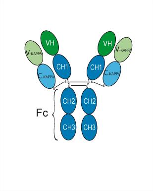
Bempikibart (ADX-914) is a fully human IgG1-κ monoclonal antibody targeting IL-7Rα. Bempikibart binds to the IL-7 receptor alpha and blocks IL-7 and TSLP signaling. Bempikibart reduces T cell populations while relatively preserving regulatory T cells. Bempikibart inhibits Th1, Th2, and Th17 immune pathways. Bempikibart can be used in research on alopecia areata and atopic dermatitis.
For research use only. We do not sell to patients.
- Purity : 98.00%
- CAS No.: 2622254-57-1
- Molecular Weight:145.745 kDa
-
Storage:
Please store the product under the recommended conditions in the Certificate of Analysis.


To place orders, for customer services and technical support, please contact: MedChemExpress USA
Tel: 609-228-6898 E-mail: [email protected] [email protected]


-
 Biological Activity
-
Chemical Information
- Protocols
- Purity & Documentation
- References
-
 Help & FAQs









Biological Activity
Description
Isotype
Human IgG1 kappa
Recommend Isotype Controls
Species Reactivity
Human
|
IL7R |
TSLP |
Th1 |
Th2 |
Th17 |
In Vitro
Bempikibart reduces T cell populations while relatively preserving regulatory T cells[1].
Bempikibart inhibits Th1, Th2, and Th17 immune pathways[2].
MedChemExpress (MCE) has not independently confirmed the accuracy of these methods. They are for reference only. Further protocols information, click here.
Clinical Trial
| NCT Number | Sponsor | Condition | Start Date |
Phase

|
|---|---|---|---|---|
| NCT01329991 | Plexxikon| | 2011-05 | PHASE1 |
Gene ID
Accession
Conjugated
Unconjugated
Reconsititution
The product can be reconstituted/diluted with sterile PBS or saline.
Format
-
Human IgG1 kappa
Application
ELISA, FACS, Functional assay
Verified Bioactivity
-
 Flow Cytometry analysis of K562 cells labelling IL-7Ra/CD127 (red) with Bempikibart (anti-IL-7Ra/CD127) (HY-P990023). Goat Anti-Human IgG (Alexa Fluor 488) (HY-P83776) at a dilution of 1/1000 was used as the secondary antibody. Blue-Human IgG1 kappa (HY-P99001). Black-Unlabelled control, cells without incubation with primary antibody.


Chemical Information
-
CAS No. 2622254-57-1
-
Appearance Liquid
-
Molecular Weight 145.745 kDa
-
Color Colorless to light yellow
-
SMILES
[Bempikibart]
-
Synonyms
ADX-914
-
Shipping
Shipping with dry ice.
-
Formulation
Please refer to the lot-specific COA for specific buffer information.
-
Storage
Please store the product under the recommended conditions in the Certificate of Analysis.

Protocols
-
Naïve CD4+ T-cell subset differentiation/polarization
Naïve CD4+ T-cell subset differentiation/polarization is an in vitro assay in which purified naïve CD4+ T cells are activated through TCR and CD28 costimulation and cultured with defined cytokines and neutralizing antibodies to generate Th0, Th1, Th2, Th17, or induced Treg-like populations. Differentiation is detected by subset-associated cytokines and transcription factors: IFN-γ/T-bet for Th1, IL-4/GATA3 for Th2, IL-17A/RORγt for Th17, and Foxp3 for induced Treg cells. The assay readout is usually generated by intracellular cytokine staining after restimulation, transcription-factor staining by flow cytometry, ELISA of secreted cytokines, or gene-expression analysis. The result reflects cytokine-directed lineage commitment or polarization rather than antigen-specific immune protection by itself.
-
TPA/Croton Oil Ear Edema and Dermatitis
The TPA (12-O-tetradecanoylphorbol-13-acetate) and croton oil-induced mouse ear edema model is a well-established acute cutaneous inflammation system used to evaluate topical anti-inflammatory activity by measuring edema formation, neutrophil infiltration, vascular permeability, and cytokine-mediated skin responses in vivo. The inflammatory response is triggered by topical application of phorbol esters (TPA) or croton oil constituents, leading to rapid activation of protein kinase C signaling, leukocyte recruitment, and increased vascular permeability, which can be quantified by ear thickness, weight, dye extravasation, and biochemical markers such as myeloperoxidase (MPO) activity and pro-inflammatory mediators in ear tissue homogenates. This model is widely used for screening anti-inflammatory agents, where reductions in edema and inflammatory biomarkers reflect suppression of acute dermal inflammation and immune cell infiltration. Histological evaluation typically confirms epidermal
-
Research Protocol for Cancer Immunology
Cancer immunology studies how the immune system recognizes, suppresses, edits, or fails to eliminate malignant cells through tumor antigen release, antigen presentation, T-cell priming, immune trafficking, tumor-cell killing, and feedback inhibition in the tumor microenvironment. The cancer-immunity cycle links tumor antigenicity, dendritic-cell priming, CD8+ T-cell infiltration, cytotoxic function, and immune-checkpoint regulation to tumor rejection or immune escape. Immune-checkpoint pathways such as PD-1/PD-L1 and CTLA-4 suppress antitumor T-cell activity and can be therapeutically blocked, but many tumors remain resistant because of poor antigen presentation, weak T-cell infiltration, suppressive myeloid cells, regulatory T cells, and tumor-intrinsic immune-exclusion programs. Unresolved questions include which immune-cell states predict response, how tumor-intrinsic pathways exclude immune cells, how myeloid suppression limits checkpoint blockade, and which combination strategies


Purity & Documentation
-
Data Sheet (272 KB)
-
SDS (251 KB)
- English - EN (251 KB)
- Français - FR (251 KB)
- Deutsch - DE (251 KB)
- Norwegian - NO (251 KB)
- Español - ES (251 KB)
- Swedish - SV (251 KB)
- Italian - IT (251 KB)
- Korean - KR (251 KB)
- Portuguese - PT (251 KB)
-
Inhibitory Antibodies User Guide (603 KB)
References

Calculators
Concentration (start) × Volume (start) = Concentration (final) × Volume (final)


Bempikibart Related Classifications
HY-P990023 Related Classifications
 Powered by Bioz