



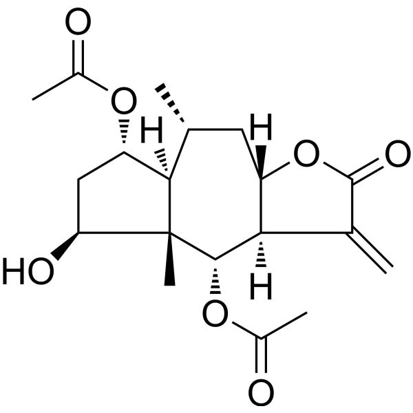
Britannin
Based on 1 publication(s) in Google Scholar
Britannin is an NLRP3 inhibitor with an IC50 of 3.630 μM, exhibiting anti-inflammatory activity. Britannin inhibits the activation and assembly of the NLRP3 inflammasome by blocking the interaction between NLRP3 and NEK7. Additionally, Britannin demonstrates antitumor activity by inhibiting the proliferation of tumor cells through blocking the interaction between HIF-1α and Myc, thereby suppressing PD-L1 expression and enhancing cytotoxic T lymphocyte activity. Britannin can also induce apoptosis and autophagy in liver cancer cells by activating ROS-regulated AMPK. Britannin holds promise for research in the fields of anti-inflammatory and antitumor therapeutics.
For research use only. We do not sell to patients.
- Purity : 99.93%
- CAS No.: 33627-28-0
- Formula: C19H26O7
- Molecular Weight:366.41
-
Storage:
4°C, protect from light
* In solvent : -80°C, 6 months; -20°C, 1 month (protect from light)

To place orders, for customer services and technical support, please contact: MedChemExpress USA
Tel: 609-228-6898 E-mail: [email protected] [email protected]


-
 Biological Activity
-
Chemical Information
-
Solvent & Solubility
- Protocols
- Purity & Documentation
- References
-
 Help & FAQs


-
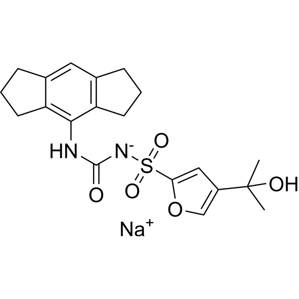
HY-12815A
MCC950 sodiumNLRP3 Inhibitor -
HY-N0633
MusconeTNF Receptor Inhibitor -
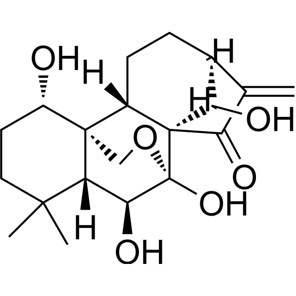
HY-N0004
OridoninBacterial Inhibitor -
HY-10074
TPCA-1IKK-2 Inhibitor -
HY-18639
Nodinitib-1NOD-like Receptor (NLR) Inhibitor -
HY-N0139
TroxerutinNOD-like Receptor (NLR) Inhibitor -
HY-16438
RRx-001Radiosensitizer/CD47 and SIRP-α Inhibitor -
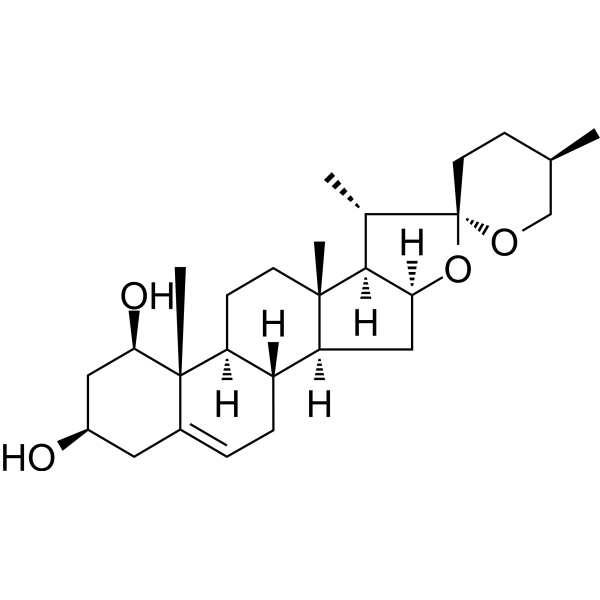
HY-N0496
RuscogeninNOD-like Receptor (NLR) Inhibitor -
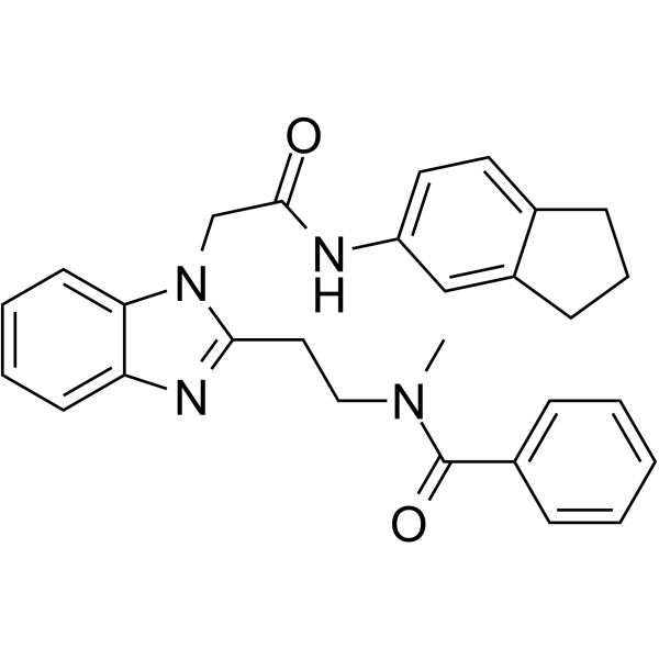
HY-136555
GSK717NOD2 Inhibitor -
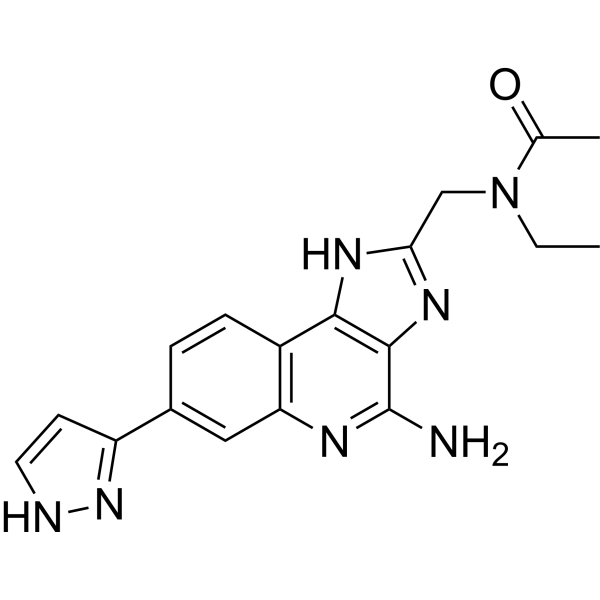
HY-139396
BMS-986299NLRP3 Agonist -
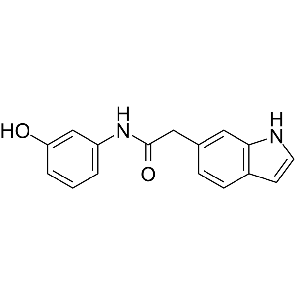
HY-144226
NLRP3/AIM2-IN-3NLRP3/AIM2 Inhibitor -
HY-100691
NOD-IN-1NLR Inhibitor
Publications Citing Use of MedChemExpress (MCE) Britannin
More Customer Validation & Images

-
Cell Proliferation/Viability Assay

-
IF

-
Cell Migration/Invasion Assay

-
WB

-
Cell Imaging/Staining


Biological Activity
Description
IC50 & Target
|
NLRP3 3.630 μM (IC50) |
Cellular Effect
|
Cell Line

|
Type | Value | Description | References |
|---|---|---|---|---|
| RAW264.7 | IC50 |
0.11 μM
Compound: 6
|
Inhibition of LPS-induced NO production in mouse RAW264.7 cells measured after 24 hrs
Inhibition of LPS-induced NO production in mouse RAW264.7 cells measured after 24 hrs
|
[PMID: 20515062] |
In Vitro
Britannin (1, 5, 10 μM, 30 min) inhibits NLRP3-mediated pyroptosis in mouse macrophages[4].
Britannin (1, 5, 10 μM, 30 min) inhibits activation of the NLRP3 inflammasome in an NF-κB-independent manner[4].
MedChemExpress (MCE) has not independently confirmed the accuracy of these methods. They are for reference only. Further protocols information, click here.
-
Cell Line:Bone marrow-derived macrophages(BMDMs)
-
Concentration:1, 5, 10 μM
-
Incubation Time:30 min
-
Result:Dose-dependently inhibited secretion of the cleaved Caspase-1 and the mature IL-1β, and suppressed NLRP3-mediated pyroptosis in murine macrophages.
In Vivo
MedChemExpress (MCE) has not independently confirmed the accuracy of these methods. They are for reference only.
-
Animal Model:Monosodium urate (HY-B2130A) -induced gouty arthritis mouse model, Lipopolysaccharides (LPS) (HY-D1056) -induced acute lung injury mouse model[4]
-
Dosage:20 mg/kg
-
Administration:Intraperitoneal injection (i.p.), inject for three consecutive days (acute lung injury mouse model) or once every other day, for a total of 3 injections (Monosodium urate (HY-B2130A) -induced gouty arthritis mouse model)
-
Result:Significantly suppressed the changes in footpad joint size, alleviated the migration of inflammatory cells in Monosodium urate (HY-B2130A) -induced gouty arthritis mouse model and nhibited the secretion of IL-1β and the activation of Caspase-1, demonstrating that Britannin prevented gouty arthritis through anti-inflammatory effects.
Suppressed pulmonary edema, breakdown of the pneumatic blood barrier, and cell infiltration in lung tissues and pathological injury and inflammatory cell infiltration in lung tissues, were ameliorated by Britannin treatment.


Chemical Information
-
CAS No. 33627-28-0
-
Appearance Solid
-
Molecular Weight 366.41
-
Formula C19H26O7
-
Color White to off-white
-
SMILES
C[C@]12[C@@]([C@@H](C[C@@](O3)([H])[C@](C(C3=O)=C)([H])[C@@H]2OC(C)=O)C)([H])[C@@H](OC(C)=O)C[C@@H]1O
-
Structure Classification
-
Shipping
Room temperature in continental US; may vary elsewhere.
-
Storage
4°C, protect from light
* In solvent : -80°C, 6 months; -20°C, 1 month (protect from light)


Publications (1)
-
Journal Impact Factor
-
Most Recent
-
Exp Ther Med
Britannin inhibits cell proliferation, migration and glycolysis by downregulating KLF5 in lung cancer. [Abstract]2024 Jan 22;27(3):109. PMID: 38361511
Britannin purchased from MedChemExpress. Usage Cited in: Exp Ther Med. 2024 Jan 22;27(3):109. [Abstract]
Cell counting kit-8 assays were used to detect (B) viability treated with Britannin (5, 10, 20 μM).
Britannin purchased from MedChemExpress. Usage Cited in: Exp Ther Med. 2024 Jan 22;27(3):109. [Abstract]
Proliferation of Britannin (5, 10, 20 μM)-treated cells was measured using EdU staining.
Britannin purchased from MedChemExpress. Usage Cited in: Exp Ther Med. 2024 Jan 22;27(3):109. [Abstract]
Migratory abilities of Britannin (5, 10, 20 μM)-treated A549 cells were detected by wound healing assays and Transwell assays.
Britannin purchased from MedChemExpress. Usage Cited in: Exp Ther Med. 2024 Jan 22;27(3):109. [Abstract]
Protein expression levels of E-cadherin, N-cadherin and Snail were detected by western blotting treated with Britannin (5, 10, 20 μM).
Britannin purchased from MedChemExpress. Usage Cited in: Exp Ther Med. 2024 Jan 22;27(3):109. [Abstract]
Proliferation of Britannin (5, 10, 20 μM)-treated cells was measured using colony formation assays.


Solvent & Solubility
In Vitro:
DMSO : 250 mg/mL (682.30 mM; Need ultrasonic; Hygroscopic DMSO has a significant impact on the solubility of product, please use newly opened DMSO)
Please refer to the solubility information to select the appropriate solvent. Once prepared, please aliquot and store the solution to prevent product inactivation from repeated freeze-thaw cycles.
Storage method and period of stock solution: -80°C, 6 months; -20°C, 1 month (protect from light). When stored at -80°C, please use it within 6 months. When stored at -20°C, please use it within 1 month.
Please refer to the solubility information to select the appropriate solvent. Once prepared, please aliquot and store the solution to prevent product inactivation from repeated freeze-thaw cycles.
Storage method and period of stock solution: -80°C, 6 months; -20°C, 1 month (protect from light). When stored at -80°C, please use it within 6 months. When stored at -20°C, please use it within 1 month.
Concentration (start) × Volume (start) = Concentration (final) × Volume (final)
In Vivo:
Select the appropriate dissolution method based on your experimental animal and administration route.
- For the following dissolution methods, please ensure to first prepare a clear stock solution using an In Vitro approach and then sequentially add co-solvents:
- To ensure reliable experimental results, the clarified stock solution can be appropriately stored based on storage conditions. As for the working solution for In Vivo experiments, it is recommended to prepare freshly and use it on the same day.
- The percentages shown for the solvents indicate their volumetric ratio in the final prepared solution. If precipitation or phase separation occurs during preparation, heat and/or sonication can be used to aid dissolution.
Add each solvent one by one: 10% DMSO 40% PEG300 5% Tween-80 45% Saline
Solubility: ≥ 2.08 mg/mL (5.68 mM); Clear solution
This protocol yields a clear solution of ≥ 2.08 mg/mL (saturation unknown).
Taking 1 mL working solution as an example, add 100 μL DMSO stock solution (20.8 mg/mL) to 400 μL PEG300, and mix evenly; then add 50 μL Tween-80 and mix evenly; then add 450 μL Saline to adjust the volume to 1 mL.
Preparation of Saline: Dissolve 0.9 g sodium chloride in ddH₂O and dilute to 100 mL to obtain a clear Saline solution.
Add each solvent one by one: 10% DMSO 90% (20% SBE-β-CD in Saline)
Solubility: ≥ 2.08 mg/mL (5.68 mM); Clear solution
This protocol yields a clear solution of ≥ 2.08 mg/mL (saturation unknown).
Taking 1 mL working solution as an example, add 100 μL DMSO stock solution (20.8 mg/mL) to 900 μL 20% SBE-β-CD in Saline, and mix evenly.
Preparation of 20% SBE-β-CD in Saline (4°C, storage for one week): 2 g SBE-β-CD powder is dissolved in 10 mL Saline, completely dissolve until clear.
In Vivo Dissolution Calculator
Please enter the basic information of animal experiments:
-
-
-
-
Recommended: Prepare an additional quantity of animals to account for potential losses during experiments.
Please enter your animal formula composition:
-
%DMSO +
Recommended: Keep the proportion of DMSO in working solution below 2% if your animal is weak.
-
%+
-
+%Tween-80 + +
-
%Saline +
The co-solvents required include: DMSO, . All of co-solvents are available by MedChemExpress (MCE). , Tween 80. All of co-solvents are available by MedChemExpress (MCE).
Working solution concentration: 0.22 mg/mL
Method for preparing stock solution: mg drug dissolved in μL DMSO. Stock solution concentration: mg/mL. * In solvent : -80°C, 6 months; -20°C, 1 month (protect from light)
1. Take μL DMSO stock solution;
2. Add μL .
μL , mix evenly;
3. Then add μL Tween 80, mix evenly;
4. Then add μL
Please ensure that the stock solution in the first step is dissolved to a clear state, and add co-solvents in sequence. You can use ultrasonic heating (ultrasonic cleaner, recommended frequency 20-40 kHz), vortexing, etc. to assist dissolution.

Protocols
-
RNA extraction experimental
By lysing cells, releasing RNA, and removing impurities such as proteins and DNA, high-purity RNA products are finally obtained. The commonly used traditional method is the guanidine isothiocyanate/phenol/chloroform method (Trizol), which is suitable for a variety of animal materials including animal tissues, microorganisms, cultured cells, etc., and most plant materials.
-
Annexin V plus membrane-impermeant dye apoptosis staining
Annexin V-based apoptosis assays rely on the detection of phosphatidylserine (PS) externalization from the inner leaflet of the plasma membrane to the outer leaflet, an early biochemical hallmark of apoptosis. Fluorescently labeled Annexin V binds PS in a calcium-dependent manner, enabling identification of early apoptotic cells by flow cytometry or fluorescence microscopy. When combined with a membrane-impermeant DNA-binding dye (e. g. , propidium iodide), this approach allows discrimination between viable (Annexin V−/dye−), early apoptotic (Annexin V+/dye−), and late apoptotic or necrotic (Annexin V+/dye+) cell populations by assessing membrane integrity and PS exposure.
-
ROS/oxidative-stress fluorescent staining
ROS/oxidative-stress fluorescent staining uses cell-permeant fluorogenic probes that become fluorescent after oxidation inside cells or tissues; commonly used examples include DCFH-DA/DCFDA for broad cellular oxidant detection, DHE for superoxide-related signal detection, MitoSOX for mitochondrial superoxide-related signal detection, and CellROX probes for oxidative-stress-associated fluorescence readouts. The assay detects probe oxidation rather than a single ROS species unless the probe and analysis method have been chemically validated for that species. DCFH-DA enters cells, is deacetylated by intracellular esterases to DCFH, and produces fluorescent DCF after oxidation, so the readout is used as an operational measure of total cellular oxidative stress rather than a species-specific ROS measurement. DHE and MitoSOX can report superoxide-related oxidation, but red fluorescence alone can include non-specific ethidium-like oxidation products; HPLC or optimized spectral approaches are
-
Research Protocol for Inflammation-related Diseases
The NLRP3 inflammasome is a cytosolic innate immune signaling platform that integrates priming signals and danger-signal activation to promote caspase-1 activation, maturation of IL-1β and IL-18, and gasdermin D-mediated pyroptotic cell death. The core experimental logic is to determine whether inflammatory disease phenotypes are driven by increased NLRP3 expression, ASC-containing inflammasome assembly, caspase-1 cleavage, GSDMD cleavage, and extracellular release of IL-1β/IL-18 rather than by nonspecific cell injury alone. The pathway is strongly linked to inflammation-related disease phenotypes because monosodium urate crystals activate NALP3/NLRP3 inflammasome signaling in gout-like crystal inflammation, cholesterol crystals activate NLRP3 inflammasomes in atherogenesis models, and DSS-induced intestinal inflammation has been reported to involve NLRP3 inflammasome activity. However, experimental colitis studies also show context-dependent protective effects of NLRP3 inflammasome co
-
Liver Cancer Modeling
Liver cancer can be classified into primary liver cancer and secondary liver cancer. Secondary liver cancer is the metastatic liver cancer. Primary liver cancer includes hepatocellular carcinoma (HCC), intrahepatic cholangiocarcinoma (ICC) and fibrolamellar HCC, of which HCC is the most common form, accounting for approximately 90% of primary liver cancers[1]. HCC mouse models include chemical agent-induced models, transplanted tumor models, and genetic engineered models.
-
Cell Viability Determination by MTT Colorimetric Assay
The following protocol uses the MTT colorimetric assay as a classic literature-established method for assessing cell viability/metabolic activity in cultured mammalian cells. MTT[3-(4,5-dimethylthiazol-2-yl)-2,5-diphenyltetrazolium bromide] is reduced by metabolically active cells to a colored formazan product; the amount of formazan is quantified spectrophotometrically and provides an indirect measure of metabolically active viable cells. Importantly, MTT reduction reflects cellular oxidoreductase/metabolic activity rather than an absolute direct count of living cells, so changes in cellular metabolism can alter the signal independently of cell number.
-
Apoptosis
Apoptosis, also called programmed cell death, is generally characterized by distinct morphological characteristics.
-
TUNEL staining for apoptotic DNA fragmentation
TUNEL staining detects DNA strand breaks by using terminal deoxynucleotidyl transferase to add labeled nucleotides to exposed 3′-OH DNA termini, generating either microscopic staining in fixed cells or tissue sections, or fluorescence/cytometric signal in cell suspensions. TUNEL positivity reflects DNA fragmentation but should not be interpreted alone as definitive apoptosis, because TUNEL can also label necrotic, autolytic, mechanically damaged, or DNA-repair-associated DNA breaks.
-
Cell Cytotoxicity Assay
Cytotoxicity assays are usually based on the assessment of cell membrane damage, which can also be indirectly detected by measuring cell viability. Detection methods include MTT assay, CKK-8 assay, LDH assay and ATP assay, etc.
-
Cotton Pellet Granuloma
Cotton pellet granuloma is a classical in vivo chronic inflammation model used to evaluate the anti-inflammatory potential of test substances by measuring their ability to inhibit granuloma tissue formation around an implanted foreign body (cotton pellet) in rodents. The method is based on the biological response to a sterile implanted material, which induces proliferative phase inflammation characterized by fibroblast proliferation and collagen-rich granuloma formation, and the final readout reflects the extent of chronic inflammatory tissue growth surrounding the pellet. In multiple preclinical pharmacological evaluations, inhibition of cotton pellet-induced granuloma formation has been used as an indicator of anti-inflammatory activity in both synthetic and natural product screening contexts.
-
Carrageenan-Induced Paw Edema
Carrageenan-induced paw edema is an acute inflammation model in which intraplantar injection of carrageenan induces localized inflammatory swelling characterized by vascular permeability, leukocyte infiltration, and production of inflammatory mediators such as prostaglandins and cytokines, making it widely used to evaluate anti-inflammatory agents in vivo. The resulting paw volume or thickness increase is quantified over time as a direct readout of inflammatory intensity and drug efficacy, typically reflecting cyclooxygenase-mediated prostaglandin-driven edema formation and immune cell recruitment in peripheral tissue[20].
-
Autophagy
Autophagy is a process in which eukaryotic cells use lysosomes to degrade their own cytoplasmic proteins and damaged organelles under the regulation of autophagy related gene (Atg). Microtubule-associated proteins light chain 3 (LC3) is recognized as autophagy marker, which transfers from cytoplasmic LC3 (LC3-I) to membrane type (LC3-II). LC3-II/I ratio could be detected by Western Blot and fluorescence microscopy.
-
Apoptosis Solutions
Apoptosis is a regulated, generally non-lytic cell-death pathway that removes unwanted, damaged, infected, or abnormal cells through coordinated morphological changes, caspase activation, DNA fragmentation, and membrane remodeling. The intrinsic apoptosis pathway is controlled mainly by mitochondrial outer membrane permeabilization, BCL-2 family proteins, cytochrome c release, apoptosome formation, caspase-9 activation, and downstream executioner caspase-3/7 activation. The extrinsic apoptosis pathway is initiated by death receptors such as Fas, TNFR, and TRAIL receptors, which recruit adaptor proteins and activate caspase-8 before engaging executioner caspases or mitochondrial amplification through BID cleavage. Apoptosis is linked to many phenotypes, including cancer cell killing, tissue homeostasis, immune regulation, neurodegeneration, infection response, and treatment-induced cytotoxicity; unresolved questions include how apoptosis interacts with necroptosis, pyroptosis, ferroptos
-
Lysosome and acidic-vesicle live-cell staining
Lysosome and acidic-vesicle live-cell staining detects acidic intracellular compartments by using membrane-permeant acidotropic probes that accumulate in low-pH vesicles, including lysosomes, late endosomes, autolysosomes, and acidic phagosomes. LysoTracker staining is commonly used as an intensity-based readout of acidic lysosomal compartment abundance or enlargement, while acridine orange produces green fluorescence in less concentrated compartments and red fluorescence after concentration-dependent accumulation in acidic vesicular organelles. Loss or reduction of acridine-orange red signal can be used as a readout of lysosomal membrane permeabilization or reduced acidic-vesicle integrity. This protocol is designed for live cultured cells and can be adapted for fluorescence microscopy, high-content imaging, plate-reader readout, or flow cytometry when the selected literature supports the readout. Because these dyes report acidotropic accumulation rather than lysosome identity alone,
-
Subcutaneous Cell-Line-Derived Xenograft
Subcutaneous cell-line-derived xenograft (CDX) models are established by implanting cultured human cancer cell lines into immunodeficient mice, where the injected cells form localized tumors that can be monitored in vivo as a measure of tumorigenic potential, growth kinetics, and treatment response. These models are widely used in oncology research because they allow reproducible tumor formation and enable comparative assessment of tumor growth between different cell lines or genetic manipulations in a controlled in vivo microenvironment. Subcutaneous implantation of cancer cells in immunodeficient mice is a standard approach for evaluating tumor growth behavior and therapeutic response across multiple cancer types, including prostate, esophageal, pancreatic, and colon cancer models.
-
Macroautophagy Solutions
Macroautophagy is a conserved lysosome-dependent degradation pathway in which cytoplasmic material is sequestered into double-membrane autophagosomes and delivered to lysosomes for degradation and recycling. The pathway supports cellular homeostasis during nutrient limitation, organelle stress, protein-aggregate accumulation, infection, differentiation, and tissue remodeling by coupling cargo sequestration, autophagosome maturation, lysosomal fusion, and degradation of cargo-derived macromolecules. The core molecular sequence includes initiation by nutrient- and stress-regulated autophagy machinery, autophagosome nucleation, LC3/ATG8-family conjugation to autophagosomal membranes, cargo selection through receptors such as SQSTM1/p62, autophagosome-lysosome fusion, and lysosomal degradation. LC3 was identified as a mammalian homolog of yeast Atg8 that localizes to autophagosomal membranes after processing, and p62/SQSTM1 was shown to connect ubiquitinated cargo with autophagic degradati
-
Pyroptosis Solutions
Pyroptosis is a lytic inflammatory cell-death pathway executed by gasdermin pores, most classically through inflammasome-mediated activation of caspase-1, cleavage of gasdermin D, membrane pore formation, LDH release, and secretion of IL-1β and IL-18. The canonical pathway is commonly modeled by priming cells with an inflammatory signal such as LPS to induce pro-IL-1β and inflammasome components, followed by an activation signal such as ATP or nigericin to activate NLRP3, ASC speck formation, caspase-1 cleavage, GSDMD cleavage, cytokine release, and pyroptotic membrane rupture. The non-canonical pathway is triggered when cytosolic LPS activates mouse caspase-11 or human caspase-4/5, leading to GSDMD cleavage and pyroptosis, and this can secondarily activate NLRP3-dependent IL-1β release. Pyroptosis is linked to inflammatory injury, infection, cancer, liver disease, ocular disease, placental inflammation, and other disease phenotypes, but unresolved questions include which gasdermin fam


Purity & Documentation
-
Data Sheet (285 KB)
-
SDS (252 KB)
- English - EN (252 KB)
- Français - FR (252 KB)
- Deutsch - DE (252 KB)
- Norwegian - NO (252 KB)
- Español - ES (252 KB)
- Swedish - SV (252 KB)
- Italian - IT (252 KB)
- Korean - KR (252 KB)
- Portuguese - PT (252 KB)
-
Handling Instructions (2659 KB)
References
[1]. Cui YQ The suppressive effects of Britannin (Bri) on human liver cancer through inducing apoptosis and autophagy via AMPK activation regulated by ROS. Biochem Biophys Res Commun. 2018 Mar 11;497(3):916-923. [Content Brief]
[2]. Park HH, et al. Britanin suppresses LPS-induced nitric oxide, PGE2 and cytokine production via NF-κB and MAPK inactivation in RAW 264.7 cells. Int Immunopharmacol. 2013 Feb;15(2):296-302. [Content Brief]
[3]. Moghadam MH, et al. Anti-proliferative activity and apoptotic potential of britannin, a sesquiterpene lactone from Inula aucheriana. Nat Prod Commun. 2012 Aug;7(8):979-80. [Content Brief]
[4]. Shao, Jj., Li, Wf., Sun, Jf. et al. Britannin as a novel NLRP3 inhibitor, suppresses inflammasome activation in macrophages and alleviates NLRP3-related diseases in mice. Acta Pharmacol Sin, 2024, 45, 803–814. [Content Brief]
[5]. Zhang YF, et al. Britannin stabilizes T cell activity and inhibits proliferation and angiogenesis by targeting PD-L1 via abrogation of the crosstalk between Myc and HIF-1α in cancer. Phytomedicine. 2021 Jan;81:153425. [Content Brief]


Complete Stock Solution Preparation Table
Please refer to the solubility information to select the appropriate solvent. Once prepared, please aliquot and store the solution to prevent product inactivation from repeated freeze-thaw cycles.
Storage method and period of stock solution: -80°C, 6 months; -20°C, 1 month (protect from light). When stored at -80°C, please use it within 6 months. When stored at -20°C, please use it within 1 month.
| Optional Solvent | Concentration Solvent Mass | 1 mg | 5 mg | 10 mg | 25 mg |
|---|---|---|---|---|---|
| DMSO | 1 mM | 2.7292 mL | 13.6459 mL | 27.2918 mL | 68.2296 mL |
| 5 mM | 0.5458 mL | 2.7292 mL | 5.4584 mL | 13.6459 mL | |
| 10 mM | 0.2729 mL | 1.3646 mL | 2.7292 mL | 6.8230 mL | |
| 15 mM | 0.1819 mL | 0.9097 mL | 1.8195 mL | 4.5486 mL | |
| 20 mM | 0.1365 mL | 0.6823 mL | 1.3646 mL | 3.4115 mL | |
| 25 mM | 0.1092 mL | 0.5458 mL | 1.0917 mL | 2.7292 mL | |
| 30 mM | 0.0910 mL | 0.4549 mL | 0.9097 mL | 2.2743 mL | |
| 40 mM | 0.0682 mL | 0.3411 mL | 0.6823 mL | 1.7057 mL | |
| 50 mM | 0.0546 mL | 0.2729 mL | 0.5458 mL | 1.3646 mL | |
| 60 mM | 0.0455 mL | 0.2274 mL | 0.4549 mL | 1.1372 mL | |
| 80 mM | 0.0341 mL | 0.1706 mL | 0.3411 mL | 0.8529 mL | |
| 100 mM | 0.0273 mL | 0.1365 mL | 0.2729 mL | 0.6823 mL |


Britannin Related Classifications
HY-N3005 Related Classifications
 Powered by Bioz
Keywords
- Britannin
- 33627-28-0
- Apoptosis
- Autophagy
- NOD-like Receptor (NLR)
- Pyroptosis
- Bone marrow-derived macrophages(BMDMs)
- Monosodium urate (HY-B2130A) -induced gouty arthritis mouse model
- Lipopolysaccharides (LPS) (HY-D1056) -induced acute lung injury mouse model
- Anti-inflammatory activity
- Anti-tumor activity
- Arthritis
- Inhibitor
- inhibitor
- inhibit